♦ Preoperative
Operative Planning
- Review imaging: magnetic resonance imaging, computed tomography (CT), radiographs, or CT myelogram
- Note location within the spinal canal: cervical location most common
- Identify ventral-to-dorsal and rostral-to-caudal extents of the lesion
- These lesions frequently extend laterally through the intervertebral foramen into the extraforaminal region to become dumb bell-shaped tumors
- Consider preoperative fluoroscopic localization of level in thoracic lesions; otherwise, intraoperative radiographs are used to identify the correct level
Equipment
- Basic spine tray
- High-speed drill (Midas Rex with AM-8 bit [Medtronic])
- Operating microscope with bridge
- Somatosensory evoked potential or direct motor evoked potential monitoring may be used in tumors causing severe spinal cord or cauda equina compression
Anesthetic Issues
- Arterial line for blood pressure monitoring
- Intravenous dexamethasone and antibiotic prophylaxis (cefazolin 1 to 2 g) administered preoperatively
- In cases of severe cord compression make sure blood pressure does not fall below baseline during induction to prevent ischemic cord injury
♦ Intraoperative
Positioning
- For posterior approaches, patient prone with pressure points well padded
- Mayfield head fixation in cervical lesions
Posterior Cervical Approach
- For lesions of the cervical cord or cervicothoracic junction
- For lesions of the thoracic cord, cervicothoracic junction, or thoracolumbar junction
Retropleural Thoracotomy, Costotransversectomy, Lateral Extracavitary, and Transthoracic Approaches
- For anteriorly and anterolaterally located thoracic tumors
Posterior Lumbar Approach
- For lesions of the lumbar cord, thoracolumbar junction, or conus
Retroperitoneal Approach
- For anteriorly and anterolaterally located thoracolumbar and lumbar tumors
Tumor Resection (Fig. 130.1)
- Perform bone removal, using one of the described approaches to expose the dura surrounding the tumor
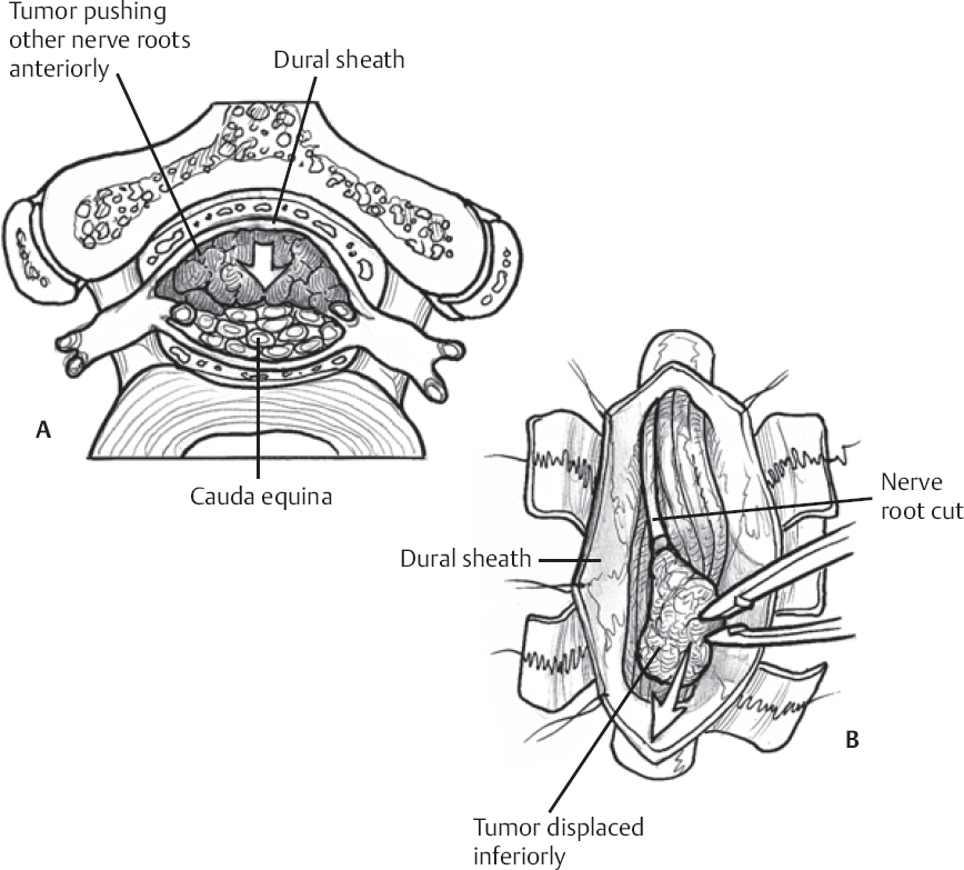
Fig. 130.1 Schematic of intradural nerve sheath tumor (A) exposure, and (B) resection.
Only gold members can continue reading. Log In or Register to continue
Related posts:

Stay updated, free articles. Join our Telegram channel

Full access? Get Clinical Tree


