Fig. 16.1
T1-weighted sagittal cervical MRI shows homogenously contrast-enhanced intramedullary lesion with regular shape at the C2 level (From Nas et al. [10], with permission)
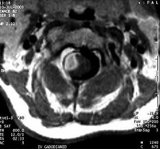
Fig. 16.2
T1-weighted axial MRI scan shows the same lesion which was located on the right side of spinal cord intramedullary and extending to epidural space (From Nas et al. [10], with permission)
Positron emission tomography combined with computed tomography (PET/CT) scan can provide additional information on the spread of brucellar spondylitis. The efficacy of treatment and the need for further antimicrobial chemotherapy could be assessed with PET/CT [8].
16.3 Incidence
Previously reported available cases of intramedullary brucellosis in the English literature are summarized in Table 16.1. As shown in Table 16.1, systemic brucellosis was present in all cases suffering from intramedullary involvement [1, 2, 6, 7, 9–11, 14, 15, 19]. The reported lesions were generally abscesses except three cases [1, 14, 15]. The most common site for involvement was the thoracic and upper cervical spinal cord. Keihani-Douste et al. [9] reported a case of multilevel cervicothoracic spinal cord lesion with concurrent multiple brain abscesses. Isolated cervical intramedullary involvement was reported by Nas et al. [10], Hendam et al. [7], Tufan et al. [15], and Talı et al. [14]. Brucella abortus and B. melitensis were the identified pathogens in all the available cases. Blood serology was positive in all cases except the cases of Cokca et al. [2] and Talı et al. [14].
Table 16.1
Previously reported cases of intramedullary neurobrucellosis
Authors/year | Patient age/sex | Type/location of the lesion | Pus culture | Blood culture | Serology | Treatment | Systemic disease |
|---|---|---|---|---|---|---|---|
Cokca et al. [2], 1994 | 17/M | Abscess/T11–L2 | B abortus | B. abortus | ? | Surgical + medical | Present |
Bingol et al. [1],1999 | 40/F | Granuloma/T5 | Not done | No growth | + | Medical | Present |
Novati et al. [11], 2002 | 24/M | Abscess/T3 | Not done | B. melitensis | + | Medical | Present |
Helvaci et al. [6], 2002 | 15/F | Abscess/T11–T12 | No growth | No growth | + | Surgical + medical | Present |
Vajramani et al. [18], 2005 | 40/F | Abscess/conus medullaris | B. melitensis | B. melitensis | + | Surgical + medical | Present |
Keihani-Douste et al. [9], 2006 | 12/M | Abscess/C1–L4 | Not done | ? | + | Surgical + medical | Present |
Nas et al. [10], 2007 | 45/F | Abscess/ C1-2 | Not done | Not done | + | Medical | Present |
Hendam et al. [7], 2014 | 57/F | Abscess/C4–C5 | Not done | Not done | + | Medical | Present |
Tufan et al. [15], 2014 | 19/M | Granuloma/C2 | Not done | Not done
Related posts:Stay updated, free articles. Join our Telegram channel
Full access? Get Clinical Tree
 Get Clinical Tree app for offline access

|


