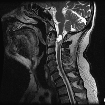
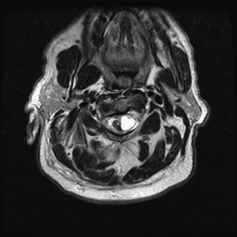
73 A 61-year-old man developed increasing quadriparesis and spasticity. He had a remote history of foreign travel. A T2-weighted cervical magnetic resonance imaging (MRI) is remarkable for a loculated, intramedullary hyperintense lesion that is widening the cord at C2 (Figs. 73-1 and 73-2). FIGURE 73-1 A loculated C2 lesion expands the cord prominently on a cervical MRI. Intramedullary cysticercosis Surgical drainage and excision with microsurgical technique and neurophysiologic monitoring were done. FIGURE 73-2 This hyperintense intramedullary lesion is again visualized on an axial slice through the area of interest. Intramedullary cystic lesions are rare occurrences. They can be related to various pathologies, including neoplastic, infectious, inflammatory, demyelinating, degenerative, vascular, or even radiation-induced causes. Parasites are lesser known causes of intramedullary cystic lesions. Yet, these infections of the central nervous system (cysticercosis and hydatid cysts) are endemic in certain areas of the world. Neurocysticercosis occurs with some frequency in Mexico, Central and South America, Africa, the Far East (India and China), and in California as well. Spinal neurocysticercosis is a rare but debilitating form of the disease. It accounts for less than 1% of reported cases of the disease. Subarachnoid cervical spine neurocysticercosis is the most common site by far and is a likely result of the ventricular to subarachnoid migration of the pathogen. Intramedullary spinal neurocysticercosis is even rarer, and is mostly reported in the thoracic cord. It is postulated to occur there because of the nature of the spinal cord vascular supply. The presentation of the spinal cord disease depends on its location and progression, with paraplegia and quadriplegia being the most common signs. Early on, progressive myelopathy with bowel/bladder dysfunction is common. Brown-Séquard syndrome (hemicord syndrome) has also been reported. Diagnosis is based on clinical, radiographic, and serologic studies, but the disease should be considered in patients with severe progressive neurologic deficit and a history of exposure to endemic areas. Imaging of the entire neuraxis is needed. Treatment of spinal cord cysticercosis often requires surgical intervention to extirpate the lesion or lesions and decompress the cord. The patients then have to take anticysticercosis drug therapy, such as flubendazole, albendazole, and praziquantel. The use of dexamethasone has been advocated in the acute phase to halt the progress of neurologic deficit. Wei GZ, Li CJ, Meng JM, Ding MC. Cysticercosis of the central nervous system: a clinical study of 1400 cases. Chin Med J (Engl) 1988; 101:493–500
Intramedullary Cervical Lesion
Presentation
Radiologic Findings

Diagnosis
Treatment

Discussion
SUGGESTED READING
< div class='tao-gold-member'>
Intramedullary Cervical Lesion
Only gold members can continue reading. Log In or Register to continue

Full access? Get Clinical Tree


