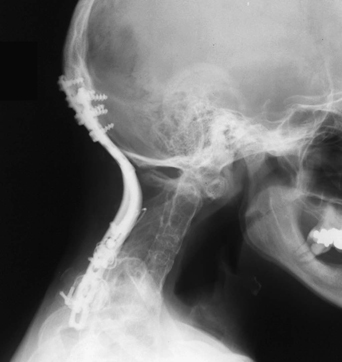
75 A 46-year-old woman with Klippel-Feil syndrome presented with gait instability and dysesthetic arm pain. She had some hand weakness. Magnetic resonance imaging (MRI) of the C-spine was notable for stenosis, mostly at the foramen magnum, and autofusion of the first five cervical vertebral bodies. Cervical x-ray taken postoperatively shows occipitocervical fusion instrumentation and vertebral body findings typical of Klippel-Feil syndrome (Fig. 75-1). Upper cervical stenosis in a patient with Klippel-Feil syndrome The patient had a suboccipital craniectomy and C1 laminectomy with occipitocervical fusion. Klippel-Feil remains a fascinating and multifaceted syndrome. The classic triad is a low posterior hairline, short neck, and limitation of neck motion due to fused cervical vertebrae. Additional findings include possible basilar impression, atlanto-occipital fusion, facial palsy, scoliosis, a raised scapula (Sprengel’s deformity), synkinesis, absence of a kidney, and deafness. The most commonly observed pathology is in the cervical spine. The involved vertebrae are typically wide and flat, with an absent or hypoplastic, immobile disc space. Often, three to five vertebrae are fused together, putting additional stress on the adjacent segments, causing accelerated degeneration. This can cause spinal canal compromise. Patients usually present with neck pain and neurologic compromise. One should also examine the lower cranial nerves and assess for other symptoms of cervicomedullary junction compromise.
Klippel-Feil Syndrome
Presentation
Radiologic Findings
Diagnosis
Treatment
Discussion
Klippel-Feil Syndrome
Only gold members can continue reading. Log In or Register to continue


Full access? Get Clinical Tree


