, Sanjay Ghosh2 and Srinath Samudrala3
(1)
University of Southern California Keck School of Medicine, Los Angeles, CA, USA
(2)
SENTA Clinic, San Diego, CA, USA
(3)
The Spine Institute at Glendale Adventist Medical Center, Glendale, CA, USA
Step 1: Positioning
The patient must be positioned to accommodate the laparoscopic surgeon (LS) at the patient’s head and to the patient’s right, the camera assistant (CA) to the patient’s left, the spine surgeon (SS) at the patient’s feet and to the right, and the anesthesiologist (A). The operating theater must allow for appropriate placement of television monitors, fluoroscopy monitors, and a c-arm [1, 2]. The patient must be adequately secured to the table so that the steep Trendelenburg position may be used to assist with the exposure. The patient’s arms should be folded over the chest to allow fluoroscopic visualization of the spine (Fig. 30.1).
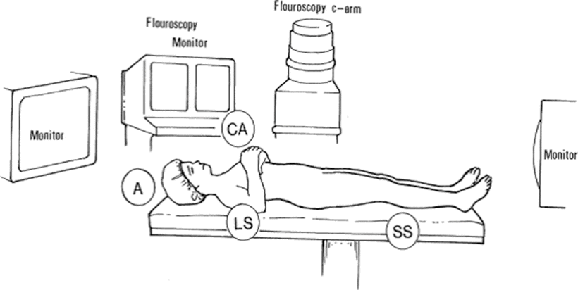
Fig. 30.1
The patient’s arms should be folded over the chest to allow fluoroscopic visualization of the spine
Step 2: Trocar Placement
A Veress needle is placed in the umbilicus (A) and the peritoneal cavity is insufflated to 15 mmHg pressure. A 10-mm trocar is then placed into the umbilicus (A), and the laparoscopic camera is placed into this port (Fig. 30.2). Two other 10-mm trocars are placed at the level of the anterosuperior iliac spines just lateral to the rectus abdominis muscles (B, C). These ports allow the laparoscopic surgeon (S) to perform the dissection with the left hand through port C and the right hand through port B. The procedure will eventually require the placement of a 5-mm port to the left of the camera to assist with retraction of the transverse mesocolon (D). A final 18-mm port is then placed in the suprapubic area to accommodate the spinal instrumentation (E). The position of the ports in the axial plane can be modified to adapt for various levels of disc dissection [3, 4].
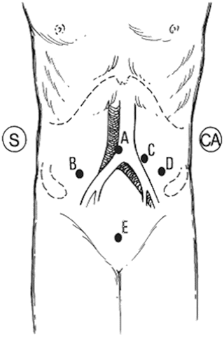
Fig. 30.2
Port positions for trocar placement
Step 3: Exposure of the Pelvic Brim
The patient is placed in steep Trendelenburg to allow the small bowel to fall out of the pelvis and away from the target site. This exposure is difficult if not impossible to achieve if the small bowel is distended. The patient should therefore have a bowel preparation preoperatively, which may involve a full liquid diet 2 days before surgery and a colon preparation the night before surgery.
Step 4: Retraction of the Sigmoid Colon
The sigmoid colon is then elevated with the mesocolon to expose the sacral promontory. Staples can be placed to secure the pericolic fat to the anterolateral abdominal wall to keep the sigmoid colon out of the field. Care must be taken to ensure that the pericolic fat alone is stapled [5].
Step 5: Dissection Through the Peritoneum
The aorta and inferior vena cava can be visualized through the peritoneum. The ureters can also be visualized lateral to the lumbar vertebral bodies overlying the psoas muscles. The ureters are lateral to the disc space and can generally be avoided during the approach to the intervertebral disc space.
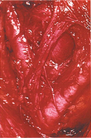
The peritoneum is incised using sharp dissection (Fig. 30.3). At this point, only ligatures and pressure should be used to control bleeding. Monopolar electrocautery in this area may lead to injury of the hypogastric plexus of the sympathetic nerves, causing the complication of retrograde ejaculation in men [6, 7].
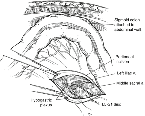
Fig. 30.3
The peritoneum is incised using sharp dissection. Only ligatures and pressure should be used at this point to control bleeding. Electrocautery in this area may lead to injury of the hypogastric plexus
Step 6: Mobilization of the Left llliac Artery and Vein
The aortic bifurcation is identified (Figs. 30.4, 30.5). In the crotch of the bifurcation lies the middle sacral artery. This vessel is ligated with vascular clips and divided, which allows mobilization of the left iliac artery. A vessel loop should be placed around the left iliac artery and gently retracted to the left (Figs. 30.6).