41 A 46-year-old man had sudden-onset low back pain, with a right S1 radiculopathy that progressed over 2 months, despite physical therapy. He has mild dorsiflexion and extensor hallucis longus (EHL) weakness on the right. Magnetic resonance imaging (MRI) of the lumbar spine reveals a large disc extrusion, with sequestered fragment, occupying half the canal at the L5-S1 level (Figs. 41-1 and 41-2).
Large Disc Herniation Treated Percutaneously
Presentation
Radiologic Findings
< div class='tao-gold-member'>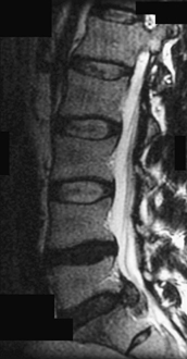
Only gold members can continue reading. Log In or Register to continue


