(1)
Marina Spine Center, Marina del Rey, CA, USA
This lateral approach is a direct approach to the spinal nerve and the vertebral artery in the intervertebral foramen [1].
1.
Position the patient supine with a slight roll under the shoulders. The head is not rotated.
2.
Incise the skin with a transverse collar-type incision for one- or two-level disease and with a longitudinal incision along the medial border sternocleidomastoid for multilevel disease. With retraction of the skin and subcutaneous tissue, the platysma muscle is divided longitudinally in line with its fibers. The superficial layers of the investing cervical fascia are opened to identify the medial border of the sternocleidomastoid muscle and the interval between sternocleidomastoid and the medial strap muscles. Angled retractors are used by the assistant to open this interval.
3.
Identify the great auricular nerve and the anterior cutaneous nerve. These cutaneous branches of the cervical plexus penetrate the deep fascia on the posterior surface of the sternocleidomastoid muscle at approximately midbelly. The great auricular nerve crosses in a cephalad direction on the surface of the sternocleidomastoid muscle toward the ear. The anterior branch of the greater auricular nerve innervates the skin over the face in the area of the parotid gland. The anterior cutaneous nerve takes a more horizontal course across the sternocleidomastoid before dividing into ascending and descending branches. The ascending branch of the anterior cutaneous nerve pierces the platysma muscle and is distributed to the skin overlying the mandible. Loss of sensation from damage to this nerve can result in decreased sensation over the mandible [2].
4.
Retract the sternocleidomastoid muscle laterally. When retraction is difficult, the sternocleidomastoid muscle is separated from its attachment to the mastoid process. In any separation of the proximal mastoid, cranial nerve XI must be identified entering the sternocleidomastoid two to three fingerbreadths below the mastoid tip and the proximal third of the muscle. The spinal accessory nerve exits the muscle obliquely caudally, passing across the posterior triangle of the neck to the ventral border of the trapezius.
5.
With lateral retraction of the sternocleidomastoid muscle, identify by finger palpation the carotid sheath. Open the middle cervical fascia with Metzenbaum scissors and dissect longitudinally with fingertips to expand the interval between the sternocleidomastoid muscle and the carotid sheath laterally and musculovisceral column medially. Retract the medial structures with a blunt-angled retractor, and palpate the carotid pulse and the anterior tubercle of the transverse process. Ligate and divide any tethering vessel. Divide the omohyoid muscle, if needed.
6.
Get Clinical Tree app for offline access

Identify the anterior tubercle of the transverse process (Figs. 8.1, 8.2). This prominent process is suitable for insertion of longus coli, longus capitis, and anterior scalene muscles, and is the key to dissection.1,3,4 Palpation of the spine under the prevertebral fascia reveals the disc spaces, the anterior tubercle of the transverse process, and, medial to the tubercle, the longitudinal groove of the costotransverse lamellae (Figs. 8.1, 8.2). It is approximately the size of a small fingernail, joins the anterior tubercle to the vertebral body, and is the roof of the foramen transversarium covering the vertebral artery [3]. The prominent Chassaignac’s tubercle, the anterior tubercle of C6, is a relatively consistent landmark, but every level should be identified with X-ray control after exposure of the anterior tubercle of the transverse process.
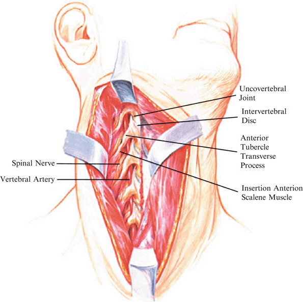
Fig. 8.1


A large expansile approach of the anterolateral aspect of the cervical spine with soft tissue removal to reveal the vertebral artery passing through each foramen transversarium. The anterior tubercle of the transverse processes with its muscular attachment and the costotransverse lamellae are a protective covering over the vertebral artery and nerve. Note the relationship of the vertebral artery anterior to the spinal nerve at each level
Related posts:


Stay updated, free articles. Join our Telegram channel
Full access? Get Clinical Tree