Limbs
reflexes and sensation
Examining reflexes
Tendon reflexes are single synapse reflexes. A rapid stretch of the muscle stimulates the muscle spindles and this message is conveyed via the sensory root to the spinal cord at the segmental level of the muscle stimulated. This synapses with the motor neurone that supplies the muscle and leads to contraction of the muscle.
If this is interrupted, by either peripheral sensory or lower motor neurone lesions, the reflex is reduced or lost. Reflexes are reduced in muscle disease. If there is an upper motor neurone lesion, the reflex is increased because inhibitory factors have been removed.
Tendon reflexes are important because they provide an objective sign indicating abnormality and some indication as to the level of the abnormality.
Reflexes can be graded as absent, obtainable with reinforcement (see below), reduced, normal, increased and increased with clonus.
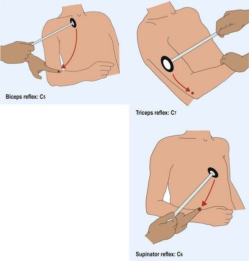
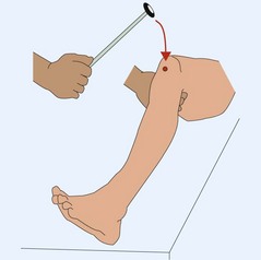
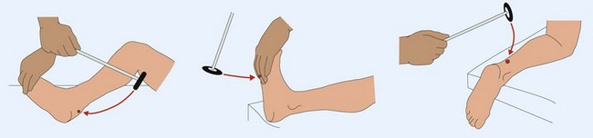
How to elicit tendon reflexes
The patient needs to be relaxed, with the muscle to be tested in the middle of its range of movement (Figs 1–3). The tendon hammer is swung to hit the tendon or, for the biceps and supinator reflexes, a finger on the tendon. The muscle is watched for movement in response to this. If the reflex is not obtainable then the patient is asked either to clench the teeth or make a fist – reinforcement – which may allow the reflex to be found. When testing the reflexes it is useful to remind yourself of the segmental level being tested.
Clonus is the rhythmic contraction of a muscle when it is stretched. This is most commonly found at the ankle (up to three beats is normal) and is occasionally found at the knee. Clonus indicates an upper motor neurone lesion.
Plantar response or Babinski response
This neurological sign has attained almost mythical status. An extensor plantar response is when the hallux extends and the other toes spread out in response to a stimulus. This stimulus is usually stroking the lateral part of the sole, though there are numerous alternatives, with different eponyms. This extensor response indicates an upper motor neurone lesion. Difficulties arise from false-positive results, especially when the patient withdraws or when there is a ‘dystonic toe’ in extrapyramidal disease, and from too great an emphasis being put on the sign. False-negative responses arise when there is sensory loss on the sole or profound weakness of toe extension. An extensor plantar response needs to be interpreted within the context of the rest of the motor examination.
Abdominal reflexes
These reflexes are not stretch reflexes. The muscles of the abdominal wall contract in response to a scratch of the skin of the same segmental innervation. The abdomen is usually considered in four quadrants. A response is normal. In upper motor neurone lesions, after multiple abdominal operations or multiple pregnancies, the response is lost. It is invisible in obesity.

Full access? Get Clinical Tree

