(1)
Marina Spine Center, Marina del Rey, CA, USA
Occasionally the need arises for maximally expanded exposure of the cervical spine. For exposure of multilevel anterior cervical disease from C1 to T2 [1]; Riley expanded the basic anterior medial Robinson approach. Various aspects of the approach, seen in Chapters 4, 6, and 9, are combined into one extensive exposure.
Get Clinical Tree app for offline access

1.
The skin incision is a modified Shoebringer incision described by Rile y.1 It begins under the mandible at the midline and extends posteriorly under the angle of the mandible to the tip of the occiput, down the posterior border of the sternocleidomastoid muscle, and swings anteriorly into a modified hemithyroid approach to the sternal notch (Fig. 11.1).
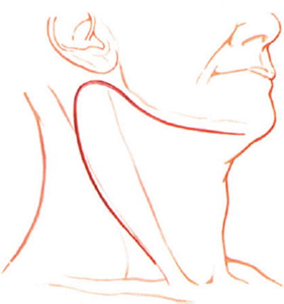

Fig. 11.1
Total exposure to the cervical spine from C1 to T2. A curvilinear incision producing a flap on the anterior lateral aspect of the neck may be used. Incise through the skin and subcutaneous tissue, divide the platysma muscle in line with the incision, and carefully retract the skin margins. When necessary, divide and ligate prominent external jugular veins
2.
Open the skin, subcutaneous tissue, and platysma muscle in line with the incision. Open the full extent of the wound through the superficial cervical fascia dissecting the flap lateral to medial.
3.
Identify the anterior border of the sternocleidomastoid muscle (Fig. 11.2). As with all the anterior medial approaches, this is the key to proper orientation at this level. Develop the interval between the medial border of sternocleidomastoid and the strap muscles (Fig. 11.3). Spread longitudinally the entire length of the anterior sternocleidomastoid border. Reware of the mandibular branch of the facial nerve in the cephaladmost extent of the exposure.
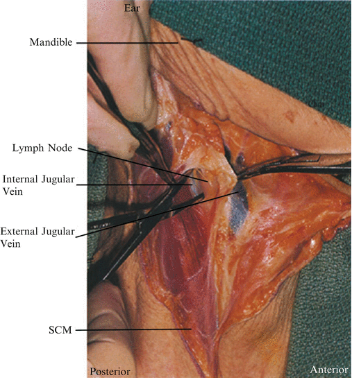
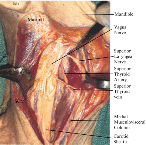

Fig. 11.2
As in the standard anteromedial approach to cervical spine (Chapter 6), identify the medial border of the sternocleidomastoid muscle (SCM), and delineate this border throughout the length of the incision

Fig. 11.3
Retract the sternocleidomastoid muscle laterally and develop the plane between the carotid sheath and the medical musculovisceral column
4.
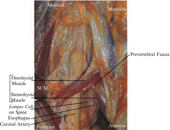
After the borders of the sternocleidomastoid and strap musculatures have been delineated, palpate for the carotid pulse. Incise the middle cervical fascial layer medial to the carotid pulse in the midportion of the neck (Fig. 11.4). Gently retract the carotid laterally and develop the interval between the medial musculovisceral column and the carotid artery. The middle thyroid vein should be identified, tied, and ligated. Continue to develop this interval, aided by deep digital palpation in the wound for the spine. It is important to attempt to dissect directly to the spine before placement of angled retractors, which could slip into the tracheal/esophageal groove and damage the recurrent laryngeal nerve. Bluntly spread longitudinally, dissection at this point should be caudad to the superior thyroid artery and cephalad to the inferior thyroid artery in the relatively avascular plane. The esophagus may be lying as a flat ribbon on the spine; this should be bluntly dissected off the prevertebral fascia with the musculovisceral column (Fig. 11.5).
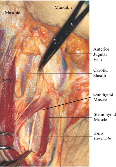

Fig. 11.4
The omohyoid muscle crosses the middle cervical fascial layer approximately at C6–C7. Fibers of the ansa cervicalis may course with the muscle. The omohyoid may be divided

