Chapter 78 Lumbar Discectomy
An estimated 12 million Americans suffer from significant degenerative lumbar disc disease. Approximately one million patients per year undergo surgeries, of which about 200,000 to 300,000 are lumbar discectomies.1–5 The combination of tremendous volume and a broad spectrum of standard care, including often vague surgical indications, makes lumbar disc pathology an object of legitimate scrutiny. However, proper patient selection and surgical technique can provide excellent and satisfying results for patients and surgeons alike. Neurosurgeons working in this field should remain scrupulously objective in patient selection, abreast of evidence-based outcomes research, and cautious in the selection of new technology.
Lumbar laminectomy for disc hernia or anular prolapse is one of the most common operations performed by North American spine surgeons; it is also one of the most potentially successful. Lumbar laminectomy for herniated lumbar disc has endured considerable analysis and the test of substantial time since its inception 75 years ago and remains a fundamental part of most spine clinical practice. It is a cost-effective procedure for carefully selected patients, being relatively moderate in cost and providing substantial improvement in quality of life.6
The incidence of lumbar disc hernia peaks between 24 and 45 years of age, with the incidence of surgery most often in patients between 30 to 39 years. Males predominate, at 1.3:1 to 2:1, possibly because of larger mechanical stresses and more tenuous nutritional and waste diffusion through the disc, as will be addressed. Other classically regarded risk factors for disc herniation have included smoking, obesity (BMI >30), sedentary lifestyles, prolonged motor vehicle driving, previous full-term pregnancies, and operating heavily vibrating machinery.1,4,7,8
The Twin Spine Study, a research program investigating various environmental factors involved in the etiology and progression of disc degeneration, looked at differing occupational exposures, driving and whole-body vibration exposure, smoking exposure, anthropomorphic factors, heredity, and the identification of genotypes associated with disc degeneration in monozygotic male twins. Although some environmental factors are relevant, as was mentioned, disc degeneration appears to be determined more significantly by genetics. There is a modest correlation with lifting and smoking but little influence from occupational and leisure-time physical loading activities such as sports and resistance training throughout adulthood. The effect of anthropometric factors, such as body weight and muscle strength, appears to be greater than the effect of occupational physical demands. Routine loading and physical demand may actually have some benefits for the disc, physical inactivity being a greater risk factor.9,10
Terminology of disc pathology is of particular importance in the venture of spine surgery and spine clinical research and has important implications for treatment options. The authors endorse the standardization of nomenclature recommended by the combination of the North American Spine Society, the American Society of Spine Radiology, and the American Society of Neuroradiology.11 Not only do diagnostic radiologists and clinicians need to recognize and utilize the same terms to communicate effectively, but vague or loosely applied terms obscure the results of clinical research. The term hernia specifically defines nucleus and/or end-plate cartilaginous tissues escaping the confines of the anulus and residing outside the apophyseal ring. It might not be possible to radiographically determine an anular defect, so the use of the term hernia is legitimately broadened to refer to displacement of nucleus, cartilage, fragmented apophyseal bone, or fragmented anulus from its normal location to lie beyond the disc space; disruption of the anulus is implied. Use of the term anular bulge or disc bulge defines localized or circumferential prominence of an otherwise radiographically intact anulus, not disconnected from the apophyseal ring. It is also useful to add descriptive terms to further define a hernia, such as protrusion (broad base), extrusion (narrow base or neck), and sequestrum (lack of continuity to the disc of origin). The use of the term rupture is discouraged as inaccurate unless there is indeed a single violent traumatic event that is clearly the origin of a defect in a previously intact anulus and of resultant disc herniation.11
History
A condition that is recognized as sciatica, although not associated with spine abnormality, was described before the time of Alexander the Great.12 In 1779, Pott13 was able to associate deformity of the spinal column with sciatic pain. However, it was Lane14 who described sciatic pain and its origin in a living patient in 1893 and Bailey and Casamajor,15 in 1911, who described a small series, complete with radiographic studies. Also in 1911, Goldthwait,16 who thought herniations of the disc were capable of producing sciatic and low back pain, presented a patient with lumbosacral disc hernia and paraplegia. In 1916, Elsberg17 (who operated on the patients of Bailey and Casamajor and often noted relief after apparently no more than decompressive laminectomy) described it as attributable to a condition of cauda equina radiculitis. Parker and Adson18 in 1925, Putti19 in 1927, Dandy20 in 1929, Mauric21 in 1933, and others attributed sciatic pain to nerve root involvement within the spinal canal and believed that adjacent vertebral structures were responsible. In 1934, Mixter and Barr22 published their milestone paper on the pathology and surgical findings associated with a ruptured nucleus pulposus, not only in the lumbar canal but also in the cervical and thoracic canals, complete with their diagnoses of the condition preoperatively.
The surgical procedure of choice for many of these pioneering surgeons was complete laminectomy, which often provided significant relief. Mixter and Barr favored a hemi-laminectomy approach, as did Love, for the cases of simple herniated disc that were amenable to preoperative localization.23 As experience accumulated, it became apparent that dural incision was unnecessary in most cases.
The complicating effect that developmental lumbar stenosis had on the pathology of disc diseases was appreciated in Verbiest’s24,25 reports from 1949 through 1955. Some of the more serious complications of surgery for lumbar disc hernia can be attributed to lack of preoperative appreciation of this anatomic variation and failure to tailor the procedure accordingly.
Working only with myelography and the power of clinical preoperative and subsequent intraoperative observation, the early surgeons were able to learn much and to steadily improve upon the surgical approach to lumbar disc disease. Currently, with the advantages of improved neurodiagnostic modalities in multiple planes, there is no longer much occasion for “surgical exploration,” and it should not be common to find an intraoperative surprise. Surgeons should be capable not only of making the preoperative diagnosis but also of adhering to a secure surgical plan, one that should accomplish the goal of radicular or cauda equina decompression with minimal risk of complication or injury. Credit for the use of magnification and small incisions, which has become standard care, is given Williams26 and Caspar.27
Outcomes
Relief of radiculopathic leg pain can be expected in the vast majority of patients who are appropriately selected for lumbar discectomy, with a 75% to 90% success rate.2,3,28–34 This variability in reported results, which ranged between 75%2 and nearly 90%3 for good to excellent results, likely is due to data quality, patient selection, short follow-up, and differing definitions of good outcome.2 The Asch paper is significant in that outcomes on over 200 patients who were operated on were prospectively determined by six parameters, including the preoperative ODI (Oswestry Disability Index), and the ODI at 1 and 10 days, 6 weeks, and 6 months and at least 12 months postoperatively. One of the most common causes for poor outcome is the poor definition of selection criteria for surgery, which varies remarkably between communities and countries, as much as fourfold or fivefold. 29,35–37The procedure can be expected, with relative certainty, to relieve radiculopathic leg pain, but relief of back pain cannot be predicted.38–42 Surgery appears to have the least measurable benefit at L5-S1, intermediate benefit at L4-5, and best results at L2-3 or L3-4, unless the conus is involved or cauda equina syndrome experienced. This may relate to the trend of conservatively treated hernias at upper lumbar levels faring worse than hernias at lower levels.43
Recurrent radiculopathy occurs in 5% to 10% of patients,2,35,40,44 which approaches the lifetime incidence of disc surgery.29 Less likely causes for failure include perineurial fibrosis and arachnoiditis. The subject of recurrence is important and is covered in depth later in the chapter.
The long-term outcomes of surgery and conservative treatment are similar, but in the short term, surgery provides the prospect of quicker relief than conservative measures do. 35,38,39,44–49Quicker relief with surgery may translate into reduced economic cost.6,50,51
It is difficult to define preoperative findings that are predictive of success or failure, even in the largest series of patients. Part of the problem arises from the instability of results over time, with as many as 40% of patients crossing over from favorable to unfavorable postoperative groups and vice versa.52 Useful clinical predictors of good outcome from surgery include good underlying health, absence of preoperative comorbidities, absence of previous nonspine surgery or of a workers’ compensation claim, young age, the presence of radicular pain to the foot, positive straight-leg raise without back pain, and reflex asymmetry, in approximately descending order of significance.28,38,39
Risk factors associated with poor outcome include time off work in excess of 3 months, psychosocial problems including poor educational level, smoking, and possibly obesity. Some authors believe that although obesity complicates anesthesia and convalescence, it might not of itself adversely affect outcome.28,53 Smoking is a risk factor for chronic low back pain4,54,55 as well as a risk factor for hernia and poorer outcomes, as has been discussed. Perhaps because of the location of the dorsal root ganglion and because of the difficulty associated with surgically approaching lateral hernias, the probability of good outcome for extraforaminal hernias is not as great as that for paramedian hernia.31,56
Radiology, Indications for Discectomy, and Clinical Correlates
Patient selection is critical to good patient outcomes. Technique may not be as important as patient selection in lumbar discectomy. The patient selection pitfalls to be encountered, recognized, and avoided in lumbar discectomy surgery are numerous. Two of the most common errors are the misinterpretation of leg pain of some other origin as being radiculopathy and correlating back pain with an unrelated neuroimaging finding. The most important determinant in favor of proceeding to surgery should be strict correlation between the distribution of the radicular leg pain and the nerve root compression seen on preoperative imaging studies. Performed carefully and correctly, clinical examination can predict findings of neuroimaging and subsequent surgery approximately 70% to 80% of the time.50,51,57–59
There has been an increasing realization that information about morphology alone is not enough to make a definitive diagnosis. Radiographic prevalence on MRI of abnormalities in an asymptomatic population is the subject of dozens of papers. MRI-documented disc bulge appears present in up to 81%, protrusion in up to 63%, extrusion in up to 24%, dark disc in up to 83%, disc height loss in up to 56%, anular tears in up to 56%, and Schmorl nodes in up to 19% of asymptomatic volunteer individuals. The numbers are similar in patient populations that are chosen without regard to symptoms.60,61 It can be said that the potential for finding false-positive indicators is universal, since they are nearly ubiquitous. As MRI scanning power and resolution improve with time, this problem will only grow larger. Also, clinicians and radiologists have different perspectives and perceptions when reading the same study; clinicians are more focused on clear description of the morphology of a particular pathologic finding than radiologists are.62
Contrasted MRI may occasionally be necessary. MRI with contrast can demonstrate inflammatory change on the periphery of the hernia, which can be of prognostic value. Contrast is useful to help differentiate nerve sheath tumor from disc material, with which it is often confused in the far lateral location, or other tumors and processes with which a hernia may be confused when it has migrated into unusual locations and into the posterior canal.63 In the postoperative setting of recurrent radiculopathic pain, contrast will display epidural scar and inflammation around a nonenhancing retained or recurrent nuclear fragment. Occasionally, it is prudent to augment the MRI with CT. CT will show bone anatomy and important bony subtleties that the MRI might not show or might not show well yet that might be important to surgical planning, such as spondylolysis and apophyseal (limbic) fracture. In the case of apophyseal ring fracture, CT is often the best method of examination, while plain radiographs and MRI might not demonstrate the bone of the apophyseal ring.
The synthesis of a decision to operate should be scrupulously clean and based upon a combination of clinical factors and radiographic findings. If MRI and CT do not provide sufficient explanation for a clinical picture, the second-line alternatives of discography and myelography remain. Proponents of discography claim that, through pain provocation, it can provide the specificity that is missing from the purely morphologic information that CT and MR imaging provide. The specificity of discography, however, is far from clear.64,65 If MR studies fail to provide clear evidence of lumbar disc hernia at the level corresponding with the clinical presentation, myelography may be useful, particularly when no neurologic deficit exists, multiple nerve roots are involved, or a centrally herniated disc affects only a single root.
Not only is there great potential for spine MRI scans and other modern neuroimaging to display abnormal findings and poorly interpreted information that have great potential to lead to inappropriate management, but they can also lead to inappropriate, expensive, and disabling behaviors from the patient. Radiologists must take some responsibility for the way in which their reports are used and interpreted. The addition of epidemiologic data and statements may be worthwhile.66 In an effort to standardize neuroradiologic readings, the North American Spine Society, American Society of Spine Radiology, and American Society of Neuroradiology established a combined task force with recommendations for readings.11
Preoperative clinical history may give clues to the anatomic severity of lumbar disc pathology, whether it is an intact anulus (negative exploration or protruding disc at surgery), a ruptured anulus (subligamentous incarceration), or a completely free disc. Vucetic et al. found that shorter duration of leg pain predicted a ruptured anulus; in their series, a 10-week period of symptoms was found in rupture versus a 50-week period found for an intact anulus. Lack of comorbidity predicted a ruptured anulus, with 18% of patients with rupture having prior medical or psychiatric treatment versus 39% of patients with an intact anulus having prior treatment for other diagnoses. Having had previous nonspine surgery was recorded in 32% of patients with a ruptured anulus, while 55% was recorded in patients with an intact anulus. The two groups differed then not only in disc pathology but also in medical, behavioral, and social factors, which undoubtedly plays a role in surgical outcomes.59
In practice and as a prerequisite for successful surgery, there should be a strong correlation between the pain, neurologic deficit, and preoperative imaging findings, and this rule should be inflexible. Much of the U.S. population obtains information from the Internet, for which there are no standards regarding quality and content, and at least a third of the available information may be of dubious value and/or distorted by potential commercial gain.67 Between the power of modern MRI scanning and the influence of the Internet on patients, contemporary spine surgeons must keep their perspectives scientifically valid and treatment goals clear.
Biology of Disc Degeneration
In the well-hydrated discs of youth, the mechanical stresses that are applied to the vertebral column are borne more upon the center of the end plates; but with desiccation, the loads become transmitted more to the periphery of the vertebral body. Ultimately, the anulus bears more load than it is capable of handling, particularly at the posterolateral segment, and hernia occurs. Nutritional support of the living tissue of the disc is dependent on a number of factors, including size of the disc, changes in the vertebral body end plates from which nutritional support diffuses, age-dependent cell density within the disc, and patterns and levels of physical activity that may encourage or discourage diffusion of nutrients to and waste from and through the tissue.68 The avascular nature of the mature disc also makes it unable to remove and replace degradation products. The mature disc is one of the most sparsely cellular tissues in the body yet with one of the densest extracellular matrices to maintain, dependent on the health of those cells.69
The chemical makeup of various tissues of the disc changes with age, potentially increasing in fragility. Collagen is widely distributed in the body, of various compositions; types I, II, III, VI, and IX are found in the nucleus and anulus in both normal and pathologically degenerated discs, but types III and VI are increased in areas of degeneration. Mutations in at least two collagen IX genes, COL9A2 and COL9A3, have been associated with higher likelihood of hernia, and the presence of childhood hernia implies genetic predisposition outside of environmental factors that are commonly held to be responsible.60,70,71 At a molecular level, collagen cross-links are important to the mechanical stability of the disc, particularly perhaps the anulus. The variety of proteoglycans in the extracellular matrix of the nucleus also change in abundance and structure through life and in their ability to retain hydration of the disc and ability to maintain the electrokinetic environment important to water and nutrient transport. Disc matrix proteins such as fibronectin and elastin throughout the anulus and nucleus also change in age and degeneration.69
The most dramatic changes in degenerating discs occur in the loss of hydrostatic pressure as maintained by the negatively charged proteoglycans, in water content, in cell populations, and consequently in cellular biosynthesis and repair. Mechanical stimuli can elicit different cellular responses from similar cells depending on whether the tissue is of the nucleus and inner anulus or outer anulus. The difference potentially amounts to an anabolic response to mechanical stress in nucleus and inner anulus tissues and a catabolic response in outer anulus tissue.72 Lack of mechanical stimuli or hypomobility of the disc produces changes that may promote degenerative change.73
Apart from the molecular changes, the anulus ages as well at a microstructural level. A significant element in the strength of the anulus comes not only from fiber orientation in alternating lamellae, but also interconnectivity between them.74 With age, decreases in the presence of pyridinoline (by age 65, 50% of that found in younger people) and increases in pentosidine occur within the disc. Decreases in pyridinoline cross-links lead to alterations in the collagen matrix of the disc. Integrity of the anulus deteriorates, perhaps beginning with the translamellar interconnections, leading to delamination of the anulus and ultimately anular tear or fissure. The presence of a tear or fissure does not imply traumatic origin.
Certain proteinases that are not normally present in the healthy disc also begin to appear in aged discs and are at least partially responsible for the degeneration of the extracellular matrix in the anulus, nucleus, and end plate.75,76 On a macroscopic radiographic level, though, while all the foregoing changes are occurring and fibrous tissue replaces the nuclear mucoid matrix of youth, disc height may yet be preserved, and disc margins remain intact.64
The anatomic composition of disc hernia changes with advancing age and perhaps to some extent with gender. In youth, a particular problem of hernia is involvement of the apophyseal ring. The movement of the hernia and anulus avulses the apophyseal ring into the canal, away from its immature attachment to the vertebral body,77 which often produces more mass effect than will more common nuclear fragments alone. In adolescents and young adults, a hernia is more likely to be composed of nucleus pulposus. As the nucleus becomes more fibrous with age, the percentage of nucleus in the fragments becomes lower, and the likelihood of finding cartilaginous end plate and anulus increases, such that by age 70, a disc hernia is unlikely to contain any nucleus. Women may be found to have higher percentages of cartilaginous end plate than men.78
Back Pain
While the topic of discogenic back pain is not the focus of this chapter, lumbar discectomy is often regarded as treatment for lumbar pain, and the association merits brief review here. Low back pain is a poor indicator for discectomy surgery. Diagnosis of the precise disc among many that might be the source of back pain can be difficult, if not impossible, in most cases because neurologic examinations in patients with only back pain and the absence of radiculopathy provide no direction and because radiologic abnormalities do not necessarily correlate with the source of pain. Something like 85% of patients with low back pain cannot be given a legitimate precise diagnosis of its anatomic origin.44,65 A plain radiographic survey of adults over age 65, examining a cohort of subjects with chronic daily lumbar pain and those without, demonstrates the ubiquity of findings in the discs and facets regardless of pain status. While higher degrees of radiographic severity on plain films may be seen in the pain group as a whole, there is no correlation with the degree of pain experienced.79
An anatomic cause is impossible to establish despite modern neuroimaging. In Western medicine, patients expect and press for plain radiographs, despite the widely known lack of correlation with back pain.66,80 The plain radiographs will often lead to more sensitive imaging studies such as MRI. MRI, in turn, is so sensitive and generally readily accessible that these virtues can in a certain way be looked upon as drawbacks. It is a rare scan that is read as normal or even normal for age, yet it is well known that sizable protrusions and extrusions exist commonly in asymptomatic patients.9,60,61,64 Therefore, interpretation of the neuroimaging studies must be made in the context of good clinical information. There is “an increasing realization that information about morphology alone is not enough to make a definitive diagnosis.”64 It is difficult for the backache patient (and perhaps even the referring doctor) to conceive that radiographs, MRI, and/or CT-myelogram showing pathology have no relationship to the pain. In fact, just the knowledge of pathology can adversely affect outcome.80 If present at multiple levels, the presence of a “dark nucleus” on MRI begins to predict a likelihood of back pain, but the pain generator remains unknown, whether it is the anulus, vertebrae, ligaments, fascia, muscles, or facets.8 The advantage of such sensitivity, of course, is that modern neuroimaging, particularly MRI, has eliminated the need for surgical exploration.
Back surgery on disc hernias as described in this chapter may do little for back pain, and the presence or absence of back pain should have little bearing on the patient’s selection for surgery.42 There are exceptions, however. If a large hernia appears to be responsible for radiographically visible elevation of the posterior longitudinal ligament off the vertebral bodies, particularly in midline,31 discectomy and the resultant relaxation of tension on the ligament might well result in relief of the resultant back pain. If the hernia is large and midline or if the lumbar spinal canal is shallow and resulting central stenosis of the lumbar canal is caused by disc herniation, the patient may develop a reflexive posture of lumbar flexion (the shopping cart position), which results in lumbar fatigue and pain. The symptoms can often be relieved by surgical decompression of the involved motion segment. In the Maine Lumbar Spine Study, an assessment of the predominant symptom, back pain or leg pain, was made, and improvement in back pain was documentable.35 However, it should be made clear to the patient without mechanical instability, facing the prospect of simple single-level surgery for degenerative disc disease, that surgery might have no impact on the lumbar pain. Lumbar pain is manageable by other interventions that are outside the scope of this chapter, such as exercises and other conservative measures, injection treatments, and surgical fusion.81
Radicular Symptoms
Radiculopathic leg pain with straight-leg raise and with Valsalva maneuver is more likely to be positive in herniations of L4-5 and L5-S1, where the compression and irritation are more likely to be at the axilla of the nerve root. The femoral stretch test is more likely to be positive at higher lumbar levels.51 Monoradiculopathic leg pain, or sciatica, is the most useful clinical correlate. It is superior to straight-leg raising, scoliosis, and sensorimotor deficits.82 Leg pain is often more severe in extraforaminal hernia than in intraforaminal or paramedian hernia, perhaps resulting from direct compression of the dorsal root ganglion by the hernia.56
Leg pain is perhaps the most common indicator, and the best indicator, for discectomy. However, conservative measures should be applied for a period of several weeks or longer from onset, if at all possible, prior to consideration for surgery, since long-term outcomes (4 years and more) are similar for conservative and operative care.38,39,46 In practice, however, the time required for spontaneous resolution of radiculopathy to occur may be more than some patients can bear, so pain becomes an important determinant for surgery. It is commonly accepted that the longer pain and numbness exist prior to decompression, the longer they will last following decompression.83 Furthermore, there is a legitimate fear that even permanent deafferentation pain can result from untreated compression.42,84 Inflammation from the disc hernia can adversely affect the nerve root over time and thus affect prognosis.52 Chronic pathologic changes can occur in the nerve root from prolonged compression, and over time (estimated at 3 to 6 months), there may be irreversible neuropathic changes.31,42,46 However, literature review and common clinical experience dictate that there is no consensus on what constitutes an appropriate conservative trial, and there is no consensus on what constitutes the factors leading to irreversible nerve root damage. Solid evidence for the hypothesis that delayed surgery impairs results may be debatable.85 Perhaps this lack of consensus is because there is selection bias to intermediate-level patients without sensorimotor deficit.
The patient’s economic imperatives become an important and valid factor in the selection of surgery in the presence of work disability and the requirement for rapid return to work. Surgery provides more rapid relief than conservative measures do.35 Also, long-term conservative care may ultimately be more expensive than surgery (in properly selected surgical candidates).50
Motor and sensory deficits are surgical indicators. It is practical to observe mild motor weakness and to follow for a period of time if stable. Motor deficit that is not improving, however, may be considered a surgical indicator,42 as should progressively worsening motor deficit31 and, of course, severe motor deficit.85
As will be seen, spontaneous resorption of disc material occurs and should be allowed to progress given the absence of severe motor deficit and the patient’s ability to comply. It is, almost paradoxically, a phenomenon that is likely to be more satisfactory and complete in larger hernias, when there is true extrusion, rather than in simple contained anular prolapse. Extrusion past the anulus marshals the processes of inflammation and phagocytosis of the mass.45,46,86–88
Disc Resorption
The spontaneous resolution of the initially agonizing symptoms of both back and leg pain as well as the radiographic findings of lumbar disc hernia with time is well established.7,35,38,39,41,44,47,75,89–95 Several mechanisms may be involved with the phenomenon of regression of disc hernia. Capillaries invade the hernia, and macrophages derived from monocytes migrate out into the hernia and begin a process of phagocytosis. Macrophages are the most commonly found cell type in both acute and chronic disc herniation. Macrophages contain enzymatic lysosomes, which degrade intracellular collagen and other substances present in disc material after phagocytosis. Macrophages also can secrete lysosomes, promoting the breakdown of extracellular substances such as collagen. Both of these mechanisms are closely involved in the regression of herniation. Apoptosis may occur at a higher rate in free disc fragments, another possible mechanism of absorption.
Macrophage activity itself can be a determinant of pain. There is a statistically significant correlation between histologically observed macrophage infiltration of intraoperative disc specimens and postoperative pain grading; patients who harbor inflammatory histology rate postoperative complaints lower than do patients with no evidence of inflammatory reactions.52,96 The periradicular inflammation that accompanies the hernia characterized by macrophage response also includes an increase in IL-1β and a release of PGE-2.75
Discs that are found to have intense perilesional gadolinium enhancement are more likely to regress spontaneously, the thickness of rim enhancement being a strong determinant of spontaneous resorption. Clinical symptom alleviation occurs concordantly with a faster resorption rate. MRI with contrast can be a useful prognostic tool for identifying patients with herniated nucleus pulposus (HNP)–induced sciatica with a benign natural course.92,93 Ninety-five percent of patients, followed out to 7 years, have decreases in the size of hernia through absorption of the disc. Progression of other disc degenerations occurs in all patients as well.90
Role of Surgery
The association between prolonged delay of treatment and poor outcome has been documented.35,38,39,83,97 In prospective studies of patients having lumbar disc hernia surgery, the duration of leg pain and duration of sick leave are found to be of prognostic value, with leg pain lasting over 8 months predicting a worse outcome, including inability to return to work.83,98
Part of the radicular symptomatology of the disc hernia is due not only to the described inflammatory changes, but also to tethering of and ischemic change in the root resulting from periradicular inflammation. As measured by laser Doppler flow, intraneural flow is improved after discectomy.99,100 Ischemic damage to the root, if present and productive of serious motor deficit, could be expected to have a poor prognosis, better avoided with decompression.
There is good objective evidence that surgery plays a legitimate role in the treatment of acute lumbar radiculopathy from lumbar disc hernia, bringing patients a faster and earlier recovery than would have occurred spontaneously. 35,38,39,44,47–49,101Nearly universally accepted indications for early surgery include significant motor deficit, unmanageable refractory pain persisting for more than 6 to 12 weeks, and of course cauda equina syndrome.7 Practicing neurosurgeons have long observed quick resolution of worrisome motor deficit in surgically treated patients.
Similarly, Peul et al. found that surgery offered short-term benefit. In a randomized prospective trial of early surgery (mean: 2 weeks) versus late surgery (mean: 18 weeks) or conservative treatment, relief of leg pain and recovery was faster in early surgery patients. By 1 year, however, there was a 95% probability of perceived recovery in both the surgically treated cohort and the delayed or conservatively treated cohorts.47 Osterman found similar results at 2 years.49 Longer-term results were found by Weber at 4 years,38,39 the SPORT (Spine Patient Outcomes Research Trial) study at 4 years,44 and the unrandomized but large and comprehensive Maine Lumbar Spine Study at 10 years.35 At 1 year and at 4 years, in the prospective randomized multi-institutional SPORT lumbar disc series, patients who were operated on (in both the randomized and nonrandomized cohorts, without regard for intent to treat) maintained greater improvement (in SF-36 Bodily Pain and Physical Function scales and in the ODI) compared to patients patients who were not operated on, although long-term work status was not significantly benefited, as was mentioned. In the short term (3 months), work status was worse in the former group than in the latter, owing to surgical convalescence.44
In the Maine lumbar spine study, there did appear to be long-term benefit for patients who received surgery compared to hernia patients who did not. Surgical patients reported resolution or substantial improvement (56% vs. 40%, P = .006) and more satisfaction with their current status (71% vs. 56%, P = .002). However, work and disability status at 10 years were comparable among those treated surgically or nonsurgically.35
With regard to work status, these two large studies agree on lack of long-term surgical benefit.35,44 On the other hand, a cost-effectiveness analysis performed in concert with the SPORT series resulted in the demonstration of a cost per quality adjusted year of life of approximately $69,000 in 2008 U.S. dollars, supporting the use of surgery as a cost-effective procedure in the short term and for markedly symptomatic patients.
In the long term, children may do significantly better than adults following lumbar disc surgery; surgery does not appear to lead to chronic complaints of back pain.102
In summary, the ideal patient for discectomy is one with severe, disabling unilateral radiculopathic leg pain without severe sensorimotor loss for whom conservative measures over a period of a few weeks to 2 months have yielded little.42 A poorer recovery can be expected in the presence of severe persistent sensorimotor loss, once pain has remitted or has acquired the burning deafferentation quality suggestive of nerve root damage. Changes that are induced in the course of back pain through discectomy are unpredictable. The relatively soft and poorly defined nature of these indication guidelines has resulted in widely variable rates of surgery: as much as a fourfold or fivefold difference between surgeons and between countries.35–37
Cauda Equina Syndrome
A somewhat separate issue is the cauda equina syndrome. Bladder and bowel sphincter dysfunction and bilateral neurologic deficits are the strongest indicators for surgery. The outcome for the cauda equina syndrome is better if there is unilateral sciatic pain, worse if there is bilateral sciatica, and very poor if there is saddle hypesthesia. Patients with complete perineal anesthesia are at risk for permanent sphincter dysfunction. The mode of onset of symptoms may also be important, the acute onset of symptoms over hours being thought to be a prognosticator of poorer outcome, particularly bladder function, than is a more insidious onset over days or weeks.103
Cauda equina compression often exists in the sensitizing presence of developmental lumbar stenosis and, in the context of this discussion, is the result of acute disc herniation rather than slowly acquired degenerative change such as anular bulge. Unlike with the lesser indications discussed previously, compression of the cauda equina constitutes a medical emergency or urgency and should be relieved as soon as possible after diagnosis.81,103 If the onset of symptoms is abrupt, the symptoms and prognosis for full recovery are worse than if the symptoms are slower in onset,103 and by inference, the urgency for decompression is greater. Other poor prognostic signs for the recovery of sphincter control in cases of cauda equina compression include saddle hypesthesia and bilateral radiculopathic leg pain.
In addition to sensitizing patients to the potential for acute cauda equina syndrome, lumbar stenosis can be responsible for unexpected or false localizing findings in the event of disc hernia, for example, producing footdrop from an L12 level.104 High lumbar hernia also can be symptomatic in the distribution of multiple roots because of their acute angles and the narrow confines of the upper lumbar canal.104–106 These patients might not fare as well as patients who have disc pathology at more caudal levels if sensorimotor deficit is present. Long-term follow-up (average, 81 months) confirmed worse outcome for patients with hernia at L1-2 and L2-3, only 33% of patients reporting an improvement in their economic or functional status, compared with an 88% rate of improvement at L3-4.105 Perhaps this is because when lumbar stenosis is present, conventional discectomy via laminotomy increases the risk of damage to the intracanalicular roots because of the narrow confines of the canal. Particularly in the case of large central and calcified hernia at upper lumbar levels, a generous central canal decompression prior to the manipulation and retraction of the lateral thecal sac must be made. An alternative, if the conus terminates above the level in question, is to retrieve a large or calcific hernia through a transdural approach with repair of the ventral and dorsal dura.106
Technique
Open laminectomy and discectomy work as well as microdiscectomy. The advantages to microdiscectomy are the smaller and more comfortable incision and the shortened hospital stay and diminished trauma to the adjacent motion segments and paraspinous musculature. Microdiscectomy requires the use of magnification. The choice of loupes and headlight or microscope is moot. Results from surgery are the same with both techniques.28,31 Loupes, with the use of a coaxial or near-coaxial headlight, offer the same or nearly the same magnification and the same or nearly the same size incision in the case of microdissection as does the operating microscope. The advantage of the microscope lies in its use by an observer or assistant; the disadvantages are some additional encumbrance and perhaps expense to the patient.
The following discussion assumes that discectomy is the planned end result of the surgery, not to be followed by fusion, in which case restrictions on bone removal would not be as significant. There is insufficient indication for routine spine arthrodesis combined with lumbar disc excision as the primary procedure.107 While primary lumbar fusion may well be successful in disc hernia,108 inclusion criteria should be strict and may include both degenerative and isthmic spondylolisthesis, complete facetectomy, and perhaps degenerative scoliosis.109,110
Primary disc replacement or dynamic stabilization, like primary fusion for HNP, is not standard care; current thought reflects conservatism. Despite early enthusiasm for disc arthroplasty,111 it has been found that there may be only a 0.5% incidence of indication in the overall population with the majority in young patients who averaged 38 years of age. Other nonfusion techniques exist with known results. With criteria for arthroplasty tightly confined and given a small number of potentially eligible patients, there is limited use of this technology.112 Alternatives such as nucleus replacement may ultimately be an option.113
Standard paramedian lumbar disc surgery is estimated to take about 75 to 80 minutes, with an average blood loss of about 65 mL, requirement for blood transfusion being a true rarity.44
Operative Positioning and Patient Preparation
The disc space is avascular and, as a result, less resistant to infection. There is evidence that the risk of disc space infection may be reduced through the use of perioperative antibiotics.114
The preoperative placement of elastic stockings to prevent thromboembolism is recommended.115 A urinary catheter is usually unnecessary unless the procedure is expected to take more than 2 hours.
For most surgeons, the prone position on a frame works well. Often, such devices are inadequate for preventing increased intra-abdominal pressure (and thus increased ventilatory, venous, and cerebrospinal fluid [CSF] pressures) in obese patients. Obese patients are better positioned in such a manner that the abdominal panniculus hangs unimpeded, without bearing any of the patient’s weight (e.g., the knee-chest position). The eyes and facial prominences should be well padded and inspected after the patient is turned to the prone position because ocular pressure, or a combination of pressure and hypotension, can lead to blindness.116 The arms, if held abducted on arm boards, must be well padded over bony prominences and over the ulnar nerves.117 To prevent costoclavicular compression of the brachial plexus, care should be taken to avoid hyperabduction over 90 degrees at the shoulders and shoulder hyperextension. The radial pulse on each side should be felt. Women should bear the weight of the chest on the ventrolateral rib cage (not on the breasts, which should be moved medially). All skin that is in contact with the frame or with pads on the frame should be protected with a layer of linens; skin should not be allowed to touch the bare occlusive surface of vinyl or silicone rubber cushions. The urinary catheter (if placed) is inspected after the patient has been turned. Male genitalia must not be subject to compression between the approximated thighs. All bony prominences of the legs must be padded; in particular, the toes must bear no weight of the foot. A diminished or absent pedal pulse may indicate femoral artery compression. This can be encountered on a conventional frame as well as in the knee-chest position.118 The patient, when positioned, should be stable so that vigorous intraoperative manipulation will not cause movement. Hardware in the room, such as IV stands, light sources, carts, tables, and anesthetic equipment, should be arranged to allow easy access for both radiograph equipment and film cassette holders.
Standard Technique and Open Laminotomy (Laminectomy)
Although the term open laminotomy may imply the absence of visual magnification, some form of low-power magnification is often used. The skin incision for laminotomy for disc herniation is conventionally about three spinous processes in length, 10 to 12 cm for a one-level operation. When performed in this manner, open laminotomy has the advantage of facilitating surgical assistance. Furthermore, illumination is not quite as problematic as with shorter incisions, and the longer incisions have been suggested to be less traumatic to the paraspinous musculature than shorter incisions (that require greater retractor pressure). After the incision has been made, hemostasis is achieved and retractors are placed. As with any incision, tension on the skin retractor should be inspected intermittently throughout the operation because pressure necrosis of the skin edge is a significant source of infectious wound complications. After the skin has been incised, the subcutaneous tissue is divided. This can be accomplished without bleeding or trauma with blunt dissection. The lumbodorsal fascia is then exposed, and for ease of subsequent closure, it is prudent to sweep the subcutaneous fat off a short distance laterally. The fascia is then incised, just lateral to the spinous process, rather than in the midline, which allows the preservation of interspinous ligaments. If the laminotomy is to be performed bilaterally, as in cases of developmental stenosis with superimposed disc hernia, a fascial incision on each side of the spinous processes results in a saved strip, the width of the spinous processes, in the midline, complete with the interspinous ligament.
With the field prepared for bone work, the overhanging caudal lip of the rostral lamina can be partially removed, allowing for further exposure of the interlaminar ligamentum flavum. The amount of bone removal is at the surgeon’s discretion. The hernia and the anulus can be accessed with little or no removal of bone if one is comfortable with the amount of force necessary to retract the nerve root medially.26,31,119,120 Wider exposure via laminotomy and medial facetectomy, flush with the medial surface of the caudal pedicle, minimizes the need to mobilize the nerve root aggressively. Paramedian discs at higher lumbar levels represent a slightly greater challenge than those at L4-5 and L5-S1 because of the lamina and facet structure. The spinal canal is smaller in caliber, the lamina and facets descend more caudally, the facets are positioned more medially, and the facet clefts are more sagittally oriented than is the case in more caudal segments. Each of these features not only makes it necessary to remove more bone, but also makes progressive removal more risky to the integrity of the pars and the facet. This anatomy becomes particularly important in addressing intraforaminal disc hernia by the midline approach (see later discussion).
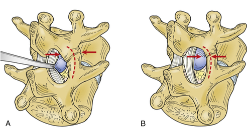
As bone removal proceeds rostrally and laterally, the lateral margin of the removal migrates caudally and laterally, in a fashion that makes the defect appear triangular or lung shaped so that the structural integrity of the pars interarticularis is not compromised. Compromise of the pars by an overly aggressive bone removal can result in a pars interarticularis fracture, either during the course of surgery or during convalescence. More than half the bone of the pars, in its lateral dimension, should be left to avoid fracture121(Fig. 78-1). As was mentioned earlier, this is particularly important at midlumbar segments, where the pars is narrower and the facet cleft is more sagittally oriented than at lower motion segments. Pars fracture isolates the facet, functionally resulting in complete facetectomy, which increases the failure rate due to back pain and instability.28
Alternatively, a minimalist approach can be taken to bone and ligamentum removal as described by Williams.26 This approach does have the disadvantage of reduced visualization and increased traction upon the nerve root during its medial mobilization. While the amount of bone removal is discretionary, as a rule, decompressive removal should be more generous in the case of concomitant developmental stenosis. At times, it can be minimal when the interlaminar space is large.
Perineurial scar has been blamed for postsurgical failure to relieve sciatica. There are many means of dealing with its development, and more discussion will follow. The ligamentum flavum is one such potential source of fibrosis if it is left in large shreds beneath the lamina. The ligamentum is therefore reasonably dealt with in one of two ways: (1) with minimal fenestration of the ligament, leaving its slick inner surface approximated to the nerve root as a natural barrier31,119,120; or (2) with its complete removal from the lateral recess. Because of impaired visualization and mobility of the underlying thecal sac and nerve root sleeve during the surgery and the variable ability to access the lateral recess and its contents with a minimal approach (dependent on the treatment level and concomitant spondylotic enlargement of the facet and lateral recess stenosis), complete flavectomy is usually preferred. With angled curettes, removal from the undersurface of the remaining rostral lamina should be thorough. If lateral ligamentum is left in place completely or partially, its raw surface may lead to a significant postoperative fibroblastic response and scar formation, and its continuation with the medial facet capsule contributes to lateral recess and foraminal stenosis, which may be of significance as the disc space narrows postoperatively and with age.
The location of the foot of the punch or the edge of the curette and the location of the nerve root sleeve should be well perceived by the operating surgeon. It is worth noting that a risk of dural tear is present in every case. If a scar is present, fixing the dura mater to overlying bone, the risk of a dural tear is increased. The chance of tear is also increased in the elderly (particularly elderly women), in whom the dura mater is thin and in whom a noncompliant scar may be present, even in the absence of prior surgery. An inflammatory response to the hernia itself is often present.45,86,88 Naturally occurring adhesion may be present, fixing the dorsal dura to overlying laminar bone and ligamentum flavum. For these reasons and as a basic element of prudent use of a bone punch, the space into which the punch foot will sit should be swept with a blunt instrument such as a Woodson or ball-ended dissector. The geometry of the instrumentation involved is crucial because the dura mater can fold over the foot of a punch or the edge of the curette that is not applied closely to the underside of the bone. This is an error that can be worsened if the dura mater is distended under increased intrathecal pressure; as with epidural venous bleeding, its risk can be reduced by careful positioning, with attention paid to intra-abdominal pressure reduction. Piecemeal bone removal is not slowed by cautious inspection. Caution simply requires keeping the eyes on the target, letting the assistant clean the punch, judiciously appreciating the tactile input, and intermittently sweeping the peripheral undersurface of the bone in the direction of the decompression with a dissector.
A dural tear is a significant problem only if it is not cared for properly. In some cases, dural tears are unavoidable, and their occurrence may even be predictable. It is a problem encountered by the best of surgeons. The risk of a tear is high in the elderly with thin and fragile dura as well as ligamentous adhesions to dura, but it is also a risk if the herniated disc fragment has been present long enough to result in dural adhesions. There is also the possibility of natural dural adhesion and of fibrous bands connecting ventral dura (Hofmann ligaments) to the posterior longitudinal ligament, particularly well developed and of potential surgical consequence in the lumbar canal.122,123
A mistake is to be cavalier about the occurrence of a CSF leak and not to repair the leak properly. Pinhole dural breaches can and often are successfully treated with fibrin glue, DuraSeal, hemostatic gelatin (Gelfoam), other adhesive substances, and/or indirectly with multilayer tight soft tissue closure. However, larger dural tears of more than 1 mm are best managed with convincing primary suture closure, as they risk spontaneously enlarging and producing CSF fistula and symptomatic pseudomeningocele.
If the tear is dorsal or lateral, it will be problematic for the remainder of the case if not attended to. The defect should be protected from further tearing or from aspiration of nerve roots by the placement of Gelfoam and a cottonoid beneath the suction tip over the tear. It should be exposed, with further bone removal, and repaired primarily with suture in a watertight fashion (preferably immediately after its occurrence). Repair can be tedious and time-consuming at a point in the operation at which much remains to be done. The temptation to delay repair until later is often best resisted. If the repair is one of the last tasks after an unusually lengthy procedure, attention might not be paid to the details of adequately repairing the leak. If the arachnoid is also involved and spinal fluid is being lost, thecal sac collapse occurs, and epidural venous bleeding intensifies because of decreased tamponade. This increased bleeding obscures visualization and blood can enter the thecal sac, possibly resulting in arachnoidal adhesions. Other possible results of unrepaired dural tear include radiculopathies with pain and deficit secondary to herniation of nerve roots through the dural defect, symptomatic pseudomeningocele, the possibility of meningitis, and persistent orthostatic headache complaints.124–128 In the modern era, with patients conventionally returning home the same or next day, primary repair becomes standard.
Confines of the field often make primary repair difficult, particularly with modern minimal exposures or tubular retractors, but only rarely is it impossible. The needle is more important than the suture size; a tapered needle not much larger than the suture should be used so as not to leave an excessive pinhole of its own. A commonly used suture is 4-0 or smaller, with 6-0 or 7-0 polypropylene suture (Prolene) being the authors’ preference (more so for BV-1 or BV-175 needle geometry). A new Ethicon (Johnson and Johnson) suture, Hemo-Seal 5-0 Prolene HS, has a sealing hydrophilic coating. The use of a small pituitary instrument to hold the needle and the use of knot pushers can significantly aid the process, even to the extent that dural repair can be effected through a tubular retractor.129
Reinforcement of the durotomy with an onlay of fat, DuraSeal, DuraGen, muscle, fascia, fibrin glue, Gelfoam, Surgicel, polyglactin acid sheet, or mesh,130 collagen mesh, or the like is prudent but might be ineffective in the long run without the underlying primary repair. Fibrin glue is very effective but short lived, lasting only several days. The authors believe that if a trustworthy repair has been provided, it is advantageous to have the patient ambulatory the day of surgery rather than enforcing bed rest; ambulation will expand the thecal sac, redistribute the caudal nerve elements, and preclude epidural bleeding. With good dural repair, only 1.8% of incidental durotomy patients must return to the operating room.131 Additional treatment such as lumbar CSF drainage should be considered second line, again being best used to reinforce a good primary suture line repair.
Repaired primarily, CSF leaks incurred intraoperatively have little or no impact on ultimate surgical outcome.127 Repaired and reinforced carefully, most will heal, but extra consideration should be given host factors that would impair wound healing, such as high CSF pressure anticipated in obesity, connective tissue disorders such as Marfan syndrome and neurofibromatosis, scar tissue, and use of steroids. On occasion, the tear may be located on the ventral aspect of the nerve root sleeve or thecal sac, in which case, while it may be impossible to repair without marked difficulty, it nevertheless does not risk the problems of a dorsal tear and will more than likely tamponade itself.
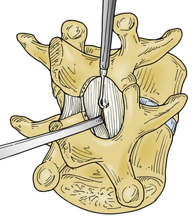
With the medial aspect of the facet joint and capsule partially removed so that the medial surface of the caudal pedicle can be observed and felt, the need for medial retraction on the nerve root to allow visualization of the disc space should be minimized (see Fig. 78-1B). Before retraction instruments are inserted, room ventral to the nerve root and dural sac must be assessed (Fig. 78-2). With a small-caliber aspiration tip (5 or 7 French), blunt hook, or ball-ended dissector, the nerve root can be mobilized medially, and while doing so, the amount of ventral fibrosis or compression can be determined. This palpation should be gentle, and if resistance to mobilization in this manner is met, the surgeon must determine the cause, as well as a solution to the problem of mobilization. The axilla of the nerve root may straddle a sharp focal prominence of the anulus fibrosus. Forcing the nerve root up and over it may invite neural injury. In such a case, the prominence can be trimmed or impacted down, medial to the nerve root in the axilla, following which the nerve root can be mobilized medially and the disc pathology better addressed. Poor ability to mobilize may reflect a nerve root anomaly such as low origin.
Related posts:
 Definition and Assessment of Dysfunctional Segmental Motion
Pathophysiology of Cervical Myelopathy: Biomechanics and Deformative Stress
Combined Ventral-Dorsal Surgery
Bone Void Fillers: Bone and Bone Substitutes
Medical Management of Neck and Low Back Pain
Posterior and Transforaminal Lumbar Interbody Fusion
Definition and Assessment of Dysfunctional Segmental Motion
Pathophysiology of Cervical Myelopathy: Biomechanics and Deformative Stress
Combined Ventral-Dorsal Surgery
Bone Void Fillers: Bone and Bone Substitutes
Medical Management of Neck and Low Back Pain
Posterior and Transforaminal Lumbar Interbody Fusion
Stay updated, free articles. Join our Telegram channel

Full access? Get Clinical Tree


