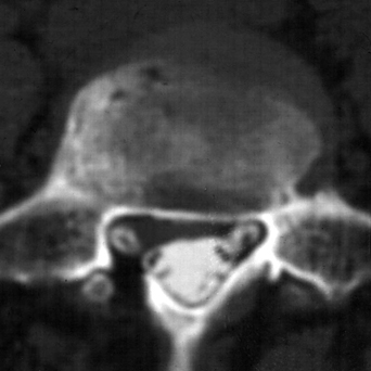
43 A 61-year-old woman with a history of rheumatoid arthritis who had four previous lumbar discectomies without fusion complains of chronic lumbar pain. She is neuro-logically intact. Magnetic resonance imaging (MRI) shows significant disc disease at L4-L5 and L5-S1 with decreased disc height and Modic end-plate changes. An axial highlight from the postmyelogram computed tomography (CT) reveals continued epidural compression on the right at the L5 level (Fig. 43-1). Recurrent degenerative disc disease This patient had a percutaneous two-level transforaminal lumbar interbody fusion (TLIF) and pedicle screw insertion (Figs. 43-2 and 43-3). Using a percutaneous pedicle screw insertion system (Sextant, Medtronic Sofamor Danek, Memphis, Tennessee), two-level lumbar fusions can be performed percutaneously through a minimal approach. Interbody fusions at L4-L5 and L5-S1 are done through small incision (Metrex tubular retractor, Medtronic Sofamor Danek). Through that same incision, pedicle screws can be inserted at L4 and S1 on that side, and through a small incision on the opposite side a similar procedure can be done. Postoperatively the patient went on to develop solid fusion and was pain free. Percutaneous pedicle screw insertion and fusion can provide an effective alternative to open fusion. In percutaneous cases, there is less muscle dissection and intraoperative blood loss, leading to better postoperative function and pain control. These techniques do have a steep learning curve, which must be overcome to optimize the results.
Lumbar Instability and Radiculopathy
Presentation
Radiologic Findings
Diagnosis
Treatment
Discussion
Lumbar Instability and Radiculopathy
Only gold members can continue reading. Log In or Register to continue


Full access? Get Clinical Tree


