Figure 2.1
Artist’s illustration of a large malignant brain tumor (green area) in the parietal region of the brain. The pressure over the normal brain is so severe that is shifting the brain from left to right (brain herniation). It is also infiltrating the surrounding brain tissue, causing severe swelling and brain dysfunction (Reprinted with permission from Barrow Neurological Institute)
Neuroepithelial Tumors (Gliomas)
Most gliomas arise from the functional tissue of the brain and are associated with swelling (edema) and sometimes bleeding (hemorrhage), which can also alter the structure and function of the normal brain. The location, growth rate, and growth pattern of the tumor determine its effects on brain function [3]. Mass effect, which is caused by the tumor compressing normal brain tissue, or brain infiltration by these tumors can cause depressive phenomenon (neurological deficits such as weakness and blindness) and irritative phenomenon (seizures). They can also occlude arteries or veins and cause strokes or interfere with the flow and absorption of cerebrospinal fluid (CSF), causing hydrocephalus. The most malignant variants of gliomas (glioblastoma multiforme) can create their own blood supply and steal blood from normal brain [3, 5].
Clinical Presentation
The most common symptoms of brain tumors are headache, seizures, and progressive neurological deficit. Headaches can be caused by increased intracranial pressure, invasion or compression of sensitive structures, and vision problems. Other worrisome symptoms include double vision with headaches, hearing loss with dizziness, and gradual onset of speech difficulty. Changes in behavior, memory loss, loss of concentration, and general confusion may all be subtle signs (Fig. 2.2). Increased intracranial pressure due to large tumors frequently causes nausea, vomiting, dizziness, vertigo, and changes in mental status. Acute deterioration is not uncommon and is caused by tumor swelling (Fig. 2.2), hemorrhage, infarction, or hydrocephalus [3, 5].


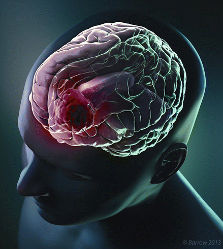
Figure 2.2
Artist’s illustration of a malignant brain tumor (red area) located at the left frontal lobe, causing mass effect and brain herniation. Tumors in this locations can cause language problems (aphasia) and personality problems, memory alterations, and in cases of large tumors, weakness and sensation abnormalities over the right side of the body (Reprinted with permission from Barrow Neurological Institute)
Clinical Symptoms According to Location
Frontal Lobe Tumors can cause problems in cognition, behavior, speech, and movement. Patients lose their energy for intellectual, physical, and social activity. They are also impulsive, lose social inhibition, and have fluctuating emotions. Motor aphasia (difficulty speaking) and agraphia (difficulty writing) may also be present (Fig. 2.2).
Temporal Lobe Tumors cause alterations in hearing, language, vision, behavior, and movement. Sounds are heard but not appropriately interpreted. Tumors in this location may cause auditory illusions or hallucinations. Patients sometimes have difficulty understanding spoken language.
Parietal Lobe Tumors produce sensory problems. Other symptoms are difficulty dressing oneself, denial of extremity weakness, lack of awareness of objects, and constructional apraxia, which is the inability to build or draw objects. Failure to recognize familiar faces is also common.
Occipital Lobe Tumors cause vision problems, including blindness in one or both eyes, visual hallucinations and illusions, and abnormal or multiple images of a single object.
Stay updated, free articles. Join our Telegram channel

Full access? Get Clinical Tree


