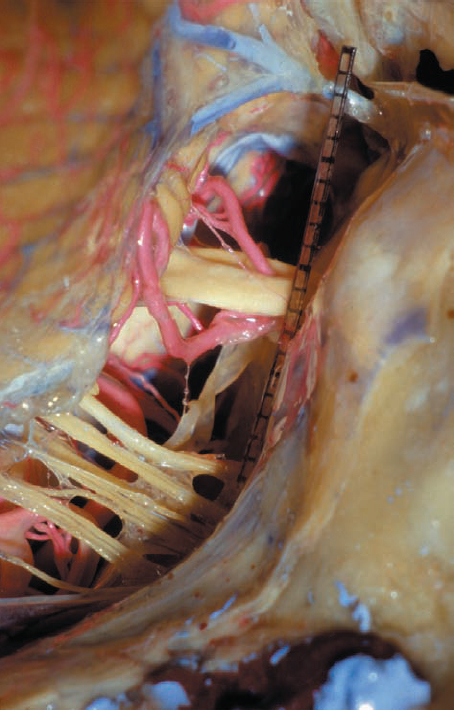
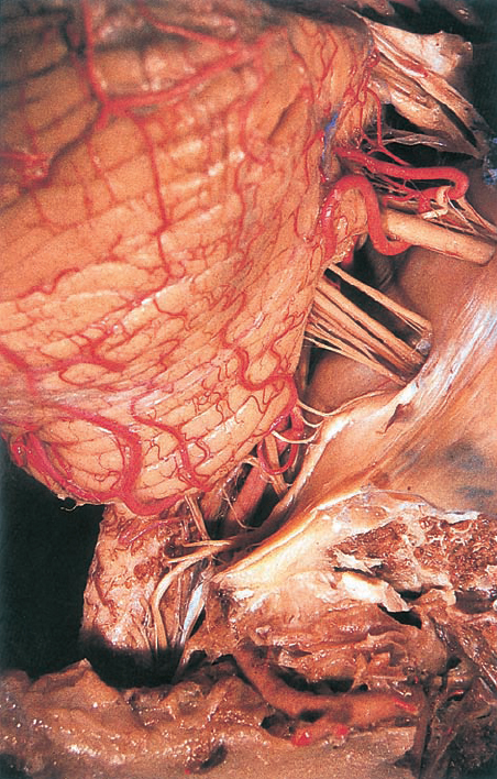
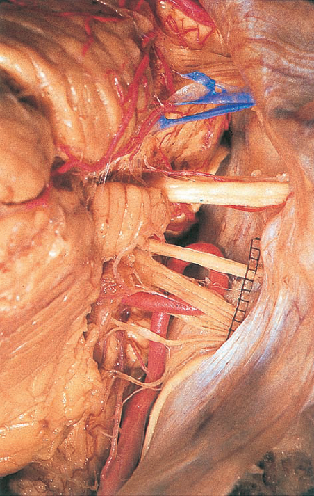
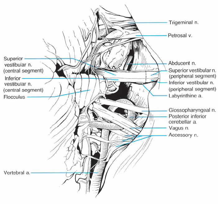
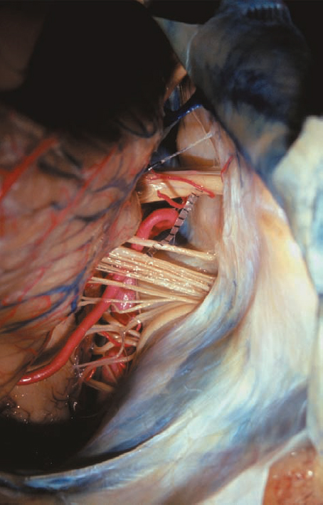
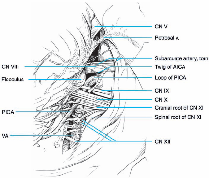
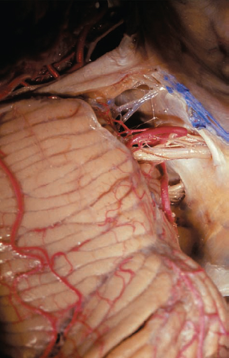
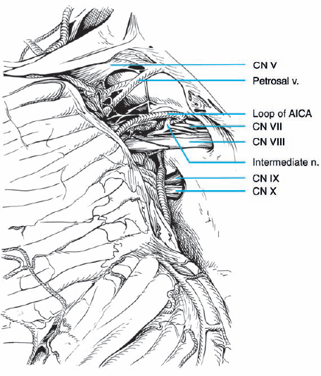
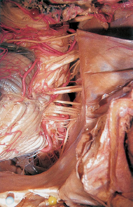
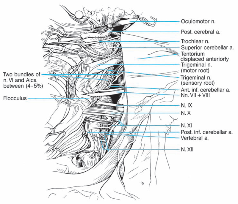
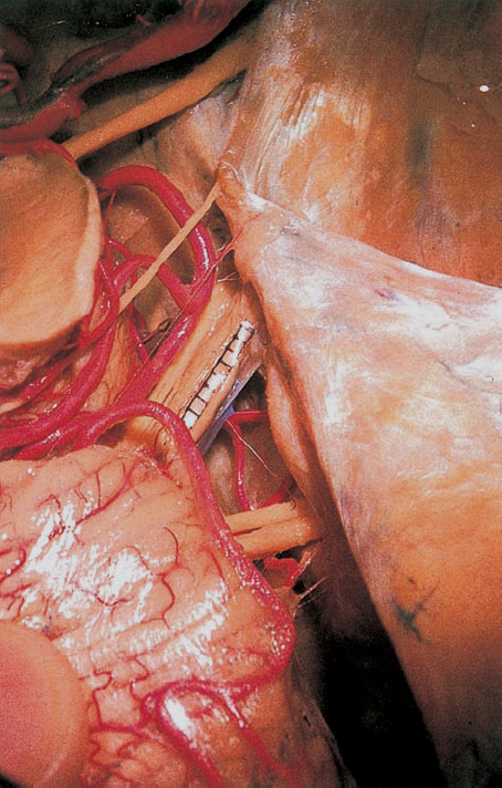
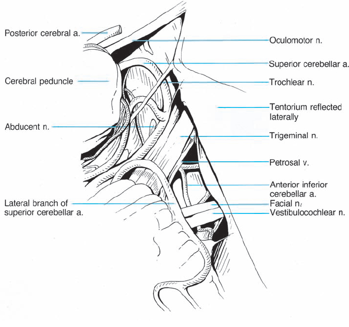
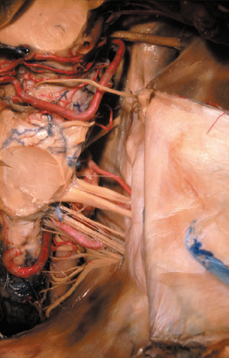
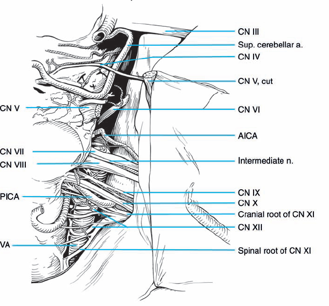
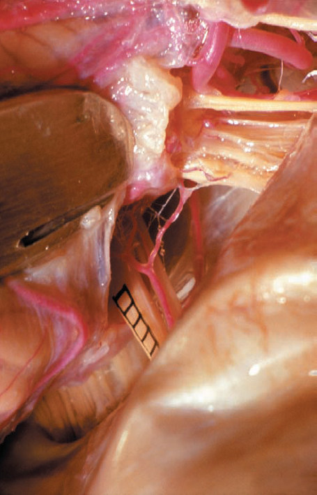
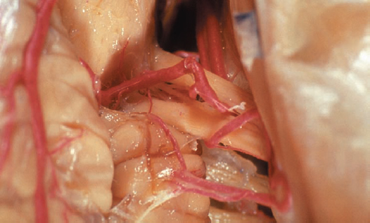
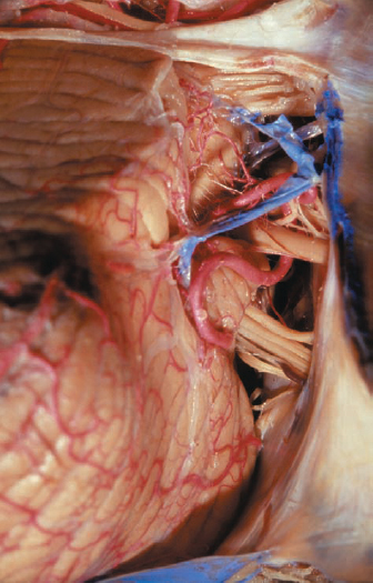
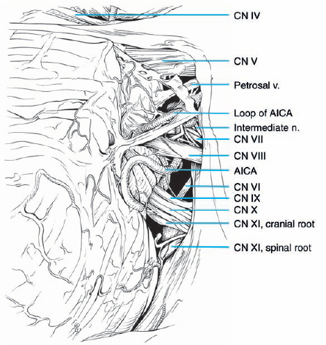
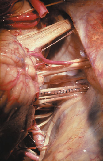
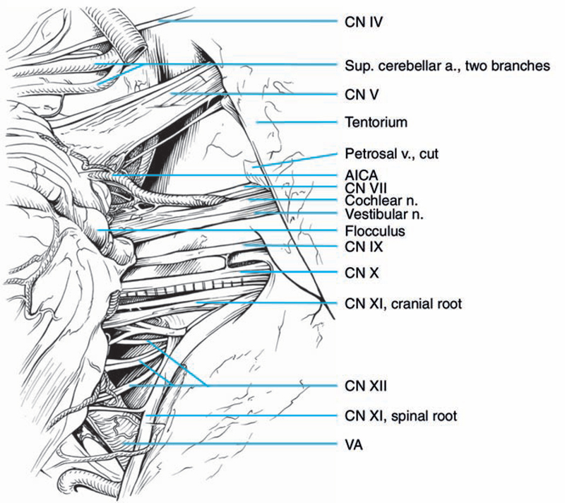
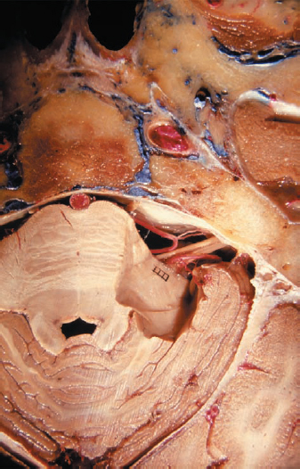
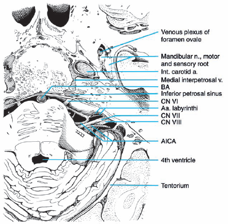
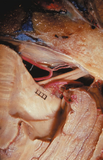
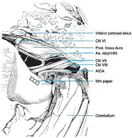
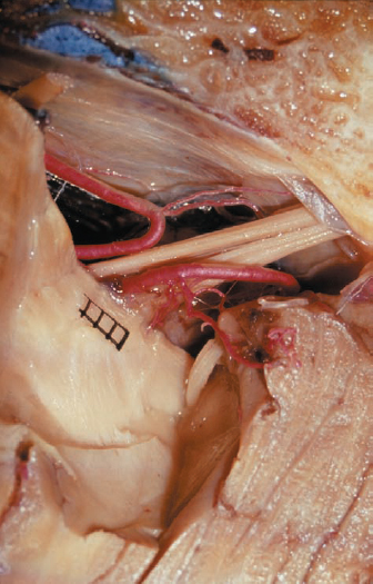
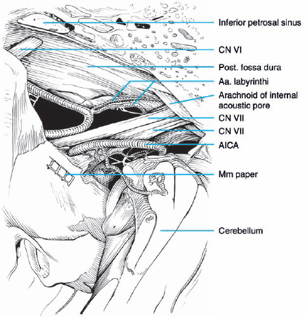
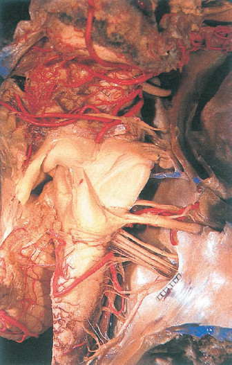
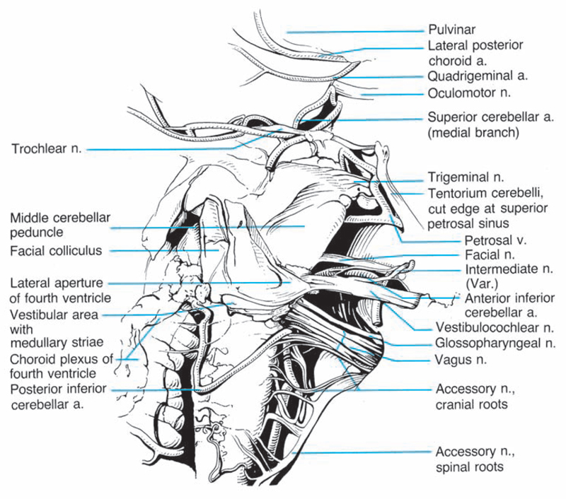
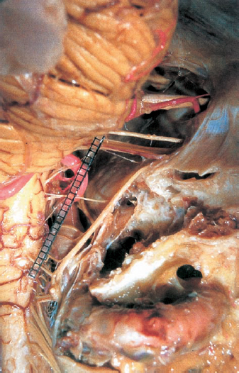
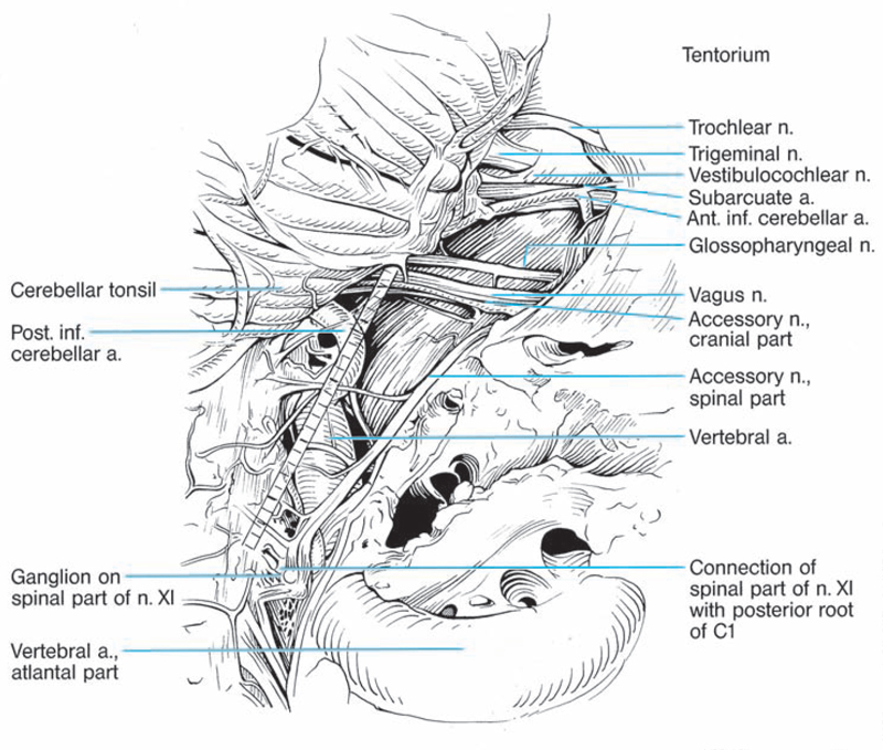
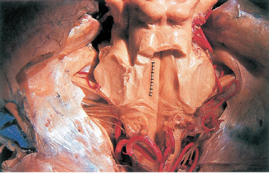
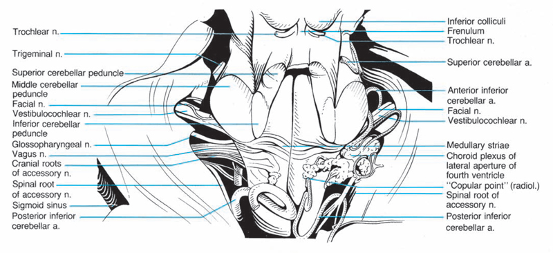
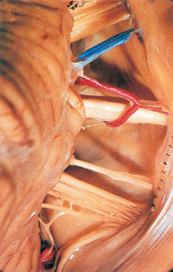
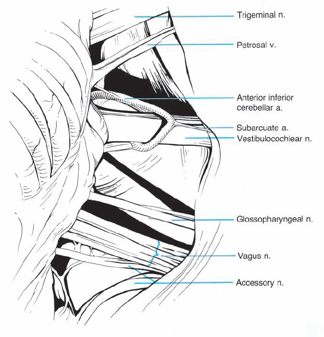
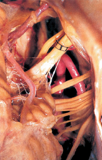
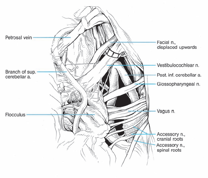
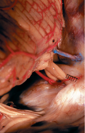
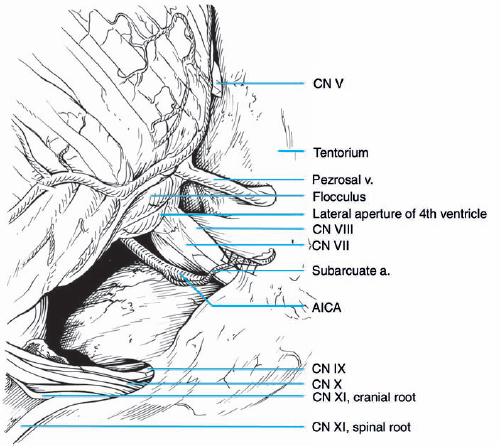
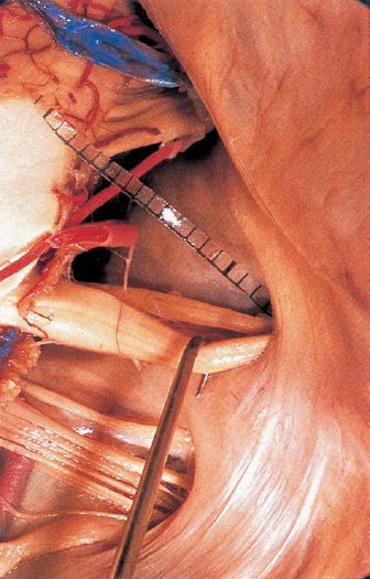
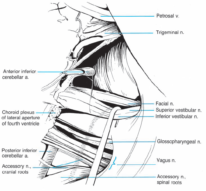
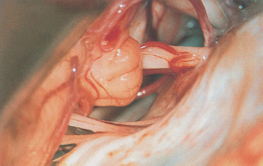
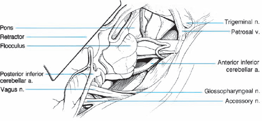
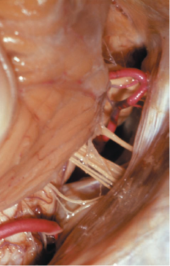
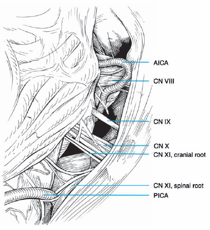
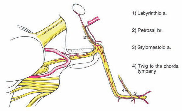
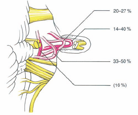
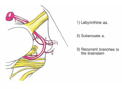
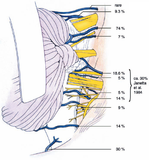
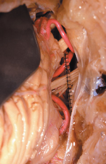
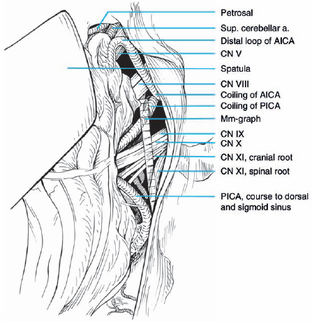
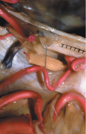
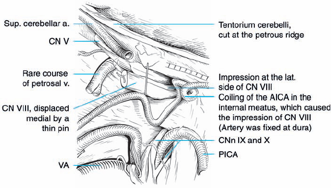
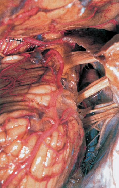
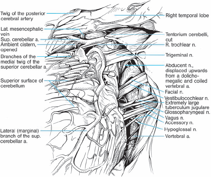
2 Microanatomy of the Cerebellopontine Angle Introduction The feasibility and safety of any microsurgical intervention depend on the surgeon’s familiarity with the neuroanatomy. The appropriate neuroanatomical preparations are therefore included here and presented in relation to the special topographic region. Special attention has been given to the microscopic anatomy. Each actual anatomical specimen is accompanied by a schematic drawing identifying the various structures that are most important. The different specimens have been chosen on the basis of their relevance to the topic of this atlas and the pathology. For identification, the arteries have mostly been injected with red latex and the veins with blue latex. By reviewing the anatomical specimens, the reader can better appreciate the neuroanatomical limits of the surgical intervention as well as the options for enlarging the surgical exposure. This chapter is subdivided into two parts. The first part starts with a general overview of the cerebellopontine region. The anatomy is presented from various visual angles to offer the reader more detail and provide a better grasp of the three-dimensional situation. The neural structures and bone structures are presented using different cross-sections through the petrous bone. The second part, on the special clinical anatomy, focuses on the different topographical relationships of the seventh and eighth nerves in large acoustic neurinomas. This is not a typical anatomical presentation, but rather a clinical one. It starts with the seventh nerve, followed by the eighth nerve, showing the different possible relationships between the anatomical structures and the pathology using real intraoperative situations. To allow the reader to follow the course in three-dimensional space, the intraoperative photographs have been modified using a computer. The courses of the seventh and eighth nerves are marked in orange and green. General Anatomy Fig. 2.1 The right cerebellopontine angle, with exposure of the nerves and arteries. The tentorium has been reflected forward, and the cerebellum has been elevated (viewed from the side and behind). Fig. 2.2 The right cerebellopontine angle, viewed from the lateral aspect. Fig. 2.3 Posterior inferior cerebellar artery, seen from the dorsolateral aspect. Fig. 2.4 The intermediate nerve has been displaced upwards by a loop of the anterior inferior cerebellar artery (a rare variation). Fig. 2.5 Posterior cranial fossa. The tentorium is reflected forward, and the cerebellum is elevated dorsally. The course of the cranial nerves in the posterior fossa en route to the clivus is shown, including the junction between the middle and posterior fossa. Seen from behind, laterally, and above. Fig. 2.6 View of the cerebellopontine angle from above. The tentorium is reflected from the side of the ambient cistern. Notice the acoustic, trigeminal, and trochlear nerves. Fig. 2.7 Nerves and vessels in the posterior cranial fossa (cerebellum sectioned), seen from the lateral aspect. Fig. 2.8 Cranial nerves IV–XI after a transtentorial approach, seen from above. Fig. 2.9 The vestibulocochlear and facial nerves from above, with the anterior inferior cerebellar artery fixed. Fig. 2.10 The intermediate nerve displaced upwards by a loop of the anterior inferior cerebellar artery (a rare variation). Fig. 2.11 The right cerebellopontine angle, seen from above, with the anterior inferior cerebellar artery between cranial nerves VII and VIII. Fig. 2.12 The labyrinthine arteries, seen from above. Fig. 2.13 The labyrinthine arteries, seen from above. Fig. 2.14 The labyrinthine arteries, seen from above, with the anterior inferior cerebellar artery (AICA) coursing between cranial nerves VII and VIII at the entry zone. Fig. 2.15 The right cerebellopontine angle, after removal of the cerebellum by dividing the cerebellar peduncle. Seen from the lateral aspect. Fig. 2.16 The cerebellopontine angle and the craniocervical junction (from behind and below, right side). Fig. 2.17 The brain stem and fourth ventricle from above, with the cerebellum resected. Fig. 2.18 Higher magnification of the course of the cranial nerves within the cerebellopontine angle. Notice also the petrosal vein and the subarcuate artery. Fig. 2.19 An anatomical specimen of the cerebellopontine angle, indicating the cranial extent of the loop of the posterior inferior cerebellar artery. Fig. 2.20 The subarcuate artery, seen from behind and below. Fig. 2.21 A lateral exposure of the right cerebellopontine angle. The eighth nerve has been retracted with a nerve hook, exposing the underlying seventh nerve. Fig. 2.22 The surgeon’s view of the cerebellopontine angle. There is a prominent flocculus overlying the origin of the seventh and eighth nerves on the brain stem. Fig. 2.23 The cerebellopontine angle, seen from the lateral aspect. Fig. 2.24 The various vessels that supply the facial nerve over its entire course. Fig. 2.25 The various positions of the meatal loop of the anterior inferior cerebellar artery. Fig. 2.26 Anatomy of the branches of the anterior inferior cerebellar artery. Fig. 2.27 Anatomy of the transcisternal veins. Fig. 2.28 The retrosigmoidal approach to the cerebellopontine angle. Fig. 2.29 Very rare nerve–vessel relationships in the posterior cranial fossa. Fig. 2.30 An overview of the clivus, brain stem, cerebellum, and cranial nerves, as seen surgically through the combined supratentorial and infratentorial approach.
 Introduction
 General Anatomy
 Special Clinical Anatomy and Topography of the Facial and Vestibulocochlear Nerves in Large Acoustic Neurinomas
Neupsy Key
Fastest Neupsy Insight Engine