(1)
University of Illinois College of Medicine, Chicago, IL, USA
Microcervical foraminotomy (MCF) is the term used by this author to specifically define a posterior microsurgical technique that has proven extremely successful for the resolution of intractable cervical radicular pain [1]. The procedure was developed in 1972 and absolutely minimizes any degree of laminectomy or facet trauma while allowing for a thorough neurolysis and bony foraminal decompression along the posterior and inferior aspect of symptomatic cervical nerve roots. As many as six foramina have been treated at one sitting. The intervertebral disc, stabilizing bony structures, and any foraminal pathology anterior to the root are not disturbed during this operation. Although primarily indicated for the treatment of intractable pain and neurologic deficit of a radicular distribution, the operation has proven to be just as successful in those individuals with persistent radicular symptoms unresponsive to previous anterior cervical or laminectomy techniques.
Clinical Rationale for This Alternative Approach
A disappointing 5-year personal experience in the consistency with which cervical radicular pain responded to accepted surgical techniques prompted this author to reassess his surgical failures in 1971. In the absence of intrinsic neural disease, persisting foraminal pathology either unrecognized or unreachable during a previous surgical operation seemed the most probable etiology. Because anterior foraminal pathology was protected by the vertebral artery and even the anterior foramen entrance could not be surgically reached without inflicting serious morbidity on the intervertebral disc, a conservative microsurgical technique was developed that allows neurolysis and decompression of any posterior irritative foraminal pathology. This technique is termed microcervical foraminotomy (MCF).
Patient Selection
Since 1972, 574 patients with 1571 symptomatic cervical nerve roots have been treated by this conservative posterior surgical approach. As many as six roots from C4 to C8 have been treated at one sitting. In the absence of intrinsic neural disease, preoperative correlation between abnormal radiodiagnostic studies and symptomatic nerve root levels has become minimally important because foraminal pathology treatable by this operation lies largely outside the spinal canal. Surgical indications for MCF are intractable pain or neurologic deficit of a radicular distribution unresponsive to either exhaustive conservative management or previous cervical operations. Regardless of any preoperative structural radiographic pathology, this operation is only applied to those foraminal levels with clinical radicular symptoms. A expert knowledge of cervical radicular neuroanatomy including dermatonal, myotomal, and reflex distributions seems all that is necessary to localize a clinically symptomatic cervical nerve root and thus its associated neural foramen [2].
Instrumentation, Anesthesia, and Positioning
Surgical Microscope and Instrumentation
A Zeiss Opmi 1 surgical microscope with straight binoculars and 300-mm lens provides excellent visibility and surgical mobility. Medium to high magnification is used. A self-retaining retractor with lateral blade (1 cm width, 5 cm depth) and dull medial hook is applied for unilateral wound retraction (Williams’ Microlumbar Discectomy Retractor; Codman and Shurtleff, Randolph, MA). Other than scalpel and electrocoagulator, instruments required are a blunt 90-degree microhook, a 45-degree, 1.5-mm rongeur, 45- and 90-degree, 1-mm rongeurs, and microcurettes (Fig. 42.1). The microcurettes best suited are one that is straight and two additional that have been smoothly angled by the author to 15 and 30 degrees, respectively. These curvatures should be placed approximately 1 cm from the tip.
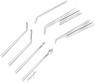
Fig. 42.1
Instrumentation for microcervical foraminotomy (MCF) is minimal. Pictured from top left clockwise are a 90-degree microhook, a 45-degree, 1.5-mm rongeur, a straight microcurette, a 90-degree, 1 -mm rongeur, and a 45-degree, 1-mm rongeur. Additional instruments not pictured are scalpel, electrocoagulator, electrocutting blade, small periosteal elevator, and the author’s 15- and 30-degree-angle microcurettes made by smoothly bending straight microcurettes 1 cm from the cup. (Redrawn with permission from Williams RW. Microcervical foraminotomy: a surgical alternative for intractable radicular pain. Spine 1983;8(7):708–716.)
Patient Preparation, Anesthesia, and Positioning
Patients receive a complete medical and pulmonary assessment. Antiseptic sponges are provided for home showers and shampoos before hospital admission. The operation is carried out with the patient in the sitting position utilizing a Mayfield pin-type head holder. The chin is slightly flexed to reduce cervical lordosis. General endotracheal anesthesia with mechanical ventilation is applied along with cardiac doppler and central venous pressure monitor for the detection and treatment of possible air emboli. The legs are wrapped, and systolic blood pressure is maintained above 120 mmHg with vasopressor as needed.
Operating Time
Wound entrance, closure, and a one-side, one-level foraminotomy require 1 hour. Each additional foraminotomy adds approximately 0.5 hour to the operating time.
Incision Placement
Accurate placement of the skin incision is paramount to ensure minimal operating time and maximal hemostasis. After palpation of a familiar spinous process, a 1-inch needle is inserted well lateral to the midline and localized by lateral radiogram. A 1.5-inch skin incision is then marked in the midline after reviewing the anatomical placement of the needle. Once the wound is open, a visible spinous process is then notched with a rongeur for future identification and grasped on its tip with a metallic clamp. A second lateral cervical X-ray is then obtained to accurately identify the vertebral level. Once the metallic clamp has been removed, the notched spinous process can be followed inward on its lateral surface by finger palpation to locate the symptomatic interlaminar level. A self-retaining retractor is then applied for direct visualization of the deeper anatomy.
This technique has proven absolutely necessary for the exact localization of foraminal levels, because it is extremely easy for even the most experienced spinal surgeon to become disorientated in the depths of a microsurgical cervical wound. If there is any doubt as to the location, check and recheck again until the appropriate interlaminar level is without question. This operation is so successful for the relief of radicular pain that any persistence in its intensity for even 24 hours following surgery likely indicates that the surgeon was in the wrong place.
Surgical Technique
Dissection to the Lamina
A 1.5-inch posterior midline wound is generous for a two-level foraminotomy. If work is to be done bilaterally, expose only one side at a time as this will ensure minimal blood loss from venous oozing in what sometimes can be a lengthy operation. Unless open veins are unattended with the patient in the sitting position, air emboli will not occur.
The technique of foraminotomy presented here is much more difficult for the surgeon when working on a foramen toward the same side as that of the surgeon’s dominant hand. That is, right-side foraminotomies are technically the most difficult for the right-handed surgeon. When working toward the dominant side, tedious wrist extension movements are required while utilizing rongeurs and microcurettes. Such maneuvers are not the case when resculpting a foramen toward the surgeon’s nondominant side. Initially, therefore, always treat those foramina that will be technically the most difficult. Proceeding in this manner will markedly reduce the surgeon’s fatigue and thus further minimize blood loss and operating time.
Once the wound is open, an electrocutting knife is applied to unilaterally separate the paravertebral fascia from the spinous processes. The lamina and ligamentum flavum are often quite superficial, so use extreme caution with the cutting current. The index finger can then be inserted through the fascia opening, applying gentle inward pressures to displace the paravertebral muscles laterally. Periosteal elevators are seldom necessary. Finger palpation allows for accurate anatomical localization of the deeper interlaminar space when correlated with the previously notched spinous process. Meticulous hemostasis is accomplished by electrocoagulation. Care is taken to avoid electrical instrument contact with the ligamentum flavum, thus preventing potential burn injury to the deeper neural structures. A unilateral self-retaining retractor, as previously described, is then inserted to maintain surgical exposure.
Foraminotomy
Once the interlaminar space associated with the symptomatic root has been accurately identified, the ligamentum flavum is separated from its most lateral attachment to the upper lamina using a straight microcurette. This area of separation is usually no more than 1 cm lateral to the midline. If the interlaminar space is quite narrow or the opposing lamina borders are indistinct as a result of osteoarthritic disease, a power bur can be applied to carefully reestablish the tissue planes.
Related posts:


Stay updated, free articles. Join our Telegram channel

Full access? Get Clinical Tree


