32 | Minimally Invasive Techniques |
 | Case Presentation |
History and Physical Examination
A 44-year-old patient had previously undergone a C5-6 anterior cervical decompression and fusion (ACDF) with allograft without instrumentation for severe neck pain and a left C6 radiculopathy. She subsequently had relief of her neck and arm pain, but 3 months later she started to experience a recurrence of her neck and arm pain. Over the next 2 years, her neck pain was worsened and was only relieved when she supported her neck with a brace. She also complained of pain in the left C6 distribution. Otherwise, she had a normal neurological examination with full strength and normal reflexes.
Radiological Findings
Dynamic films with flexion-extension were obtained that demonstrated motion and increased angulation at the C5-6 level, suggestive of pseudarthrosis. Cervical magnetic resonance imaging (MRI) was obtained that demonstrated left C5-6 foraminal stenosis. There was no spinal cord compression.
Diagnosis
Pseudarthrosis status post-anterior cervical fusion with recurrent neck pain and radiculopathy
 | Background |
Modern minimally invasive surgical (MIS) techniques to the spine have mainly focused on access to the lumbar vertebral elements. Initial experience included the use of the endoscope for visualization. More recent improvements with fiberoptic lighting and experience with dilator retractors have spawned the development of tubular retractor systems that allow direct visualization of the spinal elements.
Many reports exist on the ability to decompress the neural structures in the lumbar and thoracic spine via both an anterior and a posterior approach.1–4 Familiarity with these techniques now allows one- or two-level interbody and posterolateral fusions through MIS approaches.5–10 Direct prospective clinical studies comparing these modern MIS approaches with open techniques have not yet been published. However, advocates of MIS claim their clinical results are equal or superior to the more invasive open procedures. MIS techniques typically involve a learning curve due to the challenge in visualization of the spinal structures, and initial operating time may be increased. However, the reported advantages have included less blood loss, less muscle destruction, decreased length of stay, and smaller incisions.
In 1994, a tubular retractor system was first developed. It consisted of multiple thin-walled tubular retractors of variable length. This system was a major founding element in the minimally invasive posterior approaches. In 1997, the micro-endoscopic discectomy (MED) system was introduced and was mostly used in minimally invasive posterior lumbar approaches.11–14 The introduction of METRx system (Medtronic, Inc., Memphis, TN), which provided more working space and better illumination, improved this previous system. Although randomized prospective studies are lacking, many authors have reported their experience with MED and METRx11,15–17 demonstrating the feasibility and safety of these techniques.6,17–22
For the cervical spine, the anatomical constraints are very different and the neural structures are more critical than for the lumbar spine. However, for certain indications, the cervical spine can also be approached with MIS. Scoville et al first described the posterior cervical diskectomy and foraminotomy in 1976.23 This technique was effective in relieving radicular pain in select patients with laterally herniated disk fragments or foraminal stenosis. Compared with the anterior approach to the cervical spine, it was beneficial in avoiding a fusion. However, this standard technique carried with it the disadvantages of extensive paraspinal muscle dissection, neck pain, and potential instability. Roh, in 2000, advocated that the MED technique allowed a better decompression compared with the standard open technique in four cadaveric specimens.19 Adamson performed a microendoscopic posterior cervical laminoforaminotomy for unilateral radiculopathy on 100 patients and had excellent or good results in 97 of them with no serious complications reported.30 Use of the MED technique in posterior cervical diskectomy and foraminotomy showed excellent results with minimal disadvantages.
Minimally invasive cervical laminoplasty for cervical myelopathy secondary to canal stenosis was described by Wang.20 The exposure of six cervical levels was accomplished by creating two small incisions; the diameter of the midsagittal spinal canal was increased by a mean of 38% with this technique. Yuguchi also reported on a similar technique with both cadaveric models and clinical cases of cervical radiculopathy and myelopathy.31
Lateral mass plating with screws were first described by Roy-Camille and colleagues.24–28 Their technique provided immediate stability of the cervical spine and was feasible even when the lamina and spinous processes were damaged. The procedure was further modified and developed by Magerl et al.29 All these techniques focused on safe screw placement on the basis of anatomical landmarks and trajectories to avoid the nerve roots, the spinal cord, and the vertebral artery.
MIS cervical screw fixation has evolved from these initial advancements. There are a few cases reported in the literature.20,21 Growing indications for its use include establishing a posterior tension band for pseudarthrosis, osteomyelitis/diskitis, instability, and trauma. This technique has several advantages. Mainly, the incision size is limited and the musculature attachments to the midline can be preserved, allowing reduction of postoperative pain.20,32 A recent series of 18 patients who underwent one- or two-level cervical lateral mass screw fixation using a minimally invasive technique with tubular retractors was published. The authors concluded that it was a safe and effective technique.33
 | Authors’ Preferred Method of Surgical Management |
The patient presented with C5-6 pseudarthrosis with recurrent left C6 radiculopathy and underwent a posterior C5-6 arthrodesis with minimally invasive right unilateral C5-6 lateral mass screws and a left C5-6 foraminotomy.
Pearls
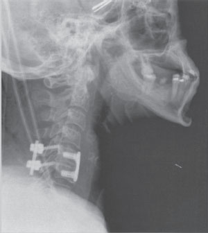
The patient is placed in the prone position with the head fixed in a three-pin head holder while keeping the cervical spine in a neutral posture. Generous use of fluoroscopy is essential to compensate for the lack of direct vision. A guide wire is used to determine the ideal position for the skin incision. A sagittal trajectory parallel to the facet joint is desired and the entry point on the skin should be at the midline approximately two levels below the desired level. Then two parallel fascial incisions just off the midline are made taking care to extend the fascial incisions in the rostral-caudal direction to accommodate the diameter of the tubular retractors. The operator may have to remove the lateral aspects of a bifid spinous process because this may force the incision too laterally. Excessively lateral incisions would result in a more medially placed lateral mass screw. Multiple tubular dilators are then inserted and docked onto the lateral masses. After exposing the surface of the lateral mass and identifying the facet joint, the synovium is cauterized or removed, and the joint can be decorticated and packed with bone graft. A 2 mm burr is then used to establish the starting points for each of the polyaxial screws under direct visualization using standard classical landmarks.24–29 A drill is used to cannulate the lateral mass. After palpation of the trajectory and tapping of the cortex, screws can be inserted. It is advisable to work in a rostral to caudal sequence. At each stage, fluoroscopy should be used to confirm the rostral-caudal trajectory and the operator should make a concerted effort to lateralize the screw, noting that a parafascial incision parallel to the midline might still force excessive medialization of the screw. Once the screws are in, rods can be placed within the heads of the screws and the construct completed in the usual manner. Currently, compressors are being developed that will be compatible with the MIS tubular retractors. Typically, endoscopy is not needed. The visualization with a fiberoptic light source and loupes is usually adequate. Generous use of fluoroscopy should be employed to follow the delivery of the hardware. A postoperative lateral radiograph reveals the implants in good position (Fig. 32–1). In a similar fashion, the same exposure can be used to perform a minimally invasive laminoforaminotomy on the contralateral side. A postoperative computed tomographic (CT) scan demonstrates the bony resection on the contralateral side (Fig. 32–2).

Figure 32–1 A postoperative lateral cervical spine radiograph demonstrating the instrumentation construct.
Stay updated, free articles. Join our Telegram channel

Full access? Get Clinical Tree


