Fig. 12.1
(a) Lateral view and (b) posterior view of the spine. The cervical, thoracic, and lumbar vertebrae are separated by a cartilaginous disc that provides cushioning and allows for movement. The sacral and coccygeal vertebrae are already fused
Each vertebra has critical functional parts. The vertebral body is the weight-bearing portion of the vertebra and is located anterior to the vertebral canal. Posterior to the vertebral body are bony projections that form the vertebral arch: bilateral pedicles, a lamina, transverse processes, facet joints, and a single posterior spinous process (Fig. 12.2). The vertebral canal contains the spinal cord or cauda equina, fat, ligaments, and blood vessels. Under each pedicle, spinal nerves exit the spinal cord and pass through the intervertebral foramen to branch out to the body. Surgeons often remove the lamina of the posterior vertebral arch to access and decompress the spinal cord or spinal nerves to treat spinal stenosis, tumors, or herniated discs.
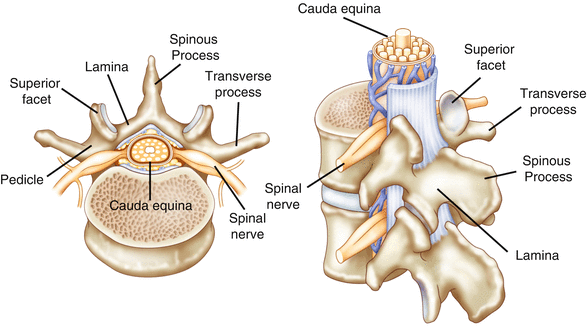
Fig. 12.2
Superior view of a vertebra showing the location of the critical functional parts relative to the spinal canal. The vertebral arch is formed from bilateral pedicles, lamina, transverse processes, facet joints, and a single posterior spinous process that protects the spinal cord and exiting nerve roots
The spinal cord extends from the foramen magnum to spinal level L1. At L1, the terminal portion of the spinal cord is called the conus medullaris. From the conus, a bundle of spinal nerves called the cauda equina further extend down to their respective vertebral level and exit.
Thirty-one pairs of spinal nerves emerge from the spinal cord (Fig. 12.3). There are 8 cervical spinal nerves, 12 thoracic nerves, 5 lumbar nerves, 5 sacral nerves, and 1 coccygeal nerve. Each spinal nerve is composed of motor and sensory fibers that pass through an intervertebral foramen between adjacent vertebrae. Cervical and thoracic spinal nerve roots exit laterally from the vertebral canal between adjacent pedicles, while lumbosacral roots extend downward as part of the cauda equina before exiting through foramina below the spinal cord. Nerve roots can be injured during surgery by electrocautery, drilling, retraction, or misplaced hardware.
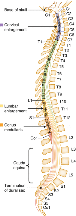
Fig. 12.3
Thirty-one pairs of spinal nerve roots branch off the spinal cord. Cervical and thoracic spinal nerve roots exit laterally from the spinal canal between adjacent pedicles, while lumbosacral roots extend downward as part of the cauda equina before exiting through foramina below the spinal cord
Procedures of the Cervical Spine
The main function of the cervical spine is to support and move the head. The first cervical vertebra (C1) is often called the atlas and contains a bony ring, without a body, that connects directly to the skull. Together with the C2 vertebra, or axis, these two vertebral joints attach the skull to the spine and allow for movement of the head. Cervical spine surgery is generally performed to treat nerve impingements (radiculopathy), spinal cord compression (myelopathy), or spinal instability that is causing pain and weakness. Common cervical procedures include anterior cervical discectomy and fusion (ACDF), posterior cervical fusion (PCF), and cervical corpectomy. Risks during surgery include but are not limited to cord contusion, motor loss or weakness, peripheral nerve injury, or vascular compromise.
Injury to the C5 nerve root is the most common injury from cervical surgery and can result in pain, paresis, or paralysis of the shoulder. Other nerve roots are subject to postoperative palsy, but most complications occur at C5 due to its shorter length and more obtuse angle of exit from the foramen [1, 2]. The C5 nerve root is the only nerve supply to the deltoid muscle of the shoulder, so injury to the nerve root leads to an obvious weakness of this muscle and difficulty raising the arm to the side [2]. The potential for C5 palsies can been detected by using EMG and Tc-MEP monitoring during spinal surgery with specific focus on the deltoid and biceps brachii muscles. Brachial plexopathy resulting from positional or traction-induced injury may mimic C5 palsy [3]. Many surgeons maintain downward traction on the shoulders during a cervical surgery. Injury to the brachial plexus may result from this traction and can be detected by monitoring SSEPs during the procedure.
Anterior Cervical Discectomy and Fusion
ACDF is a procedure often performed to remove a herniated or degenerative disc. Narrowing of the vertebral canal, a condition called spinal stenosis, can cause chronic pain, numbness, and muscle weakness in both upper and lower extremities. Bone spurs can also develop resulting in foraminal stenosis thus compressing the exiting spinal nerves.
The surgical approach during an ACDF is from the anterior, or front, of the neck. An anterior approach allows the surgeon access to the disc space without disturbing the spinal cord, spinal nerves, and posterior neck musculature. An incision is made and midline structures and musculature are retracted to expose the vertebral bodies and disc space. Bone and disc fragments are removed in order to decompress the spinal cord and nerve roots. After the disc space is cleaned out, a bone graft is placed in a metal cage and fit between the vertebral bodies. The adjacent vertebrae are held in place with a metal plate and screws. The ultimate goal of the surgery is to create a bony fusion between the adjacent vertebrae, which occurs as a result of the bone graft placement. The metal hardware simply acts as a cast, stabilizing the spine until fusion occurs (Fig. 12.4).
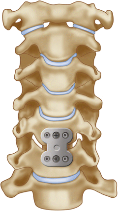
Fig. 12.4
Example of a metal plate and screws placed over the bone graft during ACDF surgery
A common complication in anterior cervical surgery is vocal cord paralysis resulting from an injury to the recurrent laryngeal nerve (RLN). Patients with RLN palsy may experience hoarseness, develop a cough, or lose their voice completely and it may take several months for the nerve to recover [4, 5]. EMG monitoring using a special endotracheal tube has been shown to be useful in detecting injury to the RLN. Monitoring the RLN is discussed in detail in another chapter of this book.
Multimodality monitoring for anterior cervical fusions should include upper and lower SSEPs and Tc-MEPs to monitor spinal cord function, as well as EMG from the myotomes at risk to provide protection for the nerve roots. Positional injury may also be detected with upper extremity SSEP monitoring.
Posterior Cervical Fusion
If spinal stenosis cannot be relieved by an anterior approach, or if the patient’s spine is not stable enough for this approach, the surgeon may opt for a posterior cervical laminectomy and fusion (PCF). The object of this procedure is decompression of the neural elements and stabilization of the cervical spine. Posterior fusions are also performed for instability of the cervical spine resulting from trauma or a degenerative pathology.
For PCF surgery, the patient is placed in a prone position (on their abdomen) with the head made immobile by placing in a special frame called a Mayfield (Fig. 12.5). The head is held in the Mayfield with pins. After the incision is made in the back of the neck, the surgeon will then dissect down through the subcutaneous tissues to the fascia overlying the spinous processes. Retractors are inserted to hold the muscle away from the spine, and the surgeon will begin to remove the lamina and other bony elements in order to decompress the spinal cord and nerve roots.
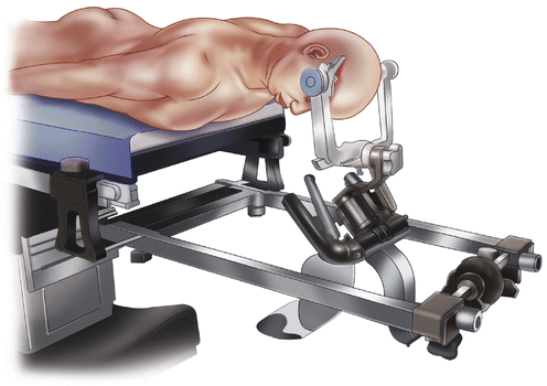
Fig. 12.5
Drawing of a patient positioned for a posterior cervical fusion using a Mayfield head frame
Various types of instrumentation are used to posteriorly fuse the cervical spine. Wiring can be used to stabilize the upper cervical vertebral segments (C1 and C2). Cervical vertebrae have anatomical structures not found elsewhere in the spine called the lateral masses, which are larger than the pedicles and are satisfactory targets for screw insertion. Placement of lateral mass screws and rods provides equal or greater biomechanical stability when compared to anterior plating or interspinous wiring techniques [6] (Fig. 12.6). Placement of lateral mass screws does not depend on the integrity of the laminae, pedicle, or spinous processes to achieve fixation as in case of cervical wiring and pedicle screws. The limitations of lateral mass fixation include risk of injury to the adjacent nerve roots, vertebral arteries, or facet joint [6, 7].
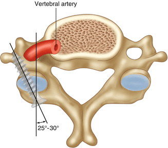
Fig. 12.6
Placement of lateral mass screws in a cervical vertebra
Similar to ACDFs, multimodality monitoring using SSEPs and Tc-MEPs to monitor the spinal cord and EMGs to monitor the nerve roots is the preferred monitoring plan for PCF.
Cervical Corpectomy
When the cervical disease involves more than just the disc space or a single level, it may be necessary to remove the vertebral body and adjacent discs in a procedure called a corpectomy. This can be necessary for multilevel stenosis, tumor removal, or vertebral infection. The approach is similar to that of an ACDF. Once a majority of the affected vertebral bodies and disc material have been removed, a graft—typically shaped bone or a titanium cage—is fitted to support the anterior vertebral column (Fig. 12.7). The cervical spine is further stabilized with a metal plate and screws similar to that used in an ACDF. If the spine appears unstable after the anterior corpectomy, a PCF may be necessary to provide long-term stability. Recommended IOM is similar to that of an ACDF [8].
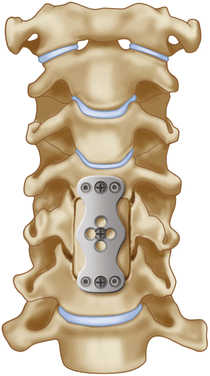
Fig. 12.7
Following removal of the vertebral body, a wedge-shaped bone graft is inserted into the space created by the corpectomy and is stabilized using a screw and plate system similar to an ACDF
Procedures of the Thoracic Spine
The thoracic vertebrae—T1 through T12—provide attachment to the rib cage. Due to the presence of the ribs and position of the spinous processes, the thoracic spine is stiff and motion is limited. This immobility can put strain on the adjacent cervical or lumbar spine, making the areas from C6 to T2 and T11 to L2 especially susceptible to injury. Most commonly trauma or metastatic lesions are the cause of thoracic spine surgery. Thoracic laminectomy and fusion and corpectomy are performed similar to other levels. Burst fractures (discussed below) are often seen in the lower thoracic segments. Among the most commonly monitored procedures of the thoracic spine are for correction of scoliosis and spinal deformity.
Surgery for Scoliosis Correction
Scoliosis describes an abnormal, side to side, curvature of the spine. The two most common forms of scoliosis are degenerative and idiopathic (adolescent). Degenerative scoliosis is caused by a deterioration of the facet joints and occurs most commonly in people over 65. Surgery is often performed to reduce pain. Older patients may have osteoporosis and may require many levels of instrumentation to achieve a complete fusion. Idiopathic or adolescent scoliosis is seen in children and teenagers and usually is discovered during routine doctor’s exams. It is necessary to prevent the curvature from progressing as the child ages. If the curve measures <20°, surgeons often choose to brace the spine or continue to observe the progression of the curvature. Surgery for adolescents with scoliosis is only recommended when the curvature is >45° and continuing to progress [9]. A high degree of curvature may put the patient at risk for cardiopulmonary compromise as the curve of the spine rotates the chest and decreases the vital capacity (ability to breathe).
Related posts:


Stay updated, free articles. Join our Telegram channel

Full access? Get Clinical Tree


