Movement Disorders

Movement Disorders
Matthew Lee Uhles
Raman K. Malhotra
In recent years, there has been a growing awareness and interest in sleep-related problems. It is being increasingly recognized that there are several different kinds of movements, behaviors, or phenomena that may occur in sleep. These can range from a variety of physiologic to pathologic conditions with a wide variety of manifestations. Some behaviors or movements are more likely to be associated with a specific state or stage of sleep. This chapter includes a basic approach to a patient who presents with abnormal events or behaviors in sleep. It focuses predominantly on the roles of polysomnography (PSG) or video-polysomnography (VPSG) and the technologist in the differential diagnostic process of “motor events in sleep.”
APPROACH TO A PATIENT WITH MOVEMENT DISORDER IN SLEEP
Because many of the disorders associated with motor movements during sleep are sporadic in nature, accurate identification and diagnosis can be clinically challenging. As a result, a detailed sleep and medical history including general medical, neurologic, psychiatric, social, and family history all form the foundation in the assessment of patients with complaints of movement disorders during sleep. Particular attention should be given to the age of onset, time of night, pattern, duration, and frequency of the abnormal motor movement or behavior (1). In most cases, the patient is asleep and unaware during the time of the motor event; therefore, it is often beneficial to include the patient’s bed partner or parent (in case of a child) at the time of the clinical interview. Often, the observer can provide more detailed observations regarding the motor event and the patient’s state of mind immediately following the event. With the wide availability of personal video recorders, capable of low light recording, home recording may be used for the purpose of providing the clinician with a detailed illustration of the motor event within the patient’s home. Vignatelli et al. reported that trained observers were able to correctly identify nocturnal frontal lobe epilepsy (NFLE) associated with major body movements using video recording alone, but video recording alone lacked the specificity in those cases associated with brief or minor body movements (2).
The International Classification of Sleep Disorders, 3rd edition (ICSD-3) lists 10 core categories of parasomnias with only one, rapid eye movement (REM) behavior disorder (RBD), requiring VPSG as an essential diagnostic requirement (3). VPSG is useful in distinguishing parasomnias such as sleepwalking or RBD from disorders such as NFLE or potentially injurious or atypical behaviors (i.e., unusual age of onset, duration, frequency, or occurrence) (1, 4). Fois et al. demonstrated that the utility of VPSG in these types of presentations was as high as 40% (5). Therefore, VPSG should be considered for patients with unusual nocturnal events that are potentially harmful or suggestive of another underlying disorder (6). If the patient is taking benzodiazepines or antidepressants, VPSG may need to be conducted after they are appropriately weaned from these medications as they may inhibit clinical manifestations during the testing (5).
Correlation of an event with the time of the night and the correct sleep stage is essential for correct diagnosis of motor events. For example, arousal disorders typically occur in the first third of the night during stage N3 slow-wave sleep (SWS); RBD occurs during REM sleep usually in the last half to third of the night; epileptic seizures are more common during nonrapid eye movement (NREM) sleep; rhythmic movement disorder usually occurs during sleep-wake transitions; and in dissociative disorders, patients may appear to be asleep, but the PSG reflects an electroencephalographic (EEG) pattern of wakefulness.
Today, most commercially available PSG systems provide digital video recording synchronized with the PSG signals, which helps time-lock the motor events to the PSG signals. It also allows for a segment of PSG to be reviewed at varying paper speeds (window durations), filters, sensitivities, and montages, which help in the detection of abnormalities and differentiation from artifacts. The infrared camera is useful for recording nighttime events. Double cameras may be used to capture the face and body simultaneously. The camera should be mounted on the wall or ceiling across from the head end of the bed. An intercom with a microphone near the patient must be available for audio recordings. The monitoring station should have a remote control, which can zoom or tilt the camera for adequate viewing. Video recordings can be played back in real time or at a slower speed to better visualize and analyze the section of interest. Digital video recordings are the preferred method for synchronized PSG and video data storage for later recall and review. Although the minimum required by the American Academy of Sleep Medicine (AASM) is 1 frame per second, the digital video should be recorded with sufficient resolution and frame rate to allow for adequate visualization during review to differentiate subtle movements and establish proper temporal relationship (3). The standard indications for PSG are outlined in the published guidelines by the AASM. Table 20-1 lists the indications for VPSG (7).
In routine PSG recordings, most laboratories utilize six EEG channels (F3-M2, F4-M1, C3-M2, C4-M1, O1-M2, and O2-M1) with electrodes placed in these locations according to the International 10-20 System of Electrode Placement (3). Although the EEG electrodes routinely used in PSG are typically insufficient to accurately identify and localize abnormal EEG activity, abnormal motor activity is often observed in cases where the patient’s clinical history did not indicate the possibility of this activity. In these cases, it may be helpful to re-reference from the standard contralateral derivation (F3 referenced to M2) to ipsilateral deviations (F3-M1). This allows for better verification and possible localization of the epileptiform activity. Extending scalp leads to include the full 10 to 20 EEG is typically required to better differentiate patients whose motor movements are due to epileptiform activity arising from other etiologies.
VPSG combines video recording with an extended EEG montage, PSG monitoring, and at times additional electromyographic (EMG) electrodes. It is superior to conventional PSG for the evaluation of parasomnias, nocturnal seizures, and other motor events in sleep because of the increased capability to identify and localize EEG abnormalities and to correlate the observed motor behavior with EEG and PSG variables (8). Watember et al. demonstrated that adding video recording to routine EEGs increases the diagnostic yield of routine EEGs in children with frequent paroxysmal events (9). The specific montage used varies depending on the indication for the study, the number of channels available for EEG, and the need to record additional PSG parameters. Table 20-2 lists a few sample recording montages that can be further customized as required (10). Foldvary et al. showed that seizure detection was better using 7 and 18 channels (sensitivity of 82% and 86%, respectively) rather than 4 EEG channels (sensitivity of 67%) in patients with temporal lobe seizures; the same was not true for frontal lobe seizures, in which accuracy was similar regardless of the number of EEG channels available (11). A follow-up study by Foldvary et al., exploring accuracy of the 8-channel montage recommended by the American Clinical Neurophysiology Society (addition of T3 and T4 to standard PSG EEG electrodes), showed the abbreviated 8-channel montage was only slightly inferior to the 18-channel montage regarding interpreter agreement distinguishing seizure activity from nonepileptic activity (78% agreement for 8 channel and 84% agreement for 18 channel) (12). The 8-channel montage, however, was not as effective in localizing the seizure activity as the 18-channel montage (27% and 49%, respectively), especially in the temporal and parieto-occipital regions.
Table 20-1 Indications for Video-Polysomnography
  • Unusual and complex arousal disorders

  • Complex behaviors suspicious of RBD but not absolutely certain based on the history

  • Behavior and motor events at night suggesting possible nocturnal seizure disorder

  • Excessive daytime sleepiness in patients with epilepsy to determine if excessive sleepiness is due to repeated nocturnal seizures, an undesirable side effect of antiepileptic medications, or an associated sleep disorder (e.g., sleep apnea)

  • Suspected psychogenic dissociative disorder

  • Other motor parasomnias which may be mistaken for nocturnal seizures, for example, rhythmic movement disorder, bruxism

  • Involuntary diurnal movement disorder persisting during sleep

  • Coexisting second sleep disorder, for example, narcolepsy, RBD, obstructive sleep apnea, or sleepwalking

  • For medicolegal purposes, when the patient presents with violent behavior during sleep, video-polysomnography studies are mandatory to evaluate such patients for making a correct diagnosis of parasomnias or seizure disorders

RBD, REM behavior disorder.

Reprinted from Chokroverty, S. (2003). Polysomnography and related procedures. In M. Hallett (Ed.), Handbook of clinical neurophysiology: Movement disorders (Vol. 1, 1st ed., p. 145). Amsterdam, The Netherlands: Elsevier. Copyright © 2003 Elsevier. With permission.

Table 20-2 Sample Electroencephalographic-Polysomnographic Montages

Indication

Montage

Arousal disorder, possible nocturnal seizures (36 channels)

F3-M2, F4-M1, C3-M2, C4-M1, O1-M2, O2-M1, Fp1-F7, F7-T3, T3-T5, T5-O1, Fp1-F3, F3-C3, C3-P3, P3-O1, Fp2-Fp8, F8-T4, T4-T6, T6-O2, Fp2-F4, F4-C4, C4-P4, P4-O2, Fz-Cz, Cz-Pz, LEOG-M2, REOG-M1, Chin EMG, LAT EMG, RAT EMG, snoring, PTAF, thermistor, thoracic effort, abdominal effort, ECG, SaO2

RBD, possible nocturnal seizures (28 channels)

F3-M2, F4-M1, C3-M2, C4-M1, O1-M2, O2-M1, Fp1-F7, F7-T3, T3-T5, T5-O1, Fp2-Fp8, F8-T4, T4-T6, T6-O2, LEOG-M2, REOG-M1, Chin EMG, LAT EMG, RAT EMG, LED EMG, RED EMG, snoring, PTAF, thermistor, thoracic effort, abdominal effort, ECG, SaO2

RBD, possible nocturnal seizures (38 channels)

F3-M2, F4-M1, C3-M2, C4-AM1, O1-M2, O2-M1, Fp1-F7, F7-T3, T3-T5, T5-O1, Fp1-F3, F3-C3, C3-P3, P3-O1, Fp2-Fp8, F8-T4, T4-T6, T6-O2, Fp2-F4, F4-C4, C4-P4, P4-O2, Fz-Cz, Cz-Pz, LEOG-M2, REOG-M1, Chin EMG, LAT EMG, RAT EMG, LFD EMG, RFD EMG, snoring, PTAF, thermistor, thoracic effort, abdominal effort, ECG, SaO2

PLMS, extended limb coverage (24 channels)

F3-M2, F4-M1, C3-M2, C4-M1, O1-M2, O2-M1, LEOG-M2, REOG-M1, Chin EMG, LAT EMG, RAT EMG, LPT EMG, RPT EMG, LED EMG, RED EMG, LFD EMG, RFD EMG, snoring, PTAF, thermistor, thoracic effort, abdominal effort, ECG, SaO2

ECG, electrocardiogram; EMG, electromyogram; EOG, electrooculogram; L, left; LAT, left anterior tibialis; LED, left extensor digitorum; LFD, left flexor digitorum; LPT, left posterior tibialis; R, right; RAT, right anterior tibilais; RED, right extensor digitorum; RFD, right flexor digitorum; RPT, right posterior tibilais; PTAF, pressure transducer airflow.

Modified from Malow, B. A., & Aldrich, M. S. (2003). Polysomnography. In S. Chokroverty, W. A. Hening, & A. S. Walters (Eds.), Sleep and movement disorders (1st ed., p. 128). Philadelphia, PA: Butterworth-Heinemann. Copyright © 2003 Elsevier. With permission.

Adjunct EMG channels to record from additional muscles are recommended in special situations. In patients with preliminary diagnosis of RBD, adding electrodes over the forearm flexor and extensor muscles aids in detecting upper extremity movements. These electrodes are placed 2 to 3 cm apart in line with the lateral edge of the belly of the forearm flexor (outside of the underside of the forearm) and belly of the forearm extensor (center of the topside of forearm) (Figs. 20-1 and 20-2) (13). Proper localization of these muscle groups can be verified by asking the patient to bend only at the base of the fingers, avoiding bending at the distal joints, and extend the fingers backward without moving the wrists, to identify the flexor and extensor muscles, respectively (3). Although bruxism can be accurately identified with standard chin EMG derivations recording mentalis and submentalis muscles, the addition of at least one masseter electrode aids in accurately identifying sleep bruxism (SB). The masseter muscle is best located by palpating the belly of the muscle several centimeters in front of the ear while the patient clenches the teeth. The recording pair can be either a single electrode placed over the center of the belly of the masseter referenced to one of the standard chin EMG leads or two masseter electrodes placed 2 to 3 cm apart in line with the direction of the muscle fibers referenced to one another (3, 13). In cases of propriospinal myoclonus (PSM), additional EMG electrodes (sternocleidomastoid, intercostal, deltoid, orbicularis oculi, and rectus abdominis) to record the progression of muscle activation can be useful to differentiate between organic and psychogenic etiologies (14, 15). Finally, placing a pair of electrodes along the back of the neck over the paraspinal muscles may be useful when rhythmic movement disorder is suspected (3).
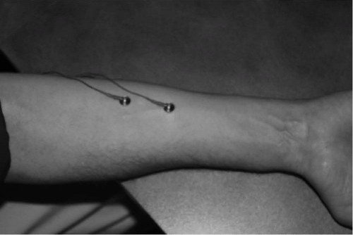
Figure 20-1 Proper placement of electrodes on the forearm flexors, 2 to 4 cm apart in line to the lateral edge of the belly of the forearm flexor (outside of the underside of the forearm).
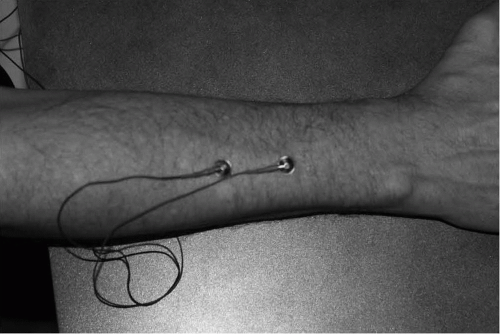
Figure 20-2 Proper placement of electrodes on the forearm extensors for evaluation of parasomnia during a sleep study.
VPSG may help define and classify abnormal nocturnal motor events and behaviors into different diagnostic entities, as listed in Table 20-3. Frequently, many nocturnal motor events may be mistaken for seizures on the basis of history alone, for example, confusional arousals, sleep terrors, bruxism, rhythmic movement disorder, or RBD. These conditions can be diagnosed and differentiated from one another on the basis of characteristic clinical features combined with the use of VPSG. The major disadvantage of VPSG is the cost of the study: the technologist time needed to place an extended EEG montage and then continuously observe the patient throughout the study. Interpretation of PSG with video and extended EEG montage requires skills in both sleep medicine and seizure recognition.
INDICATIONS FOR VPSG
Observation of the EEG and EMG patterns preceding, during, and following the events is important to enhance accurate diagnosis. Unfortunately, because of their sporadic and unpredictable occurrence, nocturnal motor spells are difficult to capture within the controlled environment of the sleep center. The chance of capturing a motor event increases with the increasing frequency of home occurrence of the abnormal motor event. For these reasons, as well as the alterations in naturally occurring sleep patterns of “first night effect,” it has been suggested that one night in the sleep center is insufficient for diagnostic purposes for suspected nocturnal motor events. Therefore, some sleep centers schedule patients for two consecutive nights to maximize the yield of recording an event. Alternatively, a few reports have suggested that one night is sufficient to confirm a diagnosis (5, 16).
Several studies have reported increasing the likelihood of capturing a nocturnal motor spell during VPSG by priming the patient using a variety of different means, including sleep deprivation, medication, alcohol, noise, and baiting (17). Sleep deprivation has long been a standard tool in increasing the yield of documenting abnormal EEG activity in seizure patients. With the high correlation of sleepwalking associated with stage N3 sleep, coupled with the SWS rebound observed on nights following sleep deprivation, several researchers have experimented with sleep deprivation to prime sleepwalking patients. Several studies with sleep deprivation periods of 24, 25, and 38 hours significantly increased the number of sleepwalking episodes in sleepwalkers (18, 19, 20, 21). Only one study failed to show an increase in sleepwalking episodes after 36 hours of sleep deprivation (22). Most of these studies involved recovery sleep periods occurring during daytime hours in patients who habitually slept at night. Given that REM rebound is often observed following sleep deprivation, this technique may also increase the likelihood of observing an RBD episode in the sleep center. Although there are no controlled studies to date using medications or alcohol to elicit motor events, there are published reports of medications and/or alcohol associated with sleepwalking (17, 23). Presentation of a loud sound during stage N3 sleep for NREM motor events and during REM for REM-related motor events has been reported to trigger motor events (17). Pilon et al. even combined the use of a loud stimulus with 25 hours of sleep deprivation to elicit sleepwalking spells in 100% of the 10 patients, in which they attempted this technique (19). In the case of sleep-related eating disorder (SRED), placing a table with the patient’s preferred food and drink beside the bedside was demonstrated to evoke episodes of documented sleep eating in 26 out of 35 patients (24).
TECHNICAL CONSIDERATIONS
Patient Safety
The increased risk of injury in patients with unexplained nocturnal motor activity requires that appropriate precautions be incorporated to ensure the safety of the patient during a PSG (25). The greatest risk of patient injury is from a sudden motor movement with the patient leaping or falling out of bed. The monitoring technologist should remain alert and attentive throughout the recording, remaining vigilant to intervene immediately if he or she perceives a potential risk to the patient. To reduce response time, it is recommended to locate the patient in the room closest to the technologist monitoring area. It is not always feasible for the technologist to react in time to prevent an injury to the patient. Therefore, universal seizure precautions are recommended. These precautions include keeping the bed as low to the floor as possible, removing any unnecessary equipment from the room, and maintaining constant observation. Some sleep centers recommend the use of bed rails or pillows to restrict the mobility of the patient during a nocturnal spell. Although these measures can impede the patient from leaving the bed, they can also trip the patient or (in case of bed rails) even increase the height from which the patient jumps out of bed. Thus, in some instances, these safety precautions may actually increase the likelihood of a patient injury (26). Individual sleep centers will need to develop their own policy regarding the use of rails or pillows. Patient injuries from striking sharp corners of furniture can be minimized by padding them with foam corners commercially available for nurseries. Any breakable room decorations (lamps, vases, etc.) that may injure the patient should be removed from the room. In extreme cases such as with a history of the patient diving out of bed, padding or a second mattress placed on the floor next to the patient’s bed may further reduce the risk of injury.
Table 20-3 Movement Disorders Stage and Characteristics

Disorder

Stage of Sleep

Other Characteristics

Confusional arousals

Predominantly N3, but any NREM

↑ Arousal out of N3/N1 theta/poorly reactive alpha

Sleep terrors

Predominantly N3, but any NREM

↑ Arousal out of N3/often associated with ≠ heart and respiratory rate. Limited recall.

Sleepwalking

Predominantly N3, but any NREM

↑ Arousal out of N3/N1 theta/poorly reactive alpha. Limited recall.

Nightmare

REM sleep

↑ REMs and mild ≠ heart and respiratory rate. Often associated dream recall.

REM behavior disorder

REM sleep

↑ Tonic or phasic activity during REM. Often associated dream recall.

Dissociative disorder

Wakefulness

Wakefulness before, during, and after event

Sleep-related eating disorder

Predominantly N3, also NREM/REM

↑ Arousal out of N3

Bruxism

All stages

Typical electromyographic artifact/audible tooth grinding

Rhythmic movement disorder

Typically transitional sleep, but any stage

Repetitive/stereotyped rhythmic movement

Epileptic seizures

NREM > REM

Sharps, spikes, or slow waves on EEG

Pseudoseizures

Wakefulness

No epileptiform activity noted

Propriospinal myoclonus

Sleep onset

Muscular jerks during transition from wakefulness to sleep

Excessive fragmentary myoclonus

All stages

Brief myoclonic potentials lasting >10 min

Sleep starts

Sleep onset

Sudden, brief, simultaneous contractions

Hypnagogic foot tremor

Sleep onset, N1 and N2

Rhythmic movement of feet and toes

Alternating leg muscle activation

All stages

Brief alternating leg movements

Myoclonus of infancy

All stages

Bilateral large myoclonic jerks in infants

Panic attacks

Typically transition from N2 to N3

↑ Sleep latency, ↑ wake after sleep onset, accompanied by other panic attack symptoms

Posttraumatic stress disorder

All stages

↓ Total sleep time, ↑ awakenings, ↓ sleep efficiency

↓ REM and ↑ REM density

EEG, electroencephalography; NREM, nonrapid eye movement; REM, rapid eye movement.

↑=increase in designated measurement or observation; ↓=decrease in designated measurement or observation.

Technologist Interactions
It is imperative for the technologist to provide detailed notes regarding his or her direct observation of the motor event. The observation notes should document the stage of sleep in which the event occurred and whether it was elicited or preceded by an arousal or any other stimulus. The technologist should refrain from drawing any conclusions in the notes but should objectively describe the observed behavior. It is helpful to describe the onset, progression, and duration of the event. Describing if the movement was repetitive, stereotypical, or unique is also helpful. If time allows, adjusting the camera to further isolate and highlight the motor event can facilitate classification. Because motor movement cases are sometimes associated with injuries to others, these cases sometimes result in medical-legal issues. Often, the sleep evaluation may precede the injury by months or even years. Therefore, the technologist should approach documentation in these cases with the same detail and rigor as if every case is a forensic one.
Often, the technologist’s assessment and documentation of the patient’s conscious state after the motor event is the key diagnostic component in accurately differentiating one motor event from another. Immediately following a motor event, the technologist should assess and document the patient’s mental status and awareness without posing leading questions that may alter the patient’s self-report. For example, following a motor event, the technologist should not ask “What were you dreaming about?” Instead the technologist could ask the more open-ended question “What thoughts or images were you just thinking before I entered the room?”
CHARACTERISTIC PSG FINDINGS IN “MOTOR EVENTS IN SLEEP”
PSG Findings in Motor Parasomnias
Disorders of Arousal (from NREM Sleep)
Disorders of arousal (DOA); confusional arousals, sleepwalking, and sleep terrors typically occur during stage N3 sleep, most commonly in the first third of nocturnal sleep, but can also occur later in the night and from other NREM sleep stages—even during daytime naps (27, 28, 29, 30). Although diagnosis of a DOA is typically based only on clinical criteria, VPSG is often indicated if the patient has a history of nocturnal injury or to differentiate them from RBD or nocturnal seizures. Patients suffering from DOA do not typically have any recollection of their motor activity and do not usually report dream content correlating with their motor activity during sleep. Complex motor activity such as talking, eating, driving, and sexual activity has been shown to occur during NREM sleep parasomnias. Eyes are usually open during the event, although the patient may appear clumsy and confused (3).
Many times during a DOA, the EEG will be difficult to interpret secondary to artifact from movement, although the event in question should be preceded by NREM sleep. If the EEG is readable, it can continue to show a slow-wave pattern (delta waves) (Fig. 20-3). The EEG may also show NREM stage N1 theta patterns, repeated microarousals, or diffuse, slow, poorly reactive alpha rhythm, all indicating incomplete awakening (see Fig. 20-4) (27, 31, 32, 33). Sleep deprivation (the night before study) and forced awakenings during stage N3 sleep have been used by some to increase the chance of precipitating an episode during an overnight sleep study (19).
Sleep terrors are sudden episodes of terror occurring out of NREM sleep initiated by a cry or loud scream and accompanied by fear, confusion, and autonomic symptoms (rapid heart rate, rapid respiratory rate, and sweating). On PSG, sleep terror, in particular, is associated with tachycardia and increased respiratory rate in addition to the above-mentioned EEG findings, which can also be found in other arousal disorders.
Even if the typical episode of motor activity does not occur during the attended sleep study, a PSG is still helpful for identifying arousal disorders by displaying NREM sleep instability or long blocks of stage N3 sleep ending in spontaneous arousals (34). Lopez et al. recently published a quantified measurement of Slow Wave Sleep Fragmentation Index (SWSFI) (35). The SWSFI is the sum of SWS interruptions per hour of SWS. They demonstrated that 6.8 or more SWS interruptions per hour correlated with a diagnosis of DOA. The PSG is also useful in evaluating for possible underlying sleep disorders (obstructive sleep apnea and periodic limb movement disorder [PLMD]) that could be fragmenting sleep and precipitating the arousal disorder (Fig. 20-5).

Only gold members can continue reading. Log In or Register to continue

Stay updated, free articles. Join our Telegram channel

Dec 12, 2019 | Posted by in NEUROLOGY | Comments Off on Movement Disorders

Full access? Get Clinical Tree

Get Clinical Tree app for offline access