Chapter 15 Multiple Sclerosis
Neurologists base a diagnosis of MS on repeated neurologic symptoms and signs disseminated in both space and time. They use recently developed diagnostic criteria in an effort to accommodate the significant advances in radiology. These criteria, named for the senior member of an international panel, Dr. W. Ian McDonald, rely on the number and location of magnetic resonance imaging (MRI) lesions as well as the number of attacks. Neurologists also use certain cerebrospinal fluid (CSF) findings and evoked potentials (see later) to support a diagnosis.
Still maintaining a high sensitivity and specificity, neurologists now diagnose MS during its first episode. They can institute therapy and blunt, although not halt, the illness early in its course. Using current criteria also allows neurologists to follow patients’ subclinical as well as clinical progression and monitor their response to treatment.
Etiology
MS – usually a chronic recurring illness – typically begins with 1-mm to 3-cm patches of inflammation developing in the oligodendrocyte-generated myelin sheaths of CNS axons. T-cell-mediated inflammation strips myelin from (demyelinates) axons and eventually leaves sclerotic (Greek, sklerosis, hard) plaques scattered throughout the CNS. Plaques disseminated throughout the myelin or “white matter” of the cerebrum, cerebellum, spinal cord, ocular motility system, and optic nerves constitute the signature of MS.
When deprived of their myelin insulation, axons transmit nerve impulses slowly or not at all. Some deficits resolve as myelin inflammation spontaneously subsides or anti-inflammatory medications, such as steroids, suppress it. As plaques recur, develop in new areas, and accumulate, MS evolves from an acute inflammatory to a chronic degenerative condition. Sooner or later the plaques leave permanent neurologic deficits.
Although MS acts primarily as a CNS demyelinating disorder, its pathology includes prominent axon degeneration. In contrast to demyelination associated with plaques, axon degeneration regularly produces permanent mental and physical disabilities.
The illness’ mean age of onset is 33 years, with 70% of cases developing between 21 and 40 years. Some studies have reported that many patients suffered their first or a subsequent MS attack after a medical insult, such as infection, childbirth, head or spine trauma, intervertebral disk surgery, or electrical injury. Other studies have reported that psychologic stress preceded the first MS or subsequent attacks. However, most carefully controlled prospective studies have shown that these insults do not play a major role in either causing or exacerbating MS.
The specific cause remains unknown, but studies suggest that genetic susceptibility and environmental factors – or more likely, their interaction – allow MS to develop. Epidemiologic studies that indicate the importance of genetic factors found that MS occurs in individuals in proportion to their relatives with MS. For example, compared to its incidence in the general population, MS occurs 20–40 times more frequently in first-degree relatives of MS patients. It occurs in 5% of dizygotic twins of MS patients, and in 25% of monozygotic twins of MS patients. It also occurs three to four times as frequently in women than men. Although some studies link several different chromosome mutations to the development of MS, none were necessary or sufficient.
Although genetic factors clearly confer susceptibility, they do not constitute the entire explanation. The concordance rates in twins, while striking, are far smaller than if the illness resulted from conventional genetic inheritance. Moreover, affected twins tend to display different symptomatology and follow a different disease course.
Epidemiologic studies have shown the powerful effect of environmental factors. In general, the prevalence of MS varies with the distance from the equator. One of the most impressive groups of studies consistently found a relatively high incidence of MS among people born and raised in cool climates. For example, the incidence of MS is higher in residents of Boston than New Orleans, states north of the 37th parallel in the United States, and Scandinavian countries compared to Italy and Spain. The reverse pattern naturally predominates in the southern hemisphere. For example, the incidence is relatively high in Australia’s cool, southern regions. Similarly, the incidence is low in the tropical areas of Asia, Latin America, and sub-Saharan Africa.
Data complementary to this gradient in the northern hemisphere linked a lack of sun exposure in late childhood to subsequently developing MS. Similar studies correlated MS with lack of ultraviolet exposure. Vitamin D has also entered the spotlight, with studies showing that vitamin D deficiency is a risk factor for MS development and increased disease severity. With its relationship to the northern latitudes, vitamin D deficiency may account for some of the geographical distribution of MS.
Related epidemiologic findings suggest that an individual’s “geographic risk” of developing MS is fixed by the age of 15 years. These studies correlate MS with the location where individuals grow up rather than their birthplace. Studies in Israel found a higher incidence of MS in individuals who emigrated from northern Europe as adults than as children. In other words, those who left Europe during childhood, before they were exposed to an environmental factor, possibly an infectious agent or relative lack of sunlight, were unlikely to contract MS. Because spouses are not particularly vulnerable, environmental factors that adults encounter are probably not the cause.
Clinical Manifestations
Course
The initial episode of MS may range from a single trivial impairment lasting several days to a group of debilitating deficits that remain for several weeks and do not fully recede. Subsequent episodes vary considerably in their manifestations, severity, and permanence. Relapse rates vary, but most untreated patients will have a clinical relapse approximately every 1–2 years. During exacerbations, the initial symptoms, accompanied by additional ones, generally reappear.
Almost all MS patients follow one of four reasonably distinct courses, disease categories, that consist of multiple attacks, steady deterioration, or several attacks followed by steady deterioration (Fig. 15-1, top). The categories reflect the clinical status as it relates to time. They do not take into account the severity or results of MRIs.

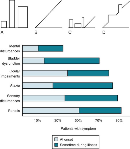
FIGURE 15-1 Top, Graphs of different clinical courses – with severity of multiple sclerosis (MS) attacks (vertical axis) plotted against time (horizontal axis) – reveal four patterns or disease categories: A, Relapsing-remitting; B, primary progressive; C, secondary progressive; D, progressive-relapsing. Bottom, This chart of initial and cumulative manifestations of MS indicates that cognitive impairment develops infrequently at the onset and ultimately less often than physical impairments.
Relapsing-remitting MS, the category that initially includes about 80% of cases, consists of discrete attacks followed by partial or complete recovery. Although deficits may accumulate following each attack, patients remain stable between them. Unfortunately, at an annual rate of about 3% per year, most patients in the relapsing-remitting category evolve into secondary progressive MS, which consists of further, steady deterioration.
Primary progressive MS, characterized by unremitting, steady deterioration from the illness’ onset, accounts for only about 10–15% of cases. Unlike the other disease categories, primary progressive MS typically develops in individuals who are in their fifth or sixth decade, rather than their third or fourth, and predominantly or exclusively affects the spinal cord. Progressive-relapsing MS, the least frequently occurring category, consists of a steady deterioration with superimposed acute attacks.
In addition to being descriptive, MS categories indicate a patient’s prognosis and likely response to immunomodulating treatments. Of the various categories, relapsing-remitting MS is the most amenable to treatment; progressive MS, the least (see later).
Frequent Symptoms
Lesions in the white-matter tracts of the CNS cause various symptoms during the course of a patient’s illness (Fig. 15-1, bottom). Moreover, simultaneous involvement of two separate CNS areas often produces combinations of disparate symptoms. For example, plaques may simultaneously develop in the cerebellum and thoracic spinal cord, which would cause ataxia and paraparesis.
Cerebellar Signs
As some of their earliest manifestations, MS patients often develop ataxia, intention tremor, and other signs of cerebellar and cerebellar outflow tract injury. When the cerebellum is involved, patients typically develop an ataxic gait (see Fig. 2-13); however, with minimal involvement, patients’ gait impairment may consist only of difficulty walking in a heel-to-toe (tandem gait) pattern. Cerebellar involvement also typically causes scanning speech, a variety of dysarthria analogous to a “speech ataxia,” characterized by irregular cadence and uneven emphasis on words. For example, when asked to repeat a pair of short syllables, such as “ba…ga…ba…ga…,” a patient might place unequal stress on different syllables, blur them together, or pause excessively. Other manifestations of cerebellar involvement include intention tremor (see Fig. 2-11), dysdiadochokinesia, and an irregular, conspicuous, head tremor (titubation).
Sensory Disturbances
Both lack of sensation and abnormal sensations occur frequently and prominently. Patients often describe hypalgesia, paresthesias, or dysesthesias in their limbs or trunk, or below a particular spinal cord level. They typically lose the ability to appreciate vibration and position sensations more than other sensations. At their onset, sensory symptoms may not conform to commonplace neurologic patterns or be unaccompanied by objective findings. Physicians may understandably mistake this situation as a psychogenic disturbance.
Ocular Impairments
Impaired visual acuity and disordered ocular motility – which neurologists lump together as “eye signs” – occur frequently not only at the onset of MS but also throughout its course. In fact, the absence of eye signs in patients believed to have MS may prompt neurologists to reconsider the diagnosis (see later).
Decreased Visual Acuity
The optic nerve is involved in MS because its covering consists entirely of CNS myelin. The acoustic nerve (cranial nerve VIII) is only partially covered by CNS myelin and is rarely, if ever, involved in MS. No other cranial nerves are covered by CNS myelin.
Neurologists usually attribute visual acuity impairment in MS to inflammation in the retrobulbar portion of the optic nerve, retrobulbar neuritis (optic neuritis) (see Fig. 12-6). Optic neuritis typically causes an irregular area of visual loss in one eye, a scotoma, that often includes the center of vision (Fig. 15-2). It also leads to color desaturation, in which colors, especially red, lose their intensity.

FIGURE 15-2 Optic or retrobulbar neuritis impairs vision in a large, irregular area (scotoma) of the affected eye.
In addition to reducing vision, optic neuritis characteristically causes pain in the affected eye. Probably because ocular movement puts traction on an inflamed optic nerve, eye pain increases when patients look from side to side.
Optic neuritis, as well as other lesions of the optic nerve, causes a readily identifiable, surprising pupillary light reaction (see Fig. 4-2). Swinging a flashlight from the normal eye to the one with optic neuritis will lead to dilation, rather than continued constriction, of both pupils. This paradoxical, unilateral reaction results from less light entering the pupillary reflex arc compared to when the light was shone into the normal eye. Sometimes called the “swinging flashing test,” this abnormality in the afferent limb of the light reflex results in an afferent pupillary defect or “Marcus Gunn” pupil, which neurologists interpret as a sign of optic nerve pathology.
Statistics vary on the relationship of optic neuritis to MS. They indicate that approximately 25% of MS patients present with optic neuritis as their initial symptom and 50% of MS patients suffer an attack during their illness. A single attack of optic neuritis with no other neurologic symptoms and an MRI showing no lesions means that the individual has only about a 25% likelihood of developing MS in the following 10 years; however, when one or more MRI lesions accompany optic neuritis, the individual’s likelihood increases to about 70% during the same period.
Ocular Motility Abnormalities
MS also causes ocular motility abnormalities, including nystagmus and the characteristic internuclear ophthalmoplegia (INO), which is also known as the medial longitudinal fasciculus (MLF) syndrome. Either brainstem or cerebellar involvement can cause nystagmus. Although it is clinically indistinguishable from nystagmus induced by other conditions (see Chapter 12), MS-induced nystagmus typically occurs in combination with dysarthria and tremor (Charcot’s triad).
In MS-induced INO, MLF demyelination interrupts nerve impulse transmission from the pontine conjugate gaze centers to the oculomotor nuclei (Figs 15-3 and 15-4). The primary symptom of INO is diplopia on lateral gaze because of paresis of the adducting eye. INO is strong evidence of MS; however, systemic lupus erythematosus (SLE or lupus [see later]) and small basilar artery strokes may also cause it. In addition, Wernicke–Korsakoff syndrome, myasthenia gravis, and botulism can produce patterns of ocular muscle weakness that mimic INO. From a physiologic viewpoint, INO is analogous to a disconnection syndrome, such as conduction aphasia, in which communicating links are severed but each neurologic center remains intact (see Chapter 8).

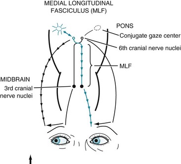
FIGURE 15-3 Under normal conditions, when looking laterally, the pontine conjugate gaze center stimulates the adjacent abducens (sixth) nerve nucleus and, through the medial longitudinal fasciculus (MLF), the contralateral oculomotor (third) nerve nucleus. For example, when looking to the right, as in this illustration, the right pontine gaze center stimulates the right abducens and the left oculomotor nuclei (see Fig. 12-12).

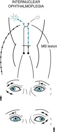
FIGURE 15-4 In internuclear ophthalmoplegia (INO), also known as the MLF syndrome, an interruption of the medial longitudinal fasciculus (MLF) prevents impulses from reaching the oculomotor (third) nuclei. Because those nuclei themselves remain intact, the pupils and eyelids are normal in both eyes. However, when looking to the right, because the left oculomotor nucleus is not stimulated, the left eye fails to adduct. The right eye abducts, but nystagmus develops. With bilateral INO, which is characteristic of multiple sclerosis (MS), neither eye adducts and abducting eyes have nystagmus.
Spinal Cord Symptoms and Signs
Patients with spinal cord involvement, typically the primary and sometimes the only source of disability in primary progressive MS, have paraparesis with hyperactive deep tendon reflexes and Babinski signs. They usually have three troublesome, common symptoms (the three Is) – incontinence, impotence, and impairment of gait. Another troublesome, often incapacitating feature of spinal cord involvement consists of spasticity of the legs. Even in the absence of paraparesis, spasticity impairs patients’ gait and causes painful leg spasms. Patients with cervical spinal cord involvement often describe electrical sensations, elicited by neck flexion, that extend from the neck down the spine (Lhermitte’s sign).
Spinal cord involvement also typically leads to urinary incontinence from a combination of spasticity, paresis, and incoordination (dyssynergia) of the bladder sphincter muscles (Fig. 15-5). MS patients initially often have incontinence during sleep and sexual intercourse. As the disease progresses, patients develop intermittent urinary retention and then complete loss of control. They often require intermittent or continuous catheterization, which leads to frequent, chronic, or recurrent urinary tract infections.

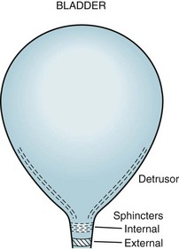
FIGURE 15-5 The urinary outflow of the bladder has two sphincters: an internal sphincter controlled by the autonomic nervous system (ANS), and an external one under voluntary control. Normal urinary bladder emptying (urination) occurs when the detrusor (wall) muscle contracts and both sphincter muscles relax. Purposefully urinating requires voluntary action (to relax the external sphincter) and reflex parasympathetic (ANS) activity (to contract the detrusor and relax the internal sphincter). Urinary retention occurs with either anticholinergic medication or excessive sympathetic activity because both inhibit detrusor contraction and internal sphincter muscle relaxation. Urinary retention also occurs with spinal cord injury because the external sphincter is unable to relax because it is spastic and paretic (dyssynergic).
Erectile dysfunction, decreased desire, and other forms of sexual impairment plague the majority of MS patients (see Chapter 16). About 40% of women with MS do not engage in sexual intercourse. Even before developing erectile dysfunction, men often experience premature or retrograde ejaculation. Sexual dysfunction, with or without urinary incontinence, is attributable to MS involving the spinal cord. With spinal cord damage severe enough to cause paraplegia, men have lowered and abnormal sperm production, but women can conceive and bear children.
Fatigue and Other Important Symptoms
An inexplicable generalized, daily sense of fatigue, which neurologists sometimes call “lassitude” (weariness of body or mind), affects about 50–80% of MS patients. This symptom, which is entirely subjective, does not correlate with patients’ age or degree of paresis. It reduces MS patients’ compliance with medical regimens, quality of life, ability to work, and compliance with medical regimens. In addition, it intensifies other MS symptoms, including depression and cognitive impairment.
MS-induced fatigue represents a physiologic cause of the chronic fatigue syndrome (see Chapter 6). Although many, but not all, studies found that depression is comorbid, antidepressants do not alleviate MS-induced fatigue.
Various pain syndromes also commonly occur in MS. For example, approximately 2% of MS patients suffer from trigeminal neuralgia (see Chapter 9) and 10% Lhermitte’s sign. Many MS patients have pain in their limbs or trunk. These pains probably arise from MS plaques irritating CNS pain-transmitting fibers in, respectively, the brainstem and cervical spinal cord. As with other forms of neuropathic pain (see Chapter 14), antiepileptic drugs, such as gabapentin and carbamazepine, provide some relief.
Because MS, in general, spares CNS structures that contain little or no myelin, symptoms that originate in gray-matter injury rarely complicate the illness. For example, MS patients seldom develop signs of focal cerebral cortical dysfunction, such as seizures or aphasia. Similarly, because the basal ganglia, like the cerebral cortex, are devoid of myelin, MS patients almost never develop involuntary movement disorders (see Chapter 18).
Pregnancy
Women with MS remain fertile. Oral contraceptives do not influence MS. If women conceive, they do not have an increased rate of miscarriages, obstetric complications, or fetal malformations. Throughout pregnancy, the rates of both first MS attacks and MS exacerbations significantly fall. In fact, during the third trimester, the exacerbation rate falls to 70% of its baseline. If MS exacerbations occur, they do not affect the pregnancy. After delivery, women who breastfeed extend the protection associated with pregnancy.
As for delivery, MS patients require cesarean sections for only the usual indications. Epidural anesthesia also has no effect on the course of MS.
Although the pregnancy and delivery pose little or no threat, during the first 3 postpartum months, mothers with MS have a 20–30% incidence of exacerbation. Postpartum exacerbations are more incapacitating than ones that strike before conception. In the long run, neither pregnancy nor parity worsens the course of MS.
Psychiatric Comorbidity in MS
Depression
Depression is the most common psychiatric comorbidity of MS. It develops more frequently in patients with MS than in patients with most other chronic, equally debilitating nonneurologic illnesses, such as rheumatoid arthritis. Depressive symptoms arise frequently at the onset of the illness, during exacerbations, and late in its course. They also correlate with cognitive and physical impairment, loss of bodily function, inability to work, and lack of family and social support. A history of depression and “trait anxiety” predisposes MS patients to depression. Depressive symptoms occur more when MS involves the cerebrum rather than only the spinal cord, and when MRIs show cerebral atrophy and a great total MS lesion area or volume (lesion load or burden).
Unlike depressive illness that occurs in families without MS, genetic influence in MS-induced depression is negligible. For example, the rate of depression in first-degree relatives of depressed MS patients is considerably lower than the rate of depression in first-degree relatives of depressed individuals who do not have MS. Also, MS-induced depression equally affects men and women.
Even when depressive symptoms do not reach the severity and duration of a major depression, which occurs in 25–50% of MS patients during their lifetime, they interfere with MS patients following their arduous regimen of self-injecting medicines, self-catheterization, and participating in rigorous physical therapy programs.
Largely reflecting the high incidence of depression, the suicide rate of patients in MS clinics is as high as seven times greater than that of comparably aged individuals. Compared to all MS patients, those who have attempted or completed suicide have been younger than 30 years and symptomatic for less than 1 year. In addition, their history includes depression in themselves or their family, alcohol abuse, and limited psychosocial support. However, MS-induced cognitive impairment is not a risk factor for suicide.
Well-controlled studies failed to prove that antidepressants improve MS patients’ mood. Moreover, antidepressants with anticholinergic side effects may precipitate urinary retention and SSRIs may increase spasticity. Nevertheless, neurologists prescribe antidepressants for patients with MS in the same regimen as for patients with other neurologic illnesses. Electroconvulsive therapy (ECT) may be effective and can be administered with only the usual precautions. In other words, MS cerebral lesions are not a counterindication to ECT. Whichever treatment physicians and their patients choose, adding psychotherapy, social services, occupational counseling, or physical therapy would benefit them.
Although bipolar disorder occurs at twice the rate in MS patients than in the general population, mania rarely develops. If it occurs, consultants must look for excessive steroid treatment of MS (see later).
Consultants may also encounter “MS-induced euphoria” – an elevation of mood clearly inappropriate to these patients’ disability. This euphoria is associated with physical deterioration, chronicity of the illness, and at least subtle intellectual impairment, as well as steroid treatment. Some euphoric patients are masking depression or protecting themselves with denial. Others simply sense relief as an MS attack subsides. Whether or not psychologic factors seem to explain the euphoria, extensive cerebral involvement usually underlies it. In particular, pseudobulbar palsy may explain pathological laughter (see Chapter 4).
Psychosis
The prevalence of psychosis, unlike the prevalence of depression, is not significantly greater in MS patients than unaffected individuals. In fact, the prevalence of psychosis in MS is less than in most other neurologic illnesses, including Alzheimer disease, head trauma, and epilepsy. Except for cases in scattered reports, MS does not present with psychosis.
Nevertheless, severely disordered thinking occurs in MS patients. On a practical level, psychiatrists should begin by assuming that, in MS patients, it reflects adverse effects of medications or concomitant physical illness, such as a urinary tract infection, i.e., delirium superimposed on cognitive impairment.
Cognitive Impairment
Almost all MS patients in the initial phase of their illness have normal cognitive capacity. They satisfactorily complete their day-to-day functions, routine mental status evaluation, and Mini-Mental State Examination (MMSE) (see Fig. 7-1). However, more demanding measures, such as the Wechsler Adult Intelligence Scale (WAIS), Selective Reminding Test, and Halstead Category Test, reveal at least clinically silent deficits in 45–65% of MS patients.
MS-induced cognitive impairment can hamper activities of daily living, prevent full compliance with medical regimens, and burden caregivers (see later). Moreover, it can precipitate thought and mood disorders.
Certain MRI abnormalities frequently occur: enlarged cerebral ventricles, corpus callosum atrophy, periventricular white-matter demyelination, and overall lesion load. Of all of them, cognitive impairment correlates most closely with the periventricular region lesion load.
Cognitive impairment in MS differs from that in Alzheimer disease in several respects. In MS, carrying an apolipoprotein E4 allele does not pose a significant risk for cognitive impairment. MS produces a subcortical rather than cortical dementia. Also, cognitive impairment typically appears late in the course of MS and long after physical disability has developed, but in Alzheimer disease, it long precedes the onset of physical disability. Also, by way of contrast, in vascular dementia, intellectual and physical deficits appear and worsen together.
Physicians attempting to reduce cognitive deficits in MS might institute cognitive rehabilitation, enhanced structure, occupational therapy, and psychotherapy. Immunomodulators possibly delay the onset or slow the progression of cognitive as well as physical disabilities (see later), but donepezil (Aricept) does not help.
Pediatric MS
Approximately 4% of cases develop in children and adolescents. Pediatric MS patients present with the same neurologic symptoms and signs and are subject to similar neuropsychiatric comorbidities as their adult counterparts. However, their course is almost always relapsing-remitting at its onset and evolves into a secondary progressive pattern only after decades.
Cognitive impairments, which typically interfere with their schoolwork, develop in 30% of pediatric MS patients. Only about 6% of pediatric MS patients develop major depression, but up to 75% develop fatigue. Despite widespread cerebral disease, they are not prone to develop attention deficit hyperactivity, autism symptoms, or specific learning disabilities.
Caregiver Stress
Distress and reduced quality of life can overwhelm caregivers of MS patients. Characteristics of both MS patients and their caregivers determine the nature and severity of the stress. Patients’ characteristics associated with high stress levels include not only their physical disabilities, but also their overall poor quality of life, presence of depression and anxiety, and degree of cognitive impairment. Caregivers’ characteristics associated with high stress levels include a change in their life’s role and the onset or exacerbation of a pre-existing, subclinical depression. To reduce the stress, the patient and caregiver require assistance from family, friends, and perhaps a support group; social and financial services; and current, valid information about all aspects of the illness.
Laboratory Tests
When patients’ clinical evaluation is equivocal, several tests are required to diagnose MS and exclude other illnesses. None is diagnostic and all yield false-negative and false-positive results.
Imaging Studies
Computed tomography (CT) can show atrophy, reveal large areas of demyelination, and exclude large mass lesions that can masquerade as MS. However, it is too insensitive and too nonspecific to be useful in diagnosing MS.
MRI – certainly the most valuable test – can readily reveal demyelinated areas indicative of MS plaques. The revised McDonald criteria call for combinations of one gadolinium-enhanced lesion or nine T2-weighted hyperintense MRI lesions located in various regions of the brain, particularly in the periventricular area (Figs 15-6 and 20-25), or spinal cord (Fig. 15-7). Although not pathognomonic, these hyperintensities are detectable in more than 90% of MS patients.

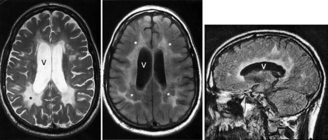
FIGURE 15-6 Left, This axial T2-weighted magnetic resonance imaging (MRI) scan through the cerebrum of a patient with multiple sclerosis (MS) shows multiple plaques (*) concentrated around the ventricles (V), particularly in the posterior regions. The MS plaques are characteristically white (hyperintense), sharply demarcated, and located in the periventricular region. Center, This axial T2-weighted, fluid-attenuated inversion recovery (FLAIR) image of the same study also shows hyperintense lesions (*) surrounding the ventricles (V). FLAIR images, in which cerebrospinal fluid remains black, highlight demyelinated areas. Right, The sagittal T2 FLAIR image of the same study shows the periventricular hyperintensities surrounding the lateral ventricle (V).

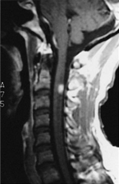
FIGURE 15-7 This magnetic resonance imaging scan of a patient with multiple sclerosis reveals a plaque – the hyperintense lesion – in the high cervical spinal cord.
MRI readily detects lesions in large, heavily myelinated tracts of the CNS, such as the corpus callosum, periventricular area, MLF and other brainstem tracts, cerebellum, optic nerves, and spinal cord. It can show asymptomatic as well as symptomatic lesions.
Because gadolinium enhances MS lesions during the first month after they arise, gadolinium-enhanced MRI can distinguish between new and old lesions. Neurologists accept the appearance of new MRI lesions, even in the absence of acute symptoms, as a marker of active disease.
In addition to showing lesions, the MRI may reveal atrophy of the corpus callosum and cerebrum. Communicating hydrocephalus or hydrocephalus ex vacuo, which occurs commonly, reflects cerebral atrophy and compensatory enlarged ventricles. The cerebral and corpus callosum atrophy correlates with chronicity and cognitive impairment; however, as previously noted, overall lesion load and, more so, periventricular white-matter demyelination correlate more closely with cognitive impairment.
Despite its reliability, MRI may be misleading. Small T2-weighted hyperintensities, “unidentified bright objects” (UBOs), appear in numerous conditions besides MS, including migraine, hypertensive cerebrovascular disease, and normal, benign age-related changes. When accompanied by neurologic symptoms, MRI UBOs may lead to a misdiagnosis. As another pitfall, the MRI reveals demyelination – although usually not multiple, large, periventricular plaques – in neurologic diseases other than MS, such as the leukodystrophies (see later).
Cerebrospinal Fluid
Routine CSF analysis during an MS attack will usually contain protein concentrations that are either normal (40 mg/100 mL) or only slightly elevated, and a mild, nonspecific gamma globulin elevation (9% or greater), but no increase in white blood cells (WBCs). One suggestive feature is that the CSF of 90% of MS patients contains CSF oligoclonal bands that consist of discrete IgG antibodies (Fig. 15-8). However, oligoclonal bands are also present in other inflammatory diseases involving the CNS, such as lupus, chronic meningitis, sarcoidosis, neurosyphilis, Lyme disease, acquired immunodeficiency syndrome (AIDS), and paraneoplastic limbic encephalitis.

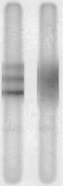
FIGURE 15-8 Electrophoresis of cerebrospinal fluid (CSF) of a patient with multiple sclerosis (left), compared to the CSF of one with no central nervous system inflammatory disease (right), shows three distinct, horizontal oligoclonal bands.
CSF myelin basic protein, another protein not normally present, is essentially a myelin breakdown product. As with oligoclonal bands, myelin basic protein occurs in many inflammatory CNS diseases. In diagnosing MS, CSF myelin basic protein carries even less weight than CSF oligoclonal bands.
Evoked Responses
Although routine electroencephalograms (EEGs) do not help in the diagnosis, related electrophysiologic testing, evoked response or evoked potential tests, can reveal characteristic interruptions in the visual, auditory, or sensory pathways. Evoked potential testing is based on repetitive stimulation of these pathways, which are heavily myelinated, and then detecting the responses with scalp electrodes similar to those used for EEGs. Normal responses are so small that they are lost in normal cerebral electrical activity and background noise. In evoked testing, hundreds of responses are computer-averaged. After canceling out normal electrical activity, computer averaging displays an otherwise undetectable composite wave pattern. MS injury slows and distorts electrophysiologic conduction. Any injury lengthens the interval between the stimulus and composite response, which is reflected in an abnormally increased latency, and distortion of the final composite wave pattern.
Evoked response tests are particularly useful in demonstrating lesions that are undetectable on neurologic examination. For example, if a patient has deficits referable only to the spinal cord, but evoked response tests reveal a subclinical optic nerve injury, the physician would know that at least two CNS areas were injured and that the illness was disseminated in space.
Visual-evoked responses (VERs) reveal visual pathway lesions. The patient stares at a rapidly flashing pattern on a television screen and a computer averages responses detected over the occipital cortex. Optic neuritis increases the latency or distorts the waveform. Because VERs can indicate the site of an interruption in the visual pathway, they are helpful in distinguishing ocular, cortical, and psychogenic blindness (see Chapter 12).
Brainstem auditory-evoked responses (BAERs

Stay updated, free articles. Join our Telegram channel

Full access? Get Clinical Tree

