7 Multiple Tumors of the Cerebellopontine Angle
Case 1
S.M., female, age 60 (Fig. 7.1)
Diagnosis | Grade 2 acoustic neurinoma and meningioma |
Approach | Retromastoid transmeatal |
Result | Total resection, CN VII preserved, CN VIII preserved |
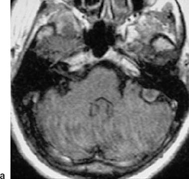
Fig. 7.1a An axial MRI scan, showing a typical small meningioma arising from the posterior surface of the petrous bone, near the entrance of the inner acoustic meatus. No additional tumor is evident in this MRI series. The patient had practically no clinical symptoms, and had good hearing on both sides.
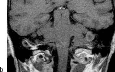
Fig. 7.1b A coronal MRI section from the same patient as in a, also showing the small meningioma.
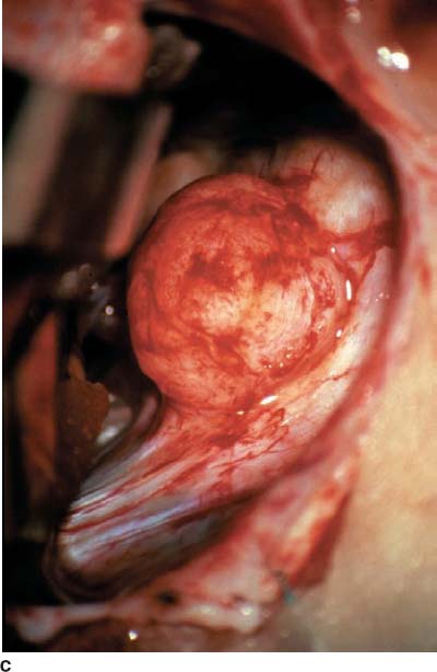
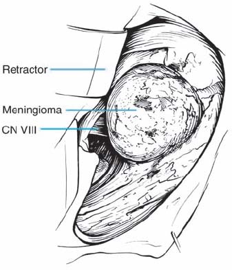
Fig. 7.1c The intraoperative view after a retromastoid approach had been performed. The tumor arises from the petrous bone behind the upper triangle, which cannot be seen at this stage of the operation.
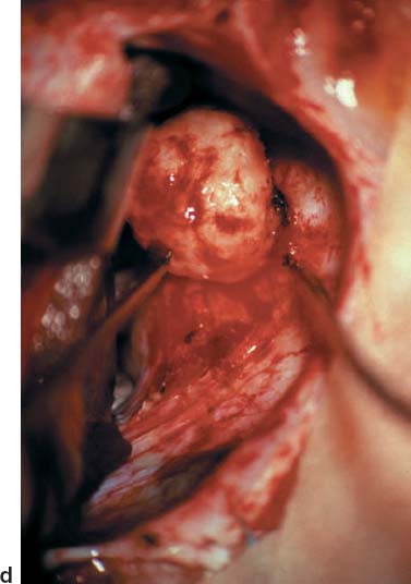
Fig. 7.1d The origin of the meningioma has been coagulated, and the rumor has been resected in toto. The seventh and eighth nerves are still not visible.
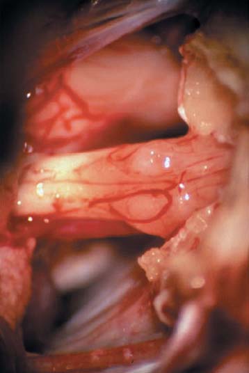
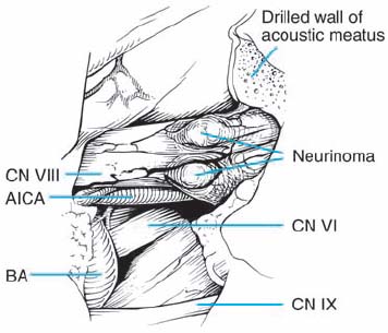
Fig. 7.1e Small neurinomas of the eighth nerve were detected during exploration of the region. The inner acoustic meatus has been opened using a diamond drill, as described in Chapter n, allowing perfect visualization of the cranial nerves.


Stay updated, free articles. Join our Telegram channel

Full access? Get Clinical Tree


