Chapter 9 Musculocutaneous Nerve
Anatomy
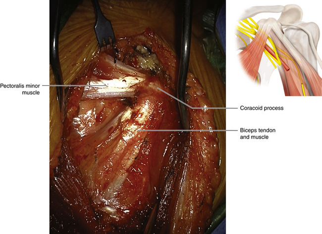
• The short head of the biceps arises from the tip of the coracoid process, lateral to its tendon of joint origin with the coracobrachialis (Figure 9-1). The long head arises from the supraglenoid tubercle. The biceps muscle, formed from this dual origin, is inserted by a tendon into the tuberosity of the radius.
• At its insertion, the tendon sends a medial fascial expansion, the bicipital aponeurosis, medially to thicken the investing fascia and gain attachment to the ulna.
• The median nerve lies under the bicipital aponeurosis, medial to the biceps tendon. The posterior interosseous nerve lies lateral to the tendon.
• The brachialis arises from the anterior aspect of the humerus and is inserted into the tuberosity of the ulna. The biceps and brachialis are strong flexors of the elbow joint. With the elbow flexed, the biceps is the key supinator of the forearm.
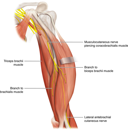
• The coracobrachialis arises from the coracoid process and is inserted into the humerus. Its importance lies in its value as a landmark in operations on the musculocutaneous nerve and not in its function (Figure 9-2).
• The radial nerve is a partial supplier of the brachialis, but not a significant one. In the absence of C6 function (conveyed by the upper trunk, anterior division, and musculocutaneous nerve to the elbow flexors), the radial nerve–supplied brachioradialis, not the trivial segment of brachialis supplied by the radial, assumes the function of weaker elbow flexion.

Figure 9-1 The tendon of the pectoralis minor is divided against the coracoid. The biceps is not disturbed.
< div class='tao-gold-member'>
Stay updated, free articles. Join our Telegram channel

Full access? Get Clinical Tree


