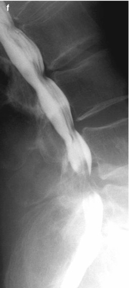
Fig. 8.1
(a–f) Waist-like contrast medium filling defect at level L2/3 (a), accentuated in upright position (b) due to degenerative lumbar spinal canal stenosis (arrow) in a patient suffering from claudicatio spinalis. (c, d) Axial post-myelo-CT at level L4/5 showing severe stenosis and consecutive slight enhancement of the thecal sac; constriction of the dural sac from anterior (medial disk protrusion; c, arrow) and dorsolateral due to thickened ligamenta flava (d, arrow) and degenerative alterations of the intervertebral articulations (d, arrowhead). (e, f) Myelography in upright position (lateral projection) exhibits severe increase of dural sac compression in retroflexion (e, f: anteflexion)
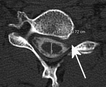
Fig. 8.2
Post-myelo-CT axial at level C6/7 illustrating a filling defect of the radicular pouch C7 left sided (arrow) due to a lateral intraforaminal disk herniation at the segment C6/7
2.
Get Clinical Tree app for offline access

Intradural extramedullary space-occupying lesions result in ovoid-shaped defects in lateral and p.a. projection and possible shift of the spinal cord. Due to the extent of such lesions, myelograms may also show a stop of contrast medium distribution going ahead with tapered margins (see Fig. 8.3).
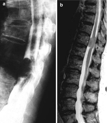
Fig. 8.3


(a, b) Dorsal shift of the lower thoracic spinal cord due to intradural extramedullary tumor, i.e., meningioma, at level Th10/11 with ventral adhesion; note ovoid-shaped contrast filling defects. However, method of choice nowadays is spinal MRI; (b): sag. T2WI showing a meningioma at level Th11
Related posts:


Stay updated, free articles. Join our Telegram channel
Full access? Get Clinical Tree