Fig. 4.1
Neonatal montage. Special montage—preterm or newborn with head circumference <35 cm
Usually, 16 channels recording are done, which should include 2 or more non-cerebral channels.
Non-cerebral electrodes include EKG, EMG, ocular, and respiratory belt. It helps in staging the sleep cycle, abnormal body movements, and also to identify the artifacts.
Duration of the recording: 60-min recording is mandatory in an attempt to capture EEG in various behavioral states. EEG abnormalities are usually the most prominent in quiet sleep. In a term neonate, the total sleep cycle lasts anywhere between 45 and 60 min (active sleep: ~25 min; quiet sleep: ~20 min; transitional sleep: ~15 min).
Timing of the Neonatal EEG
EEG is ideally done within 24 h following the insult.
EEG changes are very dynamic, and significant changes in background activity may happen rapidly (in hours). Serial EEGs provide valuable information for therapy and prognostication.
Normal Developmental Landmarks
Normal developmental landmarks on a newborn’s EEG provide information regarding the functional maturity and reflect the PCA of the baby. EEG findings in acute encephalopathy in newborns frequently show the patterns that may be normal for a baby of a lesser PCA. Such patterns are described as dysmature patterns and indicate encephalopathy in the appropriate setting. This may improve rapidly with the improvement in encephalopathy. Developmental landmarks are best categorized in 3 phases: <30 weeks, 30–37 weeks, and >37 weeks.
PCA <30 Weeks of Gestation
EEG may be indistinguishable from awake and sleep in extremely premature infants of PCA <30 weeks. A discontinuous EEG pattern termed trace discontinue is noted in all behavioral states.
Trace Discontinue
Tracé discontinue pattern is a predominant pattern seen in preterm neonates with PCA <30 weeks. It is characterized by high-amplitude (50–300 µV) bursts of mixed frequency (theta and alpha riding over delta) which are seen simultaneously on both the hemispheres followed by low-amplitude periods of quiescence (<25 µV) which can last anywhere between few seconds and 1 min (inter-burst interval—IBI) (Fig. 4.2).
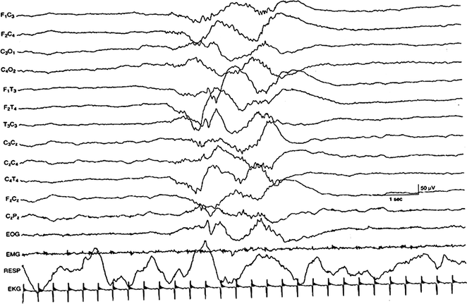
Fig. 4.2
Trace discontinue. Trace discontinue pattern seen in 26–28 weeks of PCA (Picture source Atlas of neonatal encephalography—Third Edition Lippincott Williams and Wilkins A Wolters Kluwer Company)
Inter-burst interval (IBI)
The key factor that determines the IBI in healthy babies is the postconceptional age. As the PCA increases, the IBI decreases.
Approximately 30 weeks or older, median IBI of 8 s or less has higher survival than those with longer median IBI.
Approximately IBI lasts anywhere between 20 and 35 for neonates with PMA <30–33 weeks with <25 µV. After 34 weeks, it significantly reduces, and closer to the term, it ranges anywhere between 6 and 10 s and has higher voltage >25 µV.
Prolonged IBI is seen in acute encephalopathy secondary to any type of brain injury, but is also commonly seen in moderate-to-severe hypoxic ischemic encephalopathy.
PCA Between 30 and 37 Weeks
EEG starts showing variation in different behavioral states around 35 weeks of PCA. Identifying different sleep stages in a newborn’s EEG is critical for optimal interpretation. EEG maturational changes occur first in active sleep; after a lag of about 2 weeks, the awake periods start showing more mature patterns. Quiet sleep is the last stage to show more mature changes. For these reasons, the abnormalities are most frequently seen in the quiet sleep phase. 
Trace alternant: These are characterized by high-amplitude (50–150 µV) synchronous burst of mixed frequencies predominantly delta activity for 3–10 s followed by 3–5 s of low-amplitude burst (25–50 µV) of predominant theta activity. This patterns replaces the trace discontinue with advanced PCA. This pattern is seen only in quiet sleep in neonates between PCA of 36 and 38 weeks (Fig. 4.3).

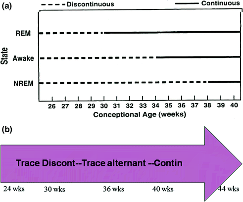
TA usually starts appearing in neonates >35 weeks and can be seen until 42 weeks but no longer than 44 weeks of PCA.
In active sleep (and later in wakefulness), TA is replaced by more continuous pattern characterized by uninterrupted, low-to-medium amplitude, mixed EEG activity with <2 s of voltage attenuation (<25 µV), and seen in mostly neonates with PCA >30 weeks.
TA pattern is replaced by medium-to-high voltage delta slow wave sleep after PCA of 44–48 weeks (Fig. 4.4a, b).
Fig. 4.3
Trace alternant. Trace alternant pattern seen in quiet sleep in 38–30 weeks of PCA (Picture source Atlas of neonatal encephalography—Third Edition Lippincott Williams and Wilkins A Wolters Kluwer Company)
Fig. 4.4
a Development of continuous activity (Picture source Levin and Luders Comprehensive Clinical Neurophysiology W.B. Saunders Company). b Evolution of the background activity
Abnormalities in Voltage
Excessive discontinuity: Abnormal discontinuous tracing separated by prolonged IBI for PCA. This is usually seen in neonates with severe hypoxic ischemic encephalopathy, meningitis, encephalitis, and severe intraventricular hemorrhage.
Burst suppression: Burst suppression pattern is a pattern with high-amplitude burst (50–300 µV) of delta and theta frequency with sharp waves and spikes followed by severe background suppression (<5 µV). Usually, the background shows no reactivity to stimuli.
In certain conditions like non-ketotic hyperglycinemia, the EEG bursts may be accompanied by myoclonic jerks (Fig. 4.5).
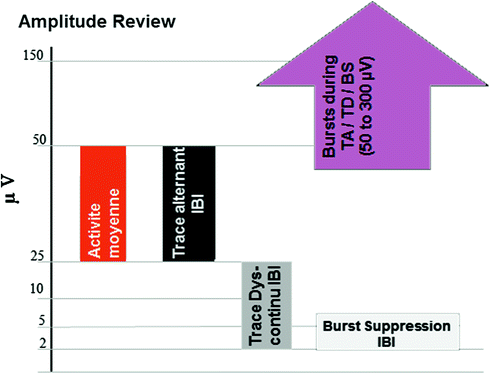
Fig. 4.5
Amplitude review
Synchrony: Burst of morphological similar activity in the homologous head regions is separated by <1.5 s.
<28 weeks: no asynchrony
28–32 weeks: 70% synchrony
>37 weeks: mostly synchronous
Abnormal finding: Asynchrony of >1.5 s may be significant if in excess for the PCA (Fig. 4.6).
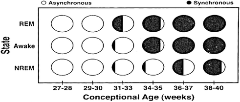
Fig. 4.6
Synchrony (Picture source Levin and Luders Comprehensive Clinical Neurophysiology W.B. Saunders Company)
Normal Graphoelements
Delta brushes: Delta brush is characterized by 0.3–1.5 Hz delta activity superimposed with beta or fast activity (18–22 Hz).
Other names are beta-delta complexes, ripples of prematurity, spindles/delta bursts.
They usually appear in the central region first followed by temporal and occipital and also disappear in the same order.
Most commonly seen during 24–34 weeks and starts to disappear first in the active sleep after 30 weeks. They can be present in the quiet sleep up to 38 weeks.
Abnormal findings: Delta brushes are often asynchronous, but when consistently absent on one side, a concern for some cerebral dysfunction is raised; if it is persistently present beyond 44 weeks then it is consistent with dysmaturity (Fig. 4.7).
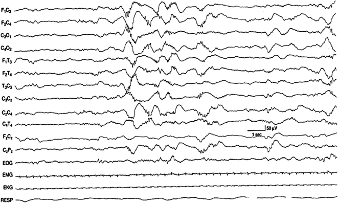
Fig. 4.7
Delta brushes in 29–30 weeks of PCA: Delta brushes seen in the bilateral central regions in a discontinuous background (Picture source Atlas of neonatal encephalography—Third Edition Lippincott Williams and Wilkins—A Wolters Kluwer Company)
Frontal sharps (Encoches Frontales): These are blunt isolated biphasic broad sharp transients (0.5–0.75 s) seen in the frontal region with initial smaller negativity followed by prominent positivity phase.
They are usually symmetric and synchronous;
Frequently seen in the transitional stage of sleep;
Usually seen between 33 and 46 weeks of PCA with peak around 35 weeks;
Mostly associated with rhythmic bi-frontal delta; and
Abnormal findings: Consistently absent on one side or any asymmetry raises concern for structural lesion on the other side (Fig. 4.8).
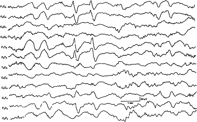
Fig. 4.8
Frontal sharps in 34–35 weeks of PCA (Picture source Atlas of neonatal encephalography—Third Edition Lippincott Williams and Wilkins—A Wolters Kluwer Company)
Temporal Theta
This graphoelement is a developmental marker that appears as a 2-s burst of 25–120 µV theta burst over the temporal region;Related posts:
 New-Generation Antiepileptic Drugs
Other Pharmacological Therapies: Investigational Antiepileptic Drugs, Animal Models of Epilepsy, Hormonal Therapy, Immunotherapy
Procedures and Outcomes in Epilepsy Surgery
New-Generation Antiepileptic Drugs
Other Pharmacological Therapies: Investigational Antiepileptic Drugs, Animal Models of Epilepsy, Hormonal Therapy, Immunotherapy
Procedures and Outcomes in Epilepsy Surgery
 Vagus Nerve Stimulation and Other Neuromodulation
 Magnetoencephalography and Magnetic Source Modeling
 Epilepsy Surgery Assessment and Testing

Stay updated, free articles. Join our Telegram channel

Full access? Get Clinical Tree


