1 Neuroanatomy
Cranial
 Vasculature
1. How can one distinguish the internal carotid artery (ICA) from the external carotid artery (ECA) in the neck?
The ICA has no branches in the neck, whereas the ECA has multiple branches.1
2. What are the major branches of the ECA?
The mnemonic SALFOPSI is very useful in remembering the branches of the ECA in ascending order (proximal to distal) (Fig. 1.1).1
1. Superior thyroid artery
2. Ascending pharyngeal artery
3. Lingual artery
4. Facial artery
5. Occipital artery
6. Posterior auricular artery
7. Superficial temporal artery
8. (Internal) maxillary artery
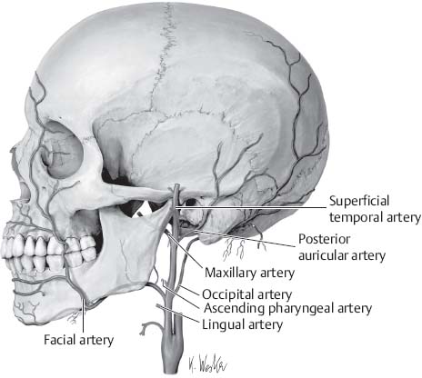
Fig. 1.1 External carotid artery and branches. (From THIEME Atlas of Anatomy, Head and Neuroanatomy, © Thieme 2007, Illustration by Karl Wesker.2)
3. From where do the common carotid arteries (CCAs) arise?
Normally, the right CCA comes off the brachiocephalic trunk, whereas the left CCA comes off straight from the aortic arch.3
4. What is referred to as the carotid siphon?
The intracavernous and the supraclinoid segment of the ICA
5. What classical clinical findings occur in an occlusion of the anterior choroidal artery?
Hemiparesis, hemianesthesia, and hemianopsia
6. The posterior cerebral artery divides into what two terminal branches?
The parietooccipital artery and calcarine artery (Fig. 1.2)4
7. What artery supplies the choroid plexus of the temporal horn? Atrium?
The posterior cerebral artery supplies both (Fig. 1.2).4
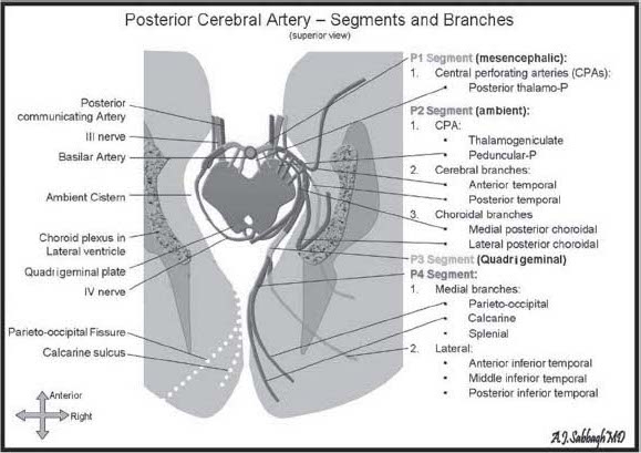
Fig. 1.2 Posterior cerebral artery—segments and branches.
8. What are Virchow-Robin spaces?
The spaces between the blood vessels and the arachnoid and pia layers within the brain and spinal cord
9. Which sinus courses within the attachment of the tentorium to the petrous ridge?
The superior petrosal sinus4
10. Which large anastomotic vein joins the superior sagittal sinus?
The vein of Trolard (also called the superior anastomotic vein) (Fig. 1.3)4
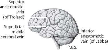
Fig. 1.3 Superficial venous anatomy of the brain. (From THIEME Atlas of Anatomy, Head and Neuroanatomy, © Thieme 2007, Illustration by Markus Voll.2)
11. Which large anastomotic vein joins the veins of the sylvian fissure with the transverse sinus?
The vein of Labbe4
12. What is the largest branch of the intracavernous portion of the carotid artery?
The meningohypophyseal trunk (Fig. 1.4)5
13. What is the most constant branch of the meningohypophyseal trunk?
The tentorial artery. It passes forward to the roof of the cavernous sinus and then posterolaterally along the free edge of the tentorium. It sends branches to cranial nerves (CNs) III and IV. Bernasconi and Cassinari first reported this artery angiographically in 1957 (Fig. 1.4).5
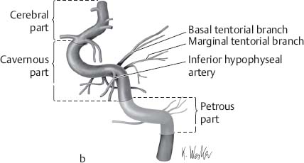
Fig. 1.4 Internal carotid artery and branches. (From THIEME Atlas of Anatomy, Head and Neuroanatomy, © Thieme 2007, Illustration by Karl Wesker.2)
14. Which branch of the intracavernous carotid artery passes between CN VI and the ophthalmic division of the trigeminal nerve?
The inferolateral trunk5
15. What is the venous angle as seen on a lateral view of a cerebral angiogram?
The angle is formed by the junction of the thalamostriate vein and the internal cerebral veins at the thalamic tubercle. This area approximates the site of the foramen of Monro.
16. What are the three main superficial cerebral veins?6
1. The superior anastomotic vein of Trolard
2. The inferior anastomotic vein of Labbé
3. The superficial middle cerebral vein
17. What is the most likely clinical symptom in a patient with a large unruptured cavernous sinus carotid aneurysm?
Ipsilateral sixth nerve palsy. The cavernous sinus contains CNs III, IV, V1 and V2, as well as CN VI. The abducens nerve has the most proximal spatial relationship to the carotid artery.7
18. Which artery is the most common cause of lateral medullary syndrome?
Also known as Wallenberg syndrome, it is most commonly due to occlusion of the vertebral artery on the ipsilateral side. This syndrome results from infarct in the region supplied by the posterior inferior cerebellar artery (PICA), which is a branch of the vertebral artery (Fig. 1.5).8
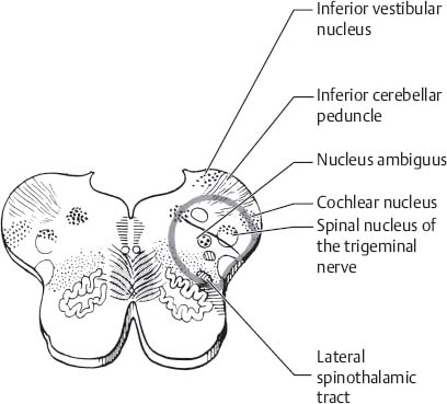
Fig. 1.5 Cross-section of the medulla with outlined blood supply of the vertebral artery. (Reprinted with permission from Mumenthaler and Matte.9)
19. What is the arterial supply of the thalamus?
Branches of the posterior communicating arteries and the perimesencephalic portion of the posterior cerebral arteries (Fig. 1.6)4
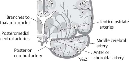
Fig. 1.6 Blood supply to internal capsule and basal ganglia. (From THIEME Atlas of Anatomy, Head and Neuroanatomy, © Thieme 2007, Illustration by Markus Voll.2)
20. What is the arterial supply of the lateral geniculate nucleus (LGN)?
The LGN has a dual arterial supply. Laterally, it receives supply from the anterior choroidal artery, and medially from the lateral posterior choroidal artery. Hence infarcts are relatively uncommon in this area.10
21. Which artery is most commonly involved in trigeminal neuralgia?
The superior cerebellar artery (SCA) (Figs. 1.7 and 1.8)11
22. Which artery is most commonly involved in hemifacial spasm?
The anterior inferior cerebellar artery (AICA) (Figs. 1.7 and 1.8)12
23. What is the most common artery involved in glossopharyngeal neuralgia?
The PICA (Figs. 1.7 and 1.8)13
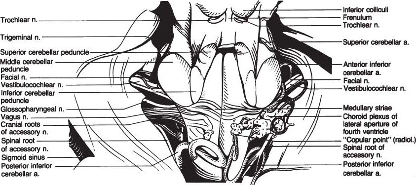
Fig. 1.7 Topographic anatomy of dorsal brainstem and tectum. (Reprinted with permission from Koos et al.14)
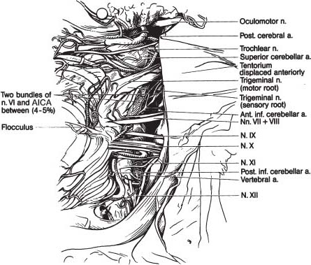
Fig. 1.8 Topographic anatomy of lateral brainstem and cerebellum. (Reprinted with permission from Koos et al.14)
24. What is the main arterial supply of the internal capsule?
This is derived from several sources. The lateral lenticulostriate branches from the middle carotid artery (MCA), the medial striate artery from the ACA, and the direct branches from the ICA. The anterior choroidal artery comes off the ICA (Fig. 1.6).10
25. Which vessel has the highest risk of injury in a Chiari decompression surgery?
The PICA13
26. Which vessels supply the superior, middle, and inferior cerebellar peduncles?
The SCA, AICA, and PICA, respectively15
 Skull
Skull
27. Where is the motor strip located in relation to the skull?
4 to 5.5 cm behind the coronal suture (Fig. 1.9)16
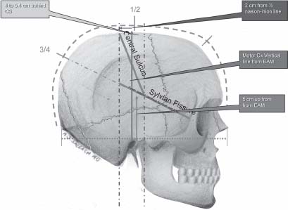
Fig. 1.9 Coronal and rolandic fissure locations. EAM, external auditory meatus.
28. What external landmark on the skull marks the lateral margin of the sphenoid ridge and sylvian fissure?
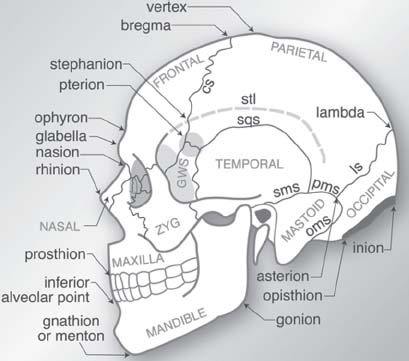
Fig. 1.10 Craniometric points and sutures. Named bones appear in all upper case letters. Abbreviations: GWS = greater wing of sphenoid bone, NAS = nasal bone, stl = superior temporal line, zyg = zygomatic. Sutures: cs = coronal, ls = lambdoid, oms = occipitomastoid, pms = parietomastoid, sms = squamomastoid, sqs = squamosal. (Reprinted with permission from Greenberg.16)
29. What part of the mandible does the temporalis muscle attach to?
The coronoid process of the mandible1
30. What muscle of mastication does the parotid duct cross?
The masseter muscle1
31. Which cranial fossa is the largest?
The posterior cranial fossa. It is also the deepest of the three cranial fossas (Fig. 1.11).
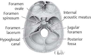
Fig. 1.11 Interior view of the skull base. (From THIEME Atlas of Anatomy, Head and Neuroanatomy, © Thieme 2007, Illustration by Karl Wesker.2)
32. What are the boundaries of the suboccipital triangle?17
The suboccipital triangle is a region bounded by the following three muscles:
1. Above and medially by the rectus capitis posterior major
2. Above and laterally by the superior oblique
3. Below and laterally by the inferior oblique
33. In a far lateral approach, it is helpful to palpate and know the location of the transverse process of the atlas. Between what landmarks can the transverse process of the atlas be palpated?
Through the skin between the mastoid process and the mandibular angle17
34. What sutures make up the asterion?
The lambdoid, parietomastoid, and occipitomastoid sutures. It is an important landmark to define the lower half of the junction of the transverse and sigmoid sinuses (Fig. 1.10).16
35. Which bones make up the osseous nasal septum?
The perpendicular plate of the ethmoid and the vomer (Fig. 1.12)1
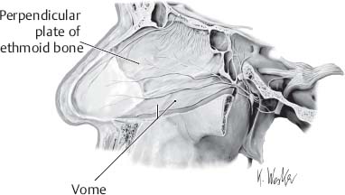
Fig. 1.12 Nasal septum innervation. (From THIEME Atlas of Anatomy, Head and Neuroanatomy, © Thieme 2007, Illustration by Karl Wesker.2)
36. What are the compartments of the jugular foramen?
Pars venosa (posterolateral), which contains the sigmoid sinus, jugular bulb, and CNs X and XI
Pars nervosa (anteromedial), which contains CN IX and Jacobson’s nerve (Fig. 1.13)18
37. What structure does the abducens nerve go through to enter the cavernous sinus?
Dorello’s canal5
38. What structures go through the internal acoustic meatus?
The facial nerve (VII), the vestibulocochlear nerve (VIII), and the labyrinthine artery (Fig. 1.13)19
39. What are the major parts of the temporal bone?
Squamous and petrous parts1
40. The cribriform plate is part of what bone?
The ethmoid bone1
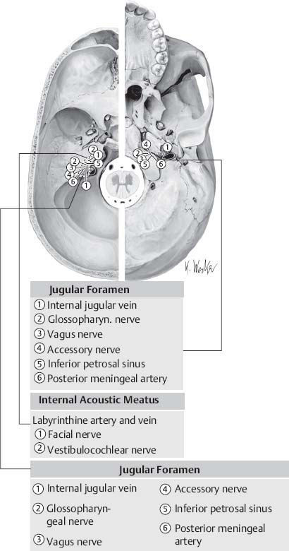
Fig. 1.13 Foramina of the skull base with exiting structures. (From THIEME Atlas of Anatomy, Head and Neuroanatomy, © Thieme 2007, Illustration by Karl Wesker.2)
41. What are the bones that form the walls of the orbit (Fig. 1.14)?20
1. Frontal
2. Zygomatic
3. Maxilla
4. Sphenoid
5. Lacrimal
6. Ethmoid
7. Palatine
42. What structures pass through the annulus of Zinn?
The optic nerve, ophthalmic artery, oculomotor nerve, abducens nerve, and nasociliary nerve (Fig. 1.14)20
43. What are the structures in the pterygopalatine fossa?
The maxillary artery, maxillary nerve, and pterygopalatine ganglion22
44. What passes through the inferior orbital fissure?
The infraorbital nerve and zygomatic nerve20
45. What bones are approximated at the pterion?
The frontal, parietal, temporal, and sphenoid bones16
46. The clivus is formed by which bones?
The occipital and the sphenoid bones
47. What structure separates the optic canal from the superior orbital fissure?
The optic strut20
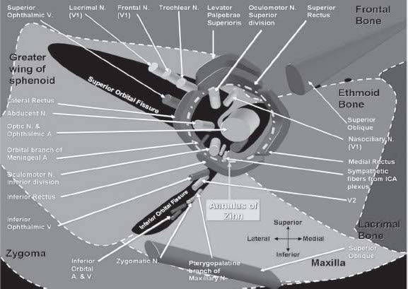
Fig. 1.14 Orbit anatomy with bony and neurovascular structures. (Reprinted with permission from Nader and Sabbagh.21)
 Ventricles
48. What are the five parts of the lateral ventricle (Fig. 1.15)?23
1. Frontal horn
2. Temporal horn
3. Occipital horn
4. Body
5. Atrium
49. Choroid plexus and flocculus protrude from which foramen?
The foramen of Luschka24
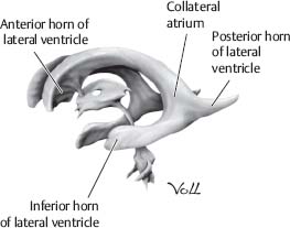
Fig. 1.15 Ventricular anatomy. (From THIEME Atlas of Anatomy, Head and Neuroanatomy, © Thieme 2007, Illustration by Markus Voll.2)
50. Which cranial nerve nuclei are positioned in the lateral recess near the foramen of Luschka?
The dorsal and ventral cochlear nuclei of CN VIII24
51. The choroid plexus of the lateral ventricle is attached along the choroidal fissure, which is the cleft between which two structures?
The fornix and thalamus (Fig. 1.16)23
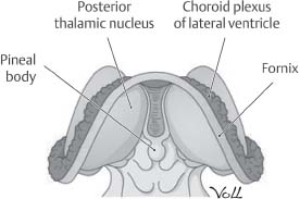
Fig. 1.16 Lateral ventricle and choroid plexus location. (From THIEME Atlas of Anatomy, Head and Neuroanatomy, © Thieme 2007, Illustration by Markus Voll.2)
52. What are the circumventricular organs?
These are areas where the blood–brain barrier is absent. Seven different such areas have been identified (Fig. 1.17):
1. Pineal gland
2. Subforniceal organ
3. Organum vasculosum of the lamina terminalis
4. Median eminence of the hypothalamus
5. Neurohypophysis
6. Area postrema
7. Subcommissural organ
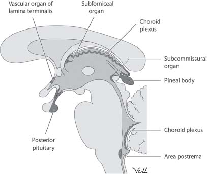
Fig. 1.17 Circumventricular organs. (From THIEME Atlas of Anatomy, Head and Neuroanatomy, © Thieme 2007, Illustration by Markus Voll.2)
53. What is the corresponding intraventricular structure at the calcarine sulcus?
The calcarine sulcus forms a prominence, the calcar avis, in the medial wall of the atrium.23
54. The atrium is deep to which cortical structure?
Supramarginal gyrus (area 40)23
55. What forms the lateral walls of the frontal horns of the lateral ventricles? Medial wall? Roof?
The caudate nucleus, septum pellucidum, and corpus callosum, respectively.23
56. Two pairs of small swellings can be seen in the floor of the 4th ventricle, the lateral and medial ridges. What do these structures represent?
The lateral ridges constitute the vagal trigone and indicate the location of the underlying dorsal motor nucleus of the vagus. The medial ridges constitute the hypoglossal trigone and indicate the location of the underlying hypoglossal nucleus (Fig. 1.18).24
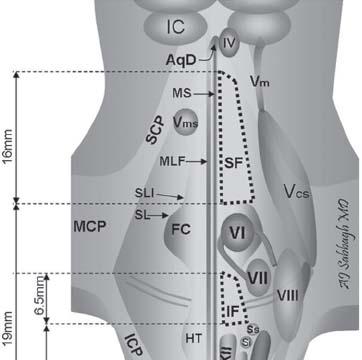
Fig. 1.18 Brainstem schematic anatomy, posterior view. AqD, Aqueduct of Sylvius; N, nucleus; IC, inferior colliculus; MS, median sulcus; Vm, mesencephalic N. of the 5th cranial nerve (V); Vcs, chief (sensory) N. of V; Vms, motor (mastication) N. of V; MLF, medial longitudinal fascicle; FC, facial colliculus; IV, trochlear N.; CTT, central tegmental tract; SL, sulcus limitans; SLI, sulcus limitans incisure; HT, hypoglossal triangle; SM, striae medullaries; SCP, MCP, and SCP, superior, middle and inferior cerebellar peduncle; VT, vagal triangle; AP, area postrema; Obx, obex; VI, abducent N.; VII, facial N. and fiber tracks and nerve; VIII, vestibular N.; XII, hypoglossal N.; Xd, dorsal vagal N.; Am, N. ambiguus of 9th and 10th cranial nerves with parasympathetics on its medial border; Ss and Si, superior and inferior salivatory Nn.; ST, spinal trigeminal tract; STT, spinothalamic tract; ML, medial lemniscus; ION, inferior olivary N.; P, pyramid; TB, trapezoid body; Pn TPF, pontine N. and transverse pontine fibers; SF, superfacial triangle; IF, intrafacial triangle. (Reprinted with permission from Nader and Sabbagh.21)
57. What is the outlet of the 4th ventricle?
There are two laterally located foramina of Luschka and one single medial foramen of Magendie.24
58. Which structure separates the chiasmatic cistern from the interpeduncular cistern?
The Liliequist membrane25
 Autonomic System
59. Where do the sympathetic fibers of the head originate from?
The hypothalamus. They descend through the brainstem and cervical spinal cord to the T1–L2 level where they exit and go back up to the head (Fig. 1.19).26
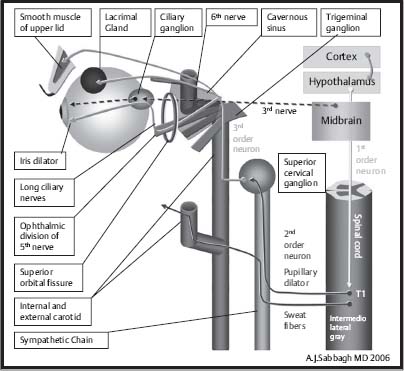
Fig. 1.19 Schematic representation of Horner syndrome. (Reprinted with permission from Nader and Sabbagh.21)
60. Where in the brain are the cholinergic neurons found?
The basal nucleus of Meynert. Abnormalities of this area have been found in patients with Alzheimer disease.27
61. Where are the norepinephrine-containing neurons found in the brain?
The locus ceruleus.28
62. What supplies the sympathetic innervation to the head and neck?
The stellate ganglion26
63. What ganglions form the stellate ganglion?
The inferior cervical ganglion fuses with the first thoracic ganglion to form the cervical thoracic (stellate) ganglion (Fig. 1.19).26
64. What type of nerve fibers does the vidian nerve carry?
Parasympathetic fibers from the greater superficial petrosal nerve and sympathetic fibers from the deep petrosal nerve around the ICA. The nerve passes in the pterygoid canal with the vidian artery.
65. What kind of fibers does the intermediate nerve (nervus intermedius) carry?
This is the sensory and parasympathetic division of the facial nerve. The intermediate nerve carries preganglionic parasympathetic fibers from the superior salivary nucleus that synapse in the pterygopalatine and submandibular ganglia. It also carries taste sensation from the anterior two-thirds of the tongue (Fig. 1.20).29
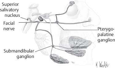
Fig. 1.20 Parasympathetic visceral innervation of the facial nerve. (From THIEME Atlas of Anatomy, Head and Neuroanatomy, © Thieme 2007, Illustration by Karl Wesker.2)
66. What provides the parasympathetics of the parotid glands?
The glossopharyngeal nerve. These parasympathetic fibers originate from the inferior salivatory nucleus and travel via CN IX. These fibers synapse in the otic ganglion before reaching the parotid gland (Fig. 1.21).30
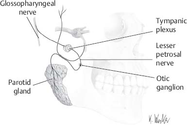
Fig. 1.21 Parasympathetic visceral innervation of the glossopharyngeal nerve. (From THIEME Atlas of Anatomy, Head and Neuroanatomy, © Thieme 2007, Illustration by Karl Wesker.2)
 The Meninges
67. What are the leptomeninges?
The arachnoid and pia mater. The pia and arachnoid layers have a common embryologic origin (ectoderm), whereas the dura is formed by the mesoderm (Fig. 1.22).
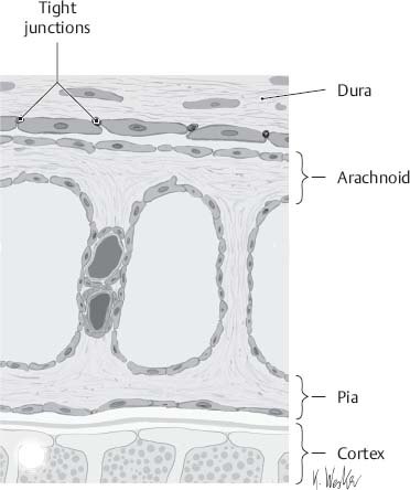
Fig. 1.22 Meningeal cross-sectional anatomy. (From THIEME Atlas of Anatomy, Head and Neuroanatomy, © Thieme 2007, Illustration by Karl Wesker.2)
68. How much cerebrospinal fluid (CSF) is produced each day?
About 450 mL. CSF is continuously reabsorbed and secreted. There is only ~150 mL of CSF in an average body at any one time; 75% of the CSF is produced by the choroid plexus.31
69. What separates the interpeduncular cistern from the chiasmatic cistern?
The Liliequist membrane. The Liliequist membrane is an arachnoidal sheet extending from the dorsum sellae to the anterior edge of the mamillary bodies.25
 Cortex
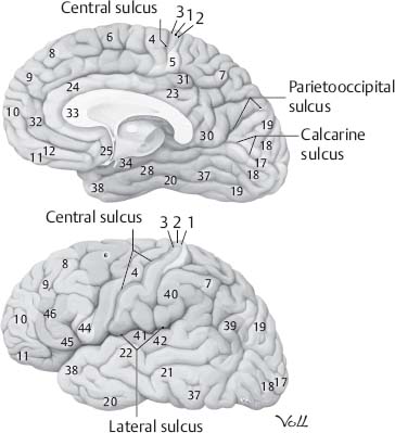
70. Which Brodmann’s area corresponds to Broca’s area? Wernicke’s area? Primary auditory cortex?
Area 44, area 22, and area 41, respectively (Fig. 1.23)32