Neuroanatomy and Physiology
 Surface Anatomy
1. Characterize the lateral cortical surface. |
| G7 p.84:65mm |
a. The pre-central sulcus is not_____. | complete |
|
b. The middle frontal gyrus connects with the_____gyrus through this_____. | precentral, isthmus |
|
c. The central sulcus is separated from the sylvian fissure_____% of the time. | 98% |
|
d. The tissue separating them is called the_____ _____. | sub-central gyrus |
|
e. The inferior and superior parietal lobules are separated by the_____sulcus. | intra-parietal |
|
f. The inferior parietal lobule is composed of |
|
|
i. the s_____m_____g_____ | supra marginal gyrus (SMG) |
|
ii. and the a_____g_____. | angular gyrus |
|
g. The sylvian fissure |
|
|
i. terminates in the_____, | SMG |
|
ii. which is the Brodmann area #_____. | 40 |
|
h. The superior temporal gyrus |
|
|
i. terminates in the_____, | AG |
|
ii. which is the Brodmann area #_____. | 39 |
|
2. Complete the following regarding surface anatomy: |
| G7 p.84:80mm |
a. The middle frontal gyrus often connects with the_____ _____. | pre-central gyrus |
|
b. The central sulcus joins the sylvian fissure in only _____%. | 2% |
|
c. A sub-central sulcus is present in_____% of patients. | 98% |
|
d. The sylvian fissure terminates in the_____ _____. | supra-marginal gyrus |
|
e. The superior temporal sulcus is capped by the_____ _____. | angular gyrus | |
3. Matching. Match the following Brodmann cortical areas and their functional significance: |
| G7 p.84:128mm |
Functional significance: |
|
|
|
|
|
Area: |
|
|
a. Area 3,1,2 |
|
|
b. Area 41,42 |
|
|
c. Area 4 |
|
|
d. Area 6 |
|
|
e. Area 44 |
|
|
f. Area 17 |
|
|
g. Area 40,39 |
|
|
h. Area 8 |
|
|
4. Complete the following regarding pars marginalis: |
| G7 p.85:18mm |
a. is the terminal part of the_____gyrus | cingulate |
|
b. is visible on axial view in >_____% | 90% |
|
c. is the_____ _____ of the midline paired grooves | most prominent |
|
d. extends_____into the hemispheres | deeper |
|
e. on axial CT is located just posterior to the line_____(the widest diameter) | 9-3 |
|
f. it curves_____in lower slices | posteriorly |
|
g. it curves_____in higher slices | anteriorly |
|
5. Complete the following regarding central sulcus: |
| G7 p.85:95mm |
a. Is visible in almost_____% | 95% |
|
b. Does it reach the midline? | no |
|
c. Terminates in the_____ _____ | para-central lobule |
|
6. True or False. The pterion is a region where each of the following bones comes together: |
| G7 p.86:110mm |
a. frontal | true |
|
b. sphenoid (greater wing) | true |
|
c. parietal | true |
|
d. temporal | true |
|
e. sphenoid (lesser wing) | false | |
7. Matching. Match the bones/sutures that form the listed craniometric points. |
|
|
Bone/suture: |
| G7 p.86:125mm |
|
|
|
a. asterion |
|
|
b. pterion |
|
|
8. True or False. The name of the junction of lambdoid, occipitomastoid, and parietomastoid sutures is |
| G7 p.86:140mm |
a. pterion | false |
|
b. asterion | true (Asterion is the junction of lambdoid, occipitomastoid suture, and parietomastoid suture.) |
|
c. lambdoid | false |
|
d. stephanion | false |
|
e. glabella | false |
|
f. opisthion | false |
|
9. The asterion junction overlies the |
| G7 p.86:160mm |
a. _____sinus and the | transverse |
|
b. _____sinus. | sigmoid |
|
10. External landmark for the sylvian fissure is a line from the lateral canthus to a spot three quarters of the way posterior along an arc running over the convexity in the mid line from the |
| G7 p.87:135mm |
a. _____to the | nasion |
|
b. _____. | inion |
|
11. True or False. In relation to external landmarks the angular gyrus is |
| G7 p.87:145mm |
a. one finger’s breadth above the zygomatic arch | false |
|
b. just above the pinna | true (The angular gyrus is just above the pinna and important as part of the Wernicke area in the dominant hemisphere.) |
|
c. a thumb’s breadth behind the frontal process of the zygomatic bone | false |
|
d. at the junction of the lambdoid and sagittal suture | false | |
12. True or False. The motor strip of the motor cortex lies |
| G7 p.87:165mm |
a. at the level of the coronal suture | false |
|
b. within 2 cm of the coronal suture | false |
|
c. 3 to 4 cm posterior to the coronal suture | false |
|
d. 4 to 5.4 cm posterior to the coronal suture | true |
|
e. 2 cm posterior to the mid-position of the | true inion-nasion arc | true |
f. 5 cm straight up from the external | auditory meatus | true |
13. True or False. In the non-hydrocephalic adult the lateral ventricles lie |
| G7 p.88:87mm |
a. 2 to 3 cm below the outer skull surface | false |
|
b. 3 to 4 cm below the outer skull surface | false |
|
c. 4 to 5 cm below the outer skull surface | true |
|
d. 5 to 6 cm below the outer skull surface | false |
|
14. True or False. In the non-hydrocephalic adult the anterior horns extend |
| G7 p.88:108mm |
a. 1 to 2 cm anterior to the coronal suture | true |
|
b. 2 to 3 cm anterior to the coronal suture | false |
|
c. 3 to 4 cm anterior to the coronal suture | false |
|
15. True or False. In the non-hydrocephalic adult the anterior horns extend |
| G7 p.88:130mm |
a. 1 to 2 cm anterior to the foramen of Monro | false |
|
b. 2.5 cm anterior to the foramen of Monro | true |
|
c. 3 to 4 cm anterior to the foramen of Monro | false |
|
16. True or False. The fastigium is located at |
| G7 p.88:145mm |
a. the midpoint of the Twinings line | false |
|
b. the floor of the fourth ventricle | false |
|
c. the apex of the fourth ventricle within the cerebellum | true (The fastigium is the apex of the fourth ventricle in the cerebellum.) |
|
d. 1 to 2 cm anterior to the coronal suture | false |
|
17. List the surface landmarks of the following cervical levels. |
| G7 p.89:35mm |
Hint: htcc |
|
|
a. C3-4_____ _____ | hyoid bone |
|
b. C4-5_____ _____ | thyroid cartilage |
|
c. C5-6_____ _____ | cricothyroid membrane |
|
d. C6-7_____ _____ | cricoid cartilage | |
18. Matching. Match the following surface landmarks and cervical levels: |
| G7 p.89:35mm |
Surface landmark: |
|
|
|
|
|
Cervical level: |
|
|
a. C1-2 |
|
|
b. C3-4 |
|
|
c. C4-5 |
|
|
d. C5-6 |
|
|
e. C6 |
|
|
f. C6-7 |
|
|
 primary motor cortex;
  Broca area (motor speech);
  Wernicke area dominant hemisphere;
  primary auditory area;
  frontal eye fields;
  primary somatosensory area;
  premotor area;
  primary visual cortex


 lambdoid suture;
  occipitomastoid suture;
  parietomastoid suture;
  frontal;
  parietal;
  temporal;
  greater wing sphenoid Craniometric point:
 ,
,  ,
, 
 ,
,  ,
,  ,
, 
 level of thyroid cartilage;
  cricoid cartilage;
  angle of mandible;
  cricothyroid membrane;
  carotid tubercle;
  1 cm above thyroid cartilage (hyoid bone)


 Cranial Foramina and Their Contents
19. Matching. Match the foramen with contents (choices may be used more than once). |
| G7 p.89:75mm |
Contents: |
|
|
|
|
|
Foramen: |
|
|
a. superior orbital fissure |
|
|
b. inferior orbital fissure |
|
|
c. foramen lacerum |
|
|
d. foramen rotundum |
|
|
e. foramen ovale |
|
|
f. foramen spinosum |
|
|
g. stylomastoid foramen |
|
|
h. jugular fora men |
|
|
20. List the cranial nerves and the three branches of one found within the superior orbital fissure (SOF). |
| G7 p.89:85mm |
a. o_____ | CN III oculomotor |
|
b. t_____ | IV trochlear |
|
c. n_____ | nasociliary nerve |
|
d. f_____ | frontal nerve ophthalmic division: all three branches |
|
e. l_____ | lacrimal nerve |
|
f. a_____ | VI abducens nerve |
|
21. Additional structures found in the SOF include the |
| G7 p.89:85mm |
a. s_____o_____v_____ | superior ophthalmic vein |
|
b. r_____m_____a_____ | recurrent meningeal artery |
|
c. which arises from the l_____artery | lacrimal |
|
d. o_____b_____ of the m_____m_____a_____ | orbital branch of the middle meningeal artery |
|
e. s_____p_____ of the ICA | sympathetic plexus of the ICA | |
22. Another name for the transverse crest is_____ _____. | crista falciformis | G7 p.89:182mm |
23. Another name for the vertical crest is_____ _____. | Bill’s bar | G7 p.89:187mm |
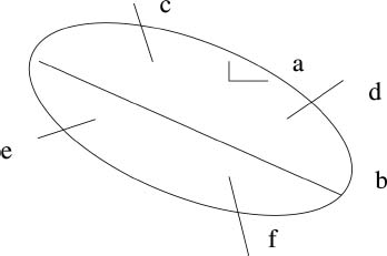
24. Draw and label the nerves in the right porus acusticus. |
| G7 p.90:22mm |
| a. Bill’s bar b. transverse crest crista falciformis c. cranial nerve VII d. SV—superior vestibular e. VIII f. IV—inferior vestibular |
|
Fig. 5.1 |
|
|
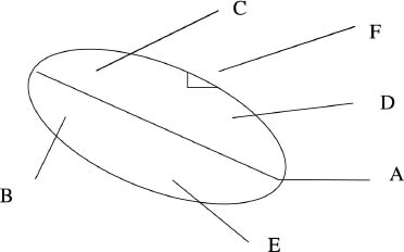
25. Label the diagram of the right internal auditory canal. |
| G7 p.90:22mm |
| a. transverse crest b. acoustic portion of CN VIII c. cranial nerve VII in facial canal d. superior vestibular nerve e. inferior vestibular nerve f. Bill’s bar—vertical crest |
|
Fig. 5.2 |
|
|
26. Matching. Match the nerves of the IAC with the areas that they serve. |
| G7 p.90:23mm |
Nerves: |
|
|
|
|
|
Areas served: |
|
|
a. Facial muscles |
|
|
b. Hair follicles |
|
|
c. Taste buds |
|
|
d. Hearing |
|
|
e. Utricle |
|
|
f. Superior semi-circular canal |
|
|
g. Lateral semi-circular canal |
|
|
h. Saccule |
|
|
 nothing;
  middle meningeal artery;
  VII facial;
  V2;
V2;  V3;
V3;  V1;
V1;  IX, X XI


 facial n.;
  nervus intermedius;
  acoustic portion of VIII n.;
  superior branch of vestibular n.;
  inferior branch of vestibular n.


 Occipitoatlantoaxial-complex Anatomy
27. Matching. Match the ligaments of the occipito-atlantoaxial complex with the statements below. |
| G7 p.91:32mm |
Ligaments: |
|
|
|
|
|
Statements: |
|
|
a. Attaches the odontoid to the foramen magnum |
|
|
b. Attaches the odontoid to the occipital condyle |
|
|
c. Attaches the odontoid to the lateral mass of C1 |
|
|
d. Attaches C1 to the clivus and to C2 |
|
|
e. Attaches odontoid to clivus |
|
|
f. Attaches C1 to C2 |
|
|
g. Traps the odontoid against the atlas |
|
|
h. Extends cephalad to become the tectorial |
|
|
i. The cephalad extension of the PLL |
|
|
j. Extends cephalad to become the anterior atlanto-occipital |
|
|
k. The cephalad extension of the anterior longitudinal |
|
|
28. The most important spinal ligaments in maintaining atlanto-occipital stability are the |
| G7 p.92:95mm |
a. _____membrane and the | tectorial |
|
b. _____ligaments. | alar |
|
 apical;
  alar;
alar;  cruciate;
  ascending portion;
  descending portion;
  transverse portion;
  posterior longitudinal;
  tectorial;
  anterior longitudinal;
  anterior atlanto occipital


 Spinal Cord Anatomy
29. The very large ascending tract closest to the dentate ligament is the _____. | lateral spinothalamic tract (LST) for pain and temperature from the opposite side of the body | G7 p.93:120mm |
30. How is the lateral spinothalamic tract (LST) somatotopically organized? |
| G7 p.93:120mm |
a. Cervical is_____. | medial |
|
b. Sacral is_____. | lateral |
|
31. Which descending motor tract facilitates |
| G7 p.92:158mm |
a. extensor tone? | vestibulospinal tract |
|
b. flexor tone? | rubrospinal tract | |
32. Matching. Match sensory function and anatomy. |
| G7 p.93:175mm |
Sensory function: |
|
|
|
|
|
Anatomy: |
|
|
a. Receptors |
|
|
i. Free nerve ending |
|
|
ii. Meissner and pacinian corpuscles |
|
|
b. First order neurons |
|
|
i. Small |
|
|
ii. Heavily myelinated |
|
|
iii. Finely myelinated |
|
|
iv. Large |
|
|
c. Soma in dorsal root ganglion |
|
|
d. Enter cord at |
|
|
i. Zone of Lissauer |
|
|
ii. Ipsilateral posterior columns |
|
|
e. Synapse in |
|
|
i. Rexed layer II |
|
|
ii. Rexed layer III and IV |
|
|
iii. Rexed layer VI and VII |
|
|
f. Second order neurons |
|
|
i. Cross obliquely in anterior white commissure |
|
|
ii. Form the internal arcuate fibers |
|
|
g. And enter the |
|
|
i. Lateral spino-thalamic tract |
|
|
ii. Medial lemniscus |
|
|
iii. Anterior spino-thalamic tract |
|
|
h. Second order neurons synapse on the ventral posterior lateral nucleus of the Thalamus |
|
|
i. Third order neurons pass through IC to post-central gyrus |
|
|
33. The major blood supply of the spinal cord vasculature |
| G7 p.95:60mm |
a. to the anterior cord arises from |
|
|
i. the vertebral artery and enters at_____ | C3 |
|
ii. the deep cervical artery and enters at_____ | C6 |
|
iii. the costo cervical trunk and enters at_____ | C8 |
|
iv. thoracic levels_____or_____ | T4 or T5 |
|
v. and from the a_____of A_____ | artery of Adamkiewicz |
|
b. to the posterior spinal cord arises from:_____to_____radicular branches | 10 to 23 |
|
c. The “watershed zone” is at the_____or_____region | T4 or T5 | |
34. List the body area with the appropriate root. |
| G7 p.95:70mm |
a. Nipple, root:_____ | T4 |
|
b. Umbilicus, root:_____ | T10 |
|
c. Inguinal crease, root:_____ | T12 |
|
d. Anterior thigh, root:_____ | L2-L3 |
|
e. Posterior thigh, root:_____ | S1 |
|
f. Lateral calf, root:_____ | L5 |
|
g. Medial calf, root:_____ | L4 |
|
h. Posterior calf, root:_____ | S1 |
|
i. Big toe, root:_____ | L5 |
|
j. Little toe, root:_____ | S1 |
|
k. Sole of foot, root:_____ | S1 |
|
l. Lateral shoulder, root:_____ | C5 |
|
m. Lateral forearm | C6 |
|
n. Thumb | C6 |
|
o. Middle finger | C7 |
|
p. Little finger | C8 |
|
q. Medial forearm | T1 |
|
Complete the following regarding upper extremity vs trunk dermatomes. Trunk sensory level is reported at T3 on a trauma patient. |
| G7 p.95:70mm |
a. This is a little the clavicle. | below |
|
b. You must check the_____dermatomes. | arm |
|
c. Dermatomes_____to_____are not represented on the trunk. | C5 to T2 |
|
36. Characterize spinal cord vasculature. The artery of Adamkiewicz serves the spinal cord from |
| G7 p.96:35mm |
a. T_____distally and from the | T8 |
|
b. _____side in | left |
|
c. _____% of the population. | 80% |
|
37. The artery of Adamkiewicz is also known as |
| G7 p.96:35mm |
a. a_____ | arteria |
|
r_____ | radicularis |
|
a_____ | anterior |
|
m_____ | magna |
|
b. Which side does it arise from? | L 80%, R 20% |
|
c. What levels does it arise from 100% inclusive? | T9 and T12 75% |
|
| T9 and L2 85% |
|
| T5 and T8 15% |
|
| T5 and L2 100% |
|
d. What is its appearance on angiography? | characterisitc hairpin shape |
|
38. An artery that has a hairpin shape on angiography is named the_____. | artery of Adamkiewicz | G7 p.96:52mm |
 pain and temperature: body;
  fine touch, deep pressure and proprioception: body;
  light (crude) touch: body
 –
–

 –
–


 –
– –
–

 –
–


 –
–


 –
– –
–
 –
– –
–
 Cerebrovascular Anatomy
39. The artery that feeds a tentorial meningioma is named after |
| G7 p.99:118mm |
a. _____.and | Bernasconi |
|
b. _____. | Cassinari |
|
40. The artery that has a bayonet-type kink is the_____ _____. | ophthalmic artery | G7 p.99:118mm |
41. Circle of Willis is intact in _____%. | 18% | G7 p.97:55mm |
42. Hypoplasia of at least one of the posterior communicating arteries occurs in_____%. | 22 to 32% | G7 p.97:55mm |
43. Absent or hypoplastic A1 occurs in_____%. | 25% | G7 p.97:55mm |
44. What are the seven segments of the internal carotid artery? |
| G7 p.98:20mm |
Hint: can Peter laugh can Charlie only clap |
|
|
a. c_____ | cervical |
|
b. p_____ | petrous |
|
c. l_____ | lacerum |
|
d. c_____ | cavernous |
|
e. c_____ | clinoid |
|
f. o_____ | ophthalmic |
|
g. c_____ | communicating |
|
What portion of the PCA traverses the ambient cistern? | P2 | G7 p.98:95mm |
46. What choroidal artery arises from it? | medial posterior choroidal artery | G7 p.98:103mm |
47. Which cistern is traversed by the P3 segment of the PCA? | quadrigeminal cistern | G7 p.98:102mm |
48. Name the segments of the carotid artery and their main branches. |
| G7 p.99:45mm |
a. C1 c_____ | cervical-carotid sheath |
|
| IJV × PGSN × vagus posterior medial to external carotid |
|
b. C2 p_____ | petrous |
|
c. C3 l_____ | lacerum |
|
d. C4 c_____ | cavernous |
|
i. m_____t_____ | meningohypophyseal trunk |
|
ii. a_____m_____a_____ | anterior meningeal artery |
|
e. C5 c_____ | clinoidal |
|
f. C6 o_____ | ophthalmic |
|
i. o_____ | ophthalmic |
|
ii. s_____h_____ | superior hypophyseal |
|
iii. p_____c_____ | posterior communicating |
|
iv. a_____ c_____ | anterior choroidal | |
g. C7 c_____d_____i_____ | communicating divides into |
|
i. A_____ | ACA |
|
ii. M_____ | MCA |
|
49. What are the branches of the meningohypophyseal trunk? |
| G7 p.99:107mm G7 p.99:100mm |
Hint: dit |
|
|
a. d_____m_____ | dorsal meningeal |
|
b. i_____h_____ | inferior hypophyseal |
|
c. t_____a_____ | tentorial artery of Bernasconi and Cassinari |
|
50. Complete the following concerning anterior circulation: |
| G7 p.99:125mm |
a. Occlusion of which artery results in Sheehan syndrome? | inferior hypophyseal artery |
|
b. It serves_____ _____ _____ | posterior lobe of pituitary |
|
c. It is a branch of the_____artery, | meningohypophyseal |
|
d. which is a branch off the_____ _____segment of carotid. | cavernous C4 |
|
e. Occlusion causes pituitary infarct in_____patients. | postpartum |
|
51. The ophthalmic artery |
| G7 p.99:145mm |
a. arises from the_____segment of the ICA. | sixth |
|
b. Is distal or inside cavernous sinus? | distal 89%, intracavernous 8% |
|
c. Has what shape on lateral angiogram? | a bayonet-type kink |
|
52. The sixth segment of the carotid artery |
| G7 p.99:150mm |
a. is known as the_____ | ophthalmic |
|
b. begins at the_____dural ring | distal |
|
c. ends just proximal to_____-_____ | P-comm |
|
d. has its branches |
|
|
i. o_____artery and the | ophthalmic |
|
ii. s_____h_____artery | superior hypophyseal |
|
53. What vessel supplies the inferior half of the posterior limb of the internal capsule? | anterior choroidal artery | G7 p.100:23mm |
54. Complete the following about the anterior choroidal artery: |
| G7 p.100:30mm |
a. The anterior choroidal artery serves six sites. (Hint: gogoup) |
|
|
i. g_____p_____ | globus pallidus |
|
ii. o_____t_____ | optic tract |
|
iii. g_____of i_____c_____ | genu of internal capsule |
|
iv. o_____r_____ | optic radiations |
|
v. u_____ | uncus |
|
vi. p_____l_____ | posterior limb of internal capsule | |
b. Occlusion may produce: Hint: 3 H _____,_____,_____ _____ | hemiplegia, hemihypesthesia, homonymous hemianopsia |
|
c. MRI shows infarct in the_____. | posterior limb of the internal capsule |
|
55. What artery enters the supracornual recess of the temporal horn to supply the choroid plexus? | plexal segment of the anterior choroidal artery | G7 p.100:30mm |
56. Complete the following regarding P-comm and the anterior choroidal artery (ACH): |
| G7 p.100:35mm |
a. They are_____ mm apart. | 2 |
|
b. The origin of the_____-_____ is proximal. | P-comm |
|
c. Is the Ach smaller or larger than the P-comm? | smaller |
|
d. Which artery has the hump of the plexal point? | Ach |
|
57. True or False. The carotid siphon |
| G7 p.100:53mm |
a. is only that part of the carotid that passes within the cavernous sinus. | false |
|
b. If an aneurysm ruptures on the siphon there is no SAH. | false |
|
58. The carotid siphon |
| G7 p.100:53mm |
a. begins at the posterior bend of the_____ carotid and | cavernous |
|
b. ends at the ICA_____. | bifurcation |
|
c. It includes the |
|
|
i. ca_____ | cavernous |
|
ii. op_____ | ophthalmic |
|
iii. co_____ | communicating |
|
59. Complete the following about vertebral artery segments: |
| G7 p.102:168mm |
a. The first segment enters the_____ foramen transversarium. | sixth |
|
b. The second ascends_____ within the foramina transversaria. | veritcally |
|
c. The second turns_____as it exits the axis. | laterally |
|
d. The third curves_____and_____. | posteriorly and medially |
|
e. The fourth pierces the_____. | dura |
|
60. The vertebral artery joins the other side at the level of the |
| G7 p.103:20mm |
a. _____ _____to form the | lower pons (pontomedullary junction) |
|
b. _____ _____. | basilar artery | |
61. The junction of the vertebral arteries is called the_____ _____. | vertebral confluens | G7 p.103:20mm |
62. What are the six branches arising from the vertebral artery? |
| G7 p.103:105mm |
Hint: A postman puts postcards away. |
|
|
a. a_____m_____ | anterior meningeal |
|
b. p_____m_____ | posterior meningeal medullary (bulbar) |
|
c. m_____ | medullary (bulbar) |
|
d. p_____s_____ | posterior spinal |
|
e. p_____ | PICA |
|
f. a_____s_____ | anterior spinal |
|
63. Complete the following statements about the PICA: |
| G7 p.103:120mm |
a. PICA arises_____mm distal to the point where VA becomes intradural. | 10 |
|
b. PICA has an extradural origin in_____to_____%. | 5 to 8% |
|
c. It includes five segments named |
|
|
i. a_____m_____ | anterior medullary |
|
ii. l_____m_____ | lateral medullary |
|
iii. t_____-m_____has_____loop | tonsillo-medullary, caudal |
|
iv. t_____-v_____has_____loop | telo-velo-tonsillar, cranial (supratonsillar) |
|
v. c_____s_____ | cortical segments |
|
d. and has three branches named |
|
|
i. c_____ | choroidal |
|
ii. t_____-h | tonsillo-hemispheric |
|
iii. i_____v_____ | inferior vermian |
|
64. The cranial loop on angio of the PICA is the_____artery. | supratonsillar (telo-velo-tonsillar) | G7 p.103:165mm |
65. The choroidal point |
| G7 p.103:173mm |
a. is the point where the_____artery | choroidal |
|
b. arises from the_____artery | supratonsillar |
|
c. which is a branch of the_____ | PICA |
|
d. enters into the_____ _____ | fourth ventricle |
|
e. to serve the_____ _____ | choroid plexus |
|
66. The copular point |
| G7 p.103:65mm |
a. is the point where the_____ _____artery | inferior vermian | G7 p.103:65mm |
b. arises from the_____. | PICA |
|
67. Name the three segments of the posterior cerebral artery. |
| G7 p.104:65mm |
a. c_____ | crural (peduncular) segment (P1) |
|
b. a_____ | ambient segment (P2) |
|
c. q_____ | quadrigeminal segment (P3) | |
68. Medial posterior choroidal artery arises from the |
| G7 p.104:84mm |
a. _____segment of PCA. | crural |
|
b. It is also called_____. | P1 |
|
69. Lateral posterior choroidal artery arises from the |
| G7 p.104:92mm |
a. _____segment of the PCA. | ambient |
|
b. It is also called_____. | P2 |
|
70. The third segment of PCA is named the_____segment. | quadrigeminal | G7 p.104:117mm |
71. Name the branches of the external carotid from proximal to distal. |
| G6 p.104:30mm |
Hint: salfops m |
|
|
a. s_____ _____ | superior thyroid |
|
b. a_____ _____ | ascending pharyngeal |
|
c. l_____ | lingual |
|
d. f_____ | facial |
|
e. o_____ | occipital |
|
f. p_____ _____ | posterior auricular |
|
g. s_____ _____ | superficial temporal |
|
h. m_____ | maxillary |
|
72. In relation to ICA, the ECA lies |
| G6 p.79:45mm |
a. _____and | anterior |
|
b. _____to ICA. | lateral |
|
73. Which internal jugular vein is usually dominant? | the right | G7 p.104:140mm |
74. Which transverse sinus is usually dominant? | the right | G7 p.104:147mm |
75. Which vertebral artery is usually dominant? | the left by 60% | G7 p.102:156mm |
76. Name the major contributors to the great cerebral vein of Galen. |
| G7 p.105:25mm |
a. p_____c_____v_____ | precentral cerebellar vein |
|
b. b_____v_____of R_____ | basal veins of Rosenthal |
|
c. i_____c_____v_____ | internal cerebral veins |
|
77. The joining of the septal vein and the thalamostriate vein with the internal cerebral vein forms an angiographic landmark called the _____ _____at the foramen of Monro. | venous angle | G7 p.105:35mm |
78. True or False. The cavernous sinus is |
| G7 p.105:140mm |
a. a large venous space with multiple trabeculations | false |
|
b. a plexus of veins | true | |
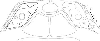
79. Draw the right and left cavernous sinus coronal view. On your drawing, label the following: |
| G7 p.106:15mm |
| 1. oculomotor (III) 2. trochlear (IV) 3. Parkinson triangle 4. ophthalmic (V1) 5. maxillary (V2) 6. abducent (VI) 7. carotid |
|
Fig. 5.3 |
|
|
80. Name six major contents of the cavernous sinus. |
| G7 p.106:15mm |
a. _____ | CN III |
|
b. _____ | CN IV |
|
c. _____ | CN V1 |
|
d. _____ | CN V2 |
|
e. _____ | CN VI |
|
f. _____ | internal carotid artery |
|
81. Complete the following regarding the cavernous sinus: |
|
|
a. Which nerve in the cavernous sinus does not also pass through the superior orbital fissure? | V2 maxillary division of trigeminal | G7 p.106:30mm |
b. Which foramen of the skull does that nerve pass through? | foramen rotundum | G7 p.106:30mm |
c. Which nerve is not attached to the wall? | VI is not attached to lateral wall (abducens) | G7 p.106:85mm |
82. With regard to the cavernous sinus, the triangular space of Parkinson is bounded by what structures? |
| G7 p.106:90mm |
a. on its superior border_____ | III and IV |
|
b. on its inferior border_____ | trigeminal V1 andV2 |
|
83. Complete the following regarding persistent fetal anastomosis: |
| G7 p.107:28mm |
a. How many are there? | 4 |
|
b. They result from a failure to_____. | involute |
|
c. Name them. |
|
|
i. t_____ | trigeminal |
|
ii. o_____ | otic |
|
iii. h_____ | hypoglossal |
|
iv. p_____ | proatlantal |
|
84. The most common persistent fetal anastomosis is the_____. | trigeminal | G7 p.107:60mm |
85. First to involute in persistent fetal anastomsosis is the_____. | otic | G7 p.107:125mm |
 Internal Capsule
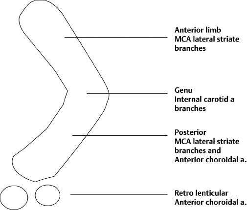
86. Name the vascular supply for the following components of the internal capsule: |
| G7 p.107:165mm |
a. anterior limb | lateral striate branches of MCA |
|
b. posterior limb | lateral striate branches of MCA |
|
c. ventral posterior limb | anterior choroidal |
|
d. genu | direct branches of ICA |
|
e. optic radiations | anterior choroidal |
|
87. Name four thalamic peduncles and where their radiations go. |
| G7 p.108:75mm |
a. a_____, f_____ l_____ | anterior, frontal lobe |
|
b. s_____, p_____ g_____ | superior, postcentral gyrus |
|
c. P_____, o_____ P_____ a_____ | posterior, occipital parietal areas |
|
d. i_____, a_____ a_____ | inferior, auditory area |
|
88. Draw the internal capsule and label which blood vessel serves which area. |
| G7 p.108:15mm |
Hint: MIMA |
|
|
| ||
89. Matching. Match the area in internal capsule with its function. |
| G7 p.108:20mm |
Area in internal capsule: |
|
|
| ||
Fig. 5.5 |
|
|
Function: |
|
|
1. Movement of face ________ | C—genu |
|
2. Movement of foot ________ | D—posterior limb |
|
3. Vision ________ | F—lateral geniculate |
|
4. Hearing ________ | G—medial geniculate |
|
 Miscellaneous
90. The Obersteiner-Redlich zone is |
| G7 p.108:130mm |
a. also known as the_____ _____ _____. | root entry zone |
|
b. It is where the central_____ and peripheral_____transition. | myelin, myelin |
|
c. It is the zone where_____tend to grow. | neoplasms |
|
d. It is located on CN VIII,_____from the brain stem. | 8 to 12 mm |
|
91. The dentate ligament |
| G7 p.108:150mm |
a. separates_____ | dorsal |
|
b. from_____roots in the spinal nerves. | ventral |
|
92. Which cranial nerve lies dorsal to the dentate ligament? | CN XI spinal accessory | G7 p.108:155mm |
 Neurophysiology
93. Answer the following concerning the blood-brain barrier (BBB): |
| G7 p.109:60mm |
a. What chemical opens the BBB? | mannitol |
|
b. What chemical closes the BBB? | steroids |
|
c. Which sites have no BBB? | pituitary | G7 p.109:70mm |
Hint: pppcta | pineal |
|
| preoptic recess |
|
| choroid plexus |
|
| tuber cinereum |
|
| area postrema |
|
d. What pathology injures BBB? | hepatic encephalopathy |
|
Hint: histt | infections |
|
| stroke |
|
| trauma |
|
| tumor |
|
94. Complete the following statements about cerebral edema: |
| G7 p.109:75mm |
a. Cytotoxic |
|
|
i. occurs with h_____ i_____ | head injury |
|
ii. occurs with h_____ | hematoma |
|
iii. shape is c_____ | circular |
|
iv. occurs with C_____ | CVA |
|
v. BBB is c_____ | closed |
|
b. Vasogenic |
|
|
i. shape is_____ | V-shaped (like fingers of white matter edema) |
|
ii. occurs with t_____ | tumors |
|
iii. occurs with m_____ | metastasis |
|
iv. treat with s_____ | steroids |
|
v. with contrast it_____ and _____ | enhances on CT and MR |
|
vi. BBB is o_____ | open |
|
95. Matching. Match the type of edema with the characteristics. |
| G7 p.109:80mm |
Type of edema: |
|
|
|
|
|
a. BBB open |
|
|
b. BBB closed |
|
|
c. Head injury |
|
|
d. Tumor |
|
|
e. Enhances |
|
|
f. Does not enhance |
|
|
g. Not appropriate to use steroids |
|
|
h. Appropriate to use steroids |
|
|
i. Circular shape on MR |
|
|
j. V-shaped finger like extensions on MR |
|
|
k. Occurs with hematoma |
|
|
l. Occurs with CVA |
| |
96. True or False. Cytotoxic edema has: |
| G7 p.109:89mm |
a. a disrupted BBB | false |
|
b. expansion of the extracellular space | false |
|
c. enhancement when contrast injected | false |
|
d. no protein extravasation | true |
|
97. Study Sheet. |
| G7 p.109:100mm |
a. Cytotoxic: |
|
|
b. Closed BBB |
|
|
c. Head injury |
|
|
d. Hematoma |
|
|
e. Circular shape |
|
|
f. CVA |
|
|
g. Cells swell then shrink |
|
|
h. Vasogenic: |
|
|
i. BBB |
|
|
j. Tumors |
|
|
k. Metastasis |
|
|
l. Steroids |
|
|
m. Protein extravasates |
|
|
n. Enhances on CT and MRI |
|
|
o. Wide extracellular space |
|
|
p. Stable cells |
|
|
98. In pituitary embryology, posterior pituitary |
| G7 p.109:110mm |
a. derives from the_____evagination | downward |
|
b. of_____ _____ cells (neuroectoderm) | neural crest |
|
c. from the_____ | floor |
|
d. of the_____ventricle. | third |
|
99. The anterior pituitary |
| G7 p.109:120mm |
a. develops from the_____ | evagination |
|
b. of_____ _____ | epithelial ectoderm |
|
c. of the_____ _____ | oropharynx |
|
d. known as_____ _____ | Rathke’s pouch |
|
100. Complete the following regarding neuroendocrinology. |
| G7 p.109:150mm |
a. The pituitary releases_____hormones | 8 |
|
b. from the anterior pituitary gland:_____hormones. | 6 |
|
c. Name them. |
|
|
Hint: pcpgtg |
|
|
i. p_____ | propriomelanocortin |
|
ii. c_____ | corticotropin |
|
iii. p_____ | prolactin |
|
iv. g_____ _____ | growth hormone |
|
v. t_____ | thyrotropin |
|
vi. g_____ | gonadotropin |
|
d. and from the posterior pituitary |
|
|
i. a_____ | antidiuretic |
|
ii. o_____ | oxytocin | |
101. The pituitary hormones that are released from the posterior pituitary are synthesized |
| G7 p109:165mm |
a. in_____neurons | neurons |
|
b. in the_____. | hypothalamus |
|
c. Are these cells glands? | no |
|
d. The hormones are conveyed by_____ | axons |
|
e. within the_____ _____ | pituitary stalk |
|
f. to the_____pituitary gland | posterior |
|
g. where they are_____. | released |
|
 cytotoxic edema;
  vasogenic edema Hint: cytotoxic—early letters of alphabet vasogenic—later letters of alphabet Characteristics:


 Regional Brain Syndromes
102. Matching. Match region with deficit. |
| G7 p.112:30mm |
Region: |
|
|
|
|
|
Deficit: |
|
|
a. Apathy abulia |
|
|
b. Disorganized thoughts |
|
|
c. Contralateral neglect |
|
|
d. Language disorders |
|
|
e. Anosognosia |
|
|
f. Dressing apraxia |
|
|
g. Homonymous hemianopsia |
|
|
h. Truncal ataxia |
|
|
i. Ipsilateral ataxia |
|
|
j. Paralysis of upward gaze |
|
|
k. Poor planning |
|
|
l. Unilateral anosmia |
|
|
103. Frontal eye fields for contra lateral gaze are |
| G7 p.112:55mm |
a. located in the_____frontal lobe | posterior |
|
b. in Broadmann area_____. | 8 |
|
c. With a destructive lesion there, the patient’s eyes look_____the lesion. | toward Hint: destructive=toward |
|
d. With an irritative lesion there, the patient’s eyes look_____ _____ the lesion. | away from Hint: irrigitative=away |
|
e. Usually the lesions are_____. | destructive | |
104. True or False. Regarding Foster-Kennedy syndrome: |
| G7 p.114:125mm |
a. usually from olfactory groove or medial third sphenoid wing tumor | true |
|
b. contralateral anosmia | false (Ipsilateral not contralateral anosmia is part of the classic triad.) |
|
c. ipsilateral central scotoma | true |
|
d. contralateral papilledema | true |
|
e. contralateral optic atrophy | false (ipsilateral optic atrophy) |
|
f. usually meningioma | true |
|
105. True or False. Regarding Weber syndrome: |
| G7 p.X:X mm |
a. Weber syndrome includes CN III palsy with contralateral hemiparesis. | true |
|
b. Weber syndrome includes CN VII palsy with contralateral hemiparesis. | false |
|
c. Weber syndrome includes CN III palsy with ipsilateral hemiparesis. | false |
|
d. Weber syndrome includes CN VI and VII palsy with contralateral hemiparesis. | false |
|
e. Weber syndrome includes |
| G7 p.114:105 |
i. Cranial nerve III palsy | false |
|
ii. Contralateral hemiparesis | false |
|
iii. Arm hyperkinesis | false |
|
iv. Ataxia | false |
|
v. Intention tremor | false |
|
106. True or False. Benedict syndrome is due to disruption of |
| G7 p.114:115mm |
a. cerebral peduncle | true |
|
b. issuing fibers of CN III | true |
|
c. red nucleus | true |
|
107. True or False. Millard-Gubler syndrome is due to disruption of |
| G7 p.114:130mm |
a. nucleus of VII | true |
|
b. nucleus of VI | true |
|
c. cortico spinal tract | true |
|
108. True or False. Regarding Parinaud syndrome: |
| G7 p.114:135mm |
a. Parinaud syndrome includes downgaze palsy. | false |
|
b. Parinaud syndrome includes lid retracion. | true |
|
c. Parinaud syndrome includes nystagmus retractorius. | true |
|
d. When Parinaud syndrome is combined with downgaze palsy it is known as the syndrome of the _____ _____. | sylvian aqueduct | |
109. True or False. The following are contents of the jugular foramen: |
| G7 p.115:70mm |
a. transverse sinus | false |
|
b. CN IX, X, and XI | true |
|
c. CN X, XI, and XII | false |
|
d. sigmoid sinus | true |
|
e. petrosal sinus | true |
|
f. branches from the ascending pharyngeal artery | true |
|
g. branches from the occipital artery | true |
|
110. Matching. Match the following numbered descriptions with the lettered syndromes. Also indicate the nerves involved and the results of the lesion. |
| G7 p.115:110mm |
Description: |
|
|
|
|
|
a. Which jugular foramen syndrome is most likely due to an intracranial lesion? |
|
|
b. Extracranial lesion? |
|
|
c. Retropharyngeal lesion? |
|
|
111. True or False. A jugular foramen syndrome that spares CN IX is |
| G7 p.115:155mm |
a. Vernet | false |
|
b. Collet-Sicard | false |
|
c. Villaret | false |
|
d. Tapia | true (Tapia X, XII vocal cords and tongue) |
|
112. True or False. The following jugular foramen syndrome also results in a Horner syndrome: |
| G7 p.115:180mm |
a. Vernet | false |
|
b. Collet-Sicard | false |
|
c. Jackson | false |
|
d. Villaret | true |
|
113. True or False. Gerstmann syndrome includes |
| G7 p.113:70mm |
a. agraphia without alexia | true |
|
b. left-right confusion | true |
|
c. digit agnosia | true |
|
d. tactile agnosia | false |
|
e. acalculia | true |
|
114. True or False. Gerstmann syndrome patients can read. | true | G7 p.113:70mm |
115. True or False. Gerstmann syndrome patients can write. | false | |
116. True or False. Cortical sensory syndrome includes |
| G7 p.113:110mm |
a. loss of position sense | true |
|
b. inability to localize tactile stimuli | true |
|
c. astereognosis | true |
|
d. loss of pain and temperature sense | false (Pain and temperature as well as vibration sense are preserved.) |
|
117. True or False. Anton Babinski syndrome includes |
| G7 p.113:155mm |
a. anosognosia | true |
|
b. apathy | true |
|
c. ipsilateral extinction to double-sided stimulation | false (contralateral extinction to double-sided stimulation) |
|
d. dressing apraxia | true |
|
118. True or False. Wernicke aphasia includes |
| G7 p.114:27mm |
a. fluent aphasia | true |
|
b. lesion is in Brodmann areas 41 and 42 | false (The lesion is in Brodmann 39 and 40.) |
|
c. speech devoid of meaning | true |
|
d. normal intonation | true |
|
119. True or False. Broca aphasia includes |
| G7 p.114:40mm |
a. dysarthria | true |
|
b. lesion is in area 44 | true |
|
c. an “apraxia” of motor sequencing | true |
|
d. similar to conduction aphasia | false (Broca is a motor aphasia—faltering dysarthric speech. Conduction aphasia is fluent speech with paraphasias.) |
|
120. Alexia without agraphia |
| G7 p.114:78mm |
a. means that the patient can_____ | write |
|
b. but cannot_____. | read |
|
c. Surprisingly, such patients can usually do what with numbers? | read and name them |
|
d. Lesion is located in the_____lobe. | parietooccipital |
|
e. On which side? | dominant (left) side |
|
f. Serves to disconnect_____ _____and | angular gyrus |
|
g. _____ _____ | occipital lobes |
|
h. also known as_____ _____ _____. | pure word blindness |
|
i. This is contrasted with what syndrome? | Gerstmann |
|
j. Where patient can_____ | read |
|
k. but can’t_____ | write |
|
l. also known as_____ _____ _____. | agraphia without alexia | |
121. Matching. Match the numbered syndromes with the lettered phrases. |
| G7 p.114:78mm |
Syndrome: |
|
|
a. alexia without agraphia |
|
|
b. agraphia without alexia |
|
|
c. where patient can’t read |
|
|
d. where patient can’t write |
|
|
 Pre-frontal lobes;
  frontal lobe;
  parietal lobe—dominant;
  parietal—non dominant;
  occipital lobe;
  cerebellum;
  brain stem;
  pineal;
  olfactory groove

 or
or 


 Vernet;
  Collet-Sicard;
  Villaret Syndrome:
 involves CN,IX,X,XI taste, vocal cords and SCM (sterno cleido mastoid muscle)
 above plus XII tongue
 above plus Horner
 Gerstmann;
  Pure word blindness Phase:


 Babinski Sign
122. Fill in the blanks to complete the details of the Babinski reflex. |
| G7 p.116:35mm |
Hint: pcrstlpt |
|
|
a. lateral_____ stimulation | plantar |
|
b. originates as a_____ _____ | cutaneous reflex |
|
c. and stimulates the_____ | receptors |
|
d. in the_____dermatome | S1 |
|
e. that travel via the_____ _____ | tibial nerve |
|
f. to the spinal cord segments number_____(_____limb) | L4-S2, afferent |
|
g. The efferent limb travels via the_____nerve (_____limb) | peroneal, efferent |
|
h. to the_____ _____ | toe extensors |
|
123. Summarize the Babinski sign. |
| G7 p.116:65mm |
a. receptor_____ | S1 dermatome |
|
b. afferent limb_____ | tibial nerve |
|
c. cord_____ | L4-S2 |
|
d. efferent limb_____ | peroneal nerve |
|
124. Fill in the blanks to complete the details of eliciting the plantar reflex. |
| G7 p.116:92mm |
Stimulate the_____ _____ surface | lateral plantar |
|
b. and the_____ _____ | transverse arch |
|
c. in a_____movement | single |
|
d. that lasts_____seconds. | 5 to 6 |
|
e. Response consists of_____of the_____ _____. | extension of the great toe |
|
f. _____of the small toes is | Fanning |
|
g. _____clinically important. | not |
|
125. True or False. The Chaddock maneuver is described as |
| G7 p. 116:108mm |
a. scratching the lateral foot | true |
|
b. pinching the Achilles tendon | false |
|
c. sliding knuckles down shin | false |
|
d. momentarily squeezing lower gastrocnemius | false | |
126. Complete the following concerning Hoffman sign: | G7 p.116:128mm | |
a. H (from Hoffman) is the_____letter of the alphabet. | eighth |
|
b. If unilaterally present Hoffman sign indicates a lesion above_____. | C8 |
|
 Bladder Neurophysiology
127. Complete the following concerning bladder physiology: |
| G7 p.116:170mm |
a. The primary coordinating center for bladder function is in the |
|
|
i. n_____ l_____ c_____ | nucleus locus coeruleus |
|
ii. of the p_____. | pons |
|
b. This center coordinates |
|
|
i. b_____ c_____ (d_____) with | bladder contraction (detrusor) |
|
ii. s_____ r_____ (e_____ s_____). | sphincter relaxation (external sphincter) |
|
128. Voluntary cortical control |
| G7 p.116:182mm |
a. inhibits the p_____ c_____. | pontine center—nucleus locus coeruleus |
|
b. It originates in the |
|
|
i. a_____ f_____ l_____ | anteromedial frontal lobes |
|
ii. and g_____of the c_____ c_____and | genu of the corpus callosum |
|
c. travels via the p_____t_____ | pyramidal tract |
|
d. to inhibit |
|
|
i. c_____of the | contraction of the |
|
ii. d_____ and contraction | detrusor and contraction |
|
iii. of the e_____ s_____. | external sphincter |
|
129. Immaturity, infarct, or cortical lesions cause |
| G7 p.117:17mm |
a. inability to s_____ | suppress |
|
b. the m_____r_____ | micturition reflex |
|
c. and results in i_____. | incontinence |
|
130. The efferents to the bladder |
| G7 p.117:28mm |
a. travel in the_____portion | dorsal |
|
b. of the_____ _____. | lateral columns |
|
131. Parasympathetic control |
| G7 p.117:48mm |
a. detrusor_____ | contracts |
|
b. internal sphincter_____ | relaxes |
|
c. travels via the p_____ s_____nerves | pelvic splanchnic |
|
132. Somatic nerve |
| G7 p.117:48mm |
a. external sphincter_____ | contracts |
|
b. maintains c_____ | continence |
|
c. travels via p_____ nerve | pudendal | |
133. Sympathetic nerve |
| G7 p.117:48mm |
a. provides bladder neck_____and | closure |
|
b. travels via the i_____ h_____plexus. | inferior hypogastric |
|
134. True or False. The detrusor muscle of the bladder contracts and the internal sphincter relaxes under |
| G7 p.117:53mm |
a. PNS stimulation | true (parasympathetic nervous system stimulation) |
|
b. somatic nerve stimulation | false |
|
c. sympathetic nervous system stimulation | false |
|
d. all of the above | false |
|
135. True or False. The following can cause detrusor hyperreflexia: |
| G7 p.117:125mm |
a. CVA | true |
|
b. spinal cord tumor | true |
|
c. chronic bladder catheterization | false (Detrusor hyperreflexia can result from interruption of efferents anywhere from cortex to sacral cord.) |
|
d. multiple sclerosis | true |
|
e. Parkinson disease | true |
|
136. True or False. Interruption of the efferents results in |
| G7 p.117:142mm |
a. atonic bladder | false—root lesion |
|
b. overflow incontinence | false—root lesion |
|
c. uncontrollable voiding | true |
|
d. reflex bladder empting | true |
|
e. voiding triggered by critical volume | true |
|
f. produced by myelopathy | true |
|
g. produced by head injury | true |
|
h. produced by certain drugs | false—detrusor areflexia |
|
i. produced by diabetes mellitus | false—automatic neuropathy |
|
137. True or False. Patients with multiple sclerosis develop voiding symptoms from demyelination primarily involving the |
| G7 p.118:127mm |
a. posterior and lateral columns of lumbar spinal cord | false |
|
b. lateral column of cervical spine | false |
|
c. posterior column of lumbar spine | false |
|
d. lateral column of lumbar spine | false |
|
e. posterior and lateral columns of cervical spinal cord | true (posterior and lateral columns of cervical spinal cord) |
|
138. True or False. Causes of urinary retention are |
| G7 p.118:145mm |
a. urethral stricture | true |
|
b. prostatic enlargement | true |
|
c. detrusor areflexia | true |
|
d. herpes zoster | true |
|

Stay updated, free articles. Join our Telegram channel

Full access? Get Clinical Tree


