Types of tumors
Japana
Koreaa
TVGHb
Australiac
N = 118
N = 125
N = 84 (8.5%)
N = 37
1–25 years
1–12 years
1–18 years
1–16 years
Pineal tumors
74 (88.1 %)
Suprasellar-pineal tumors
10 (11.9 %)
Germ cell tumor
71.2 %
80.0 %
79.8 %
54.1 %
Germinoma
46.6 %
47.2 %
42.8 %
35.1 %
NGMGCT
26.2 %
16.2 %
MT
8.3 %
2.7 %
Neuroectodermal tumors/pineoblastomas
15.2 %
16.8 %
9.5 %
16.2 %
Astrocytoma
2.4 %
8.1 %
No histological/clinical Dx
5.9 %
16.2 %
Since 1970 we have treated 176 patients with germ cell tumors and from them 85 have been pineal (Table 14.2).
Table 14.2
Types of pineal GCTs in children series of Taipei Veterans General Hospital, 1970–2007
Other sites GCTs | Pineal GCTs | Pineal GCTs seeding at Dxa | |
|---|---|---|---|
No. of patients | 91 | 85 | |
Germinoma | 58.2 % | 58.8 % | 5.6 % (V 1, IS 1) |
Mature teratoma | 1.1 % | 8.2 % | |
NGMGCT | 35.2 % | 31.2 % | 0 % |
Immature teratoma | 13.2 % | 8.2 % | |
Mixed GCT | 16.5 % | 14.1 % | |
Yolk sac tumor | 6.6 % | 4.7 % | |
Dx by tumor marker | 3.3 % | 4.7 % | |
Unclassified GCT | 1.1 % | 1.2 % |
In the majority of patients, hydrocephalus has coexisted as shown on Table 14.3. The simultaneous presence of active hydrocephalus is well reported in the literature and needs to be treated.
Table 14.3
Tumoral hydrocephalus with obstruction at posterior third ventricle, pineal, and midbrain tumors, 1970–2007
No. of patients | Hydrocephalus (%) | |
|---|---|---|
116 | 87.1 | |
Pineal tumors | 105 | 85.7 |
Germ cell tumors | 85 | 83.5 |
Astrocytic tumors | 2 | 100 |
Pineal parenchymal tumors | 11 | 90.9 |
Embryonal tumors | 2 | 100 |
No. of clinical/histological Dx | 6 | 83.3 |
Midbrain tumors | 11 | 100 |
At the pineal region a variety of lesions can occur, single or bifocal tumors, and with varying histology (pineoblastoma, pineocytoma, astrocytoma, meningioma, cyst, and also midbrain tumors) [7, 8] (Fig. 14.1).
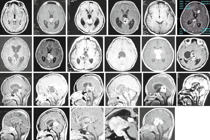

Fig. 14.1
MR scans with examples of pineal germinomas/NG-GCTs, single and bifocal tumors
14.2 Management of Pineal Region Tumors
The management of pineal region tumors can be summarized as follows:
1.
In case of suspected germinoma or pineoblastoma with accompanying hydrocephalus and normal values of AFP and β-hCG, we propose endoscopic tumor biopsy and third ventriculostomy. At the same time an estimation of the tumor vascularity can be made as well as the presence of tumor seeding to the walls of the ventricles (Fig. 14.2). If the biopsy provides tumor-type diagnosis, further oncological management is pursued as the next step.
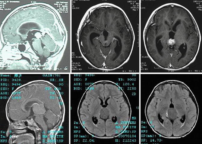
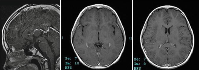


Fig. 14.2
MR scans with examples of pineoblastoma (2 first rows) and pineocytoma (third row)
2.
In case of suspected mature teratoma, non-germinomatous malignant germ cell tumor (NGMGCT), or other tumors, we propose radical excision of the tumor, endoscopically assisted when needed and navigation assisted if necessary (Figs. 14.3 and 14.4).
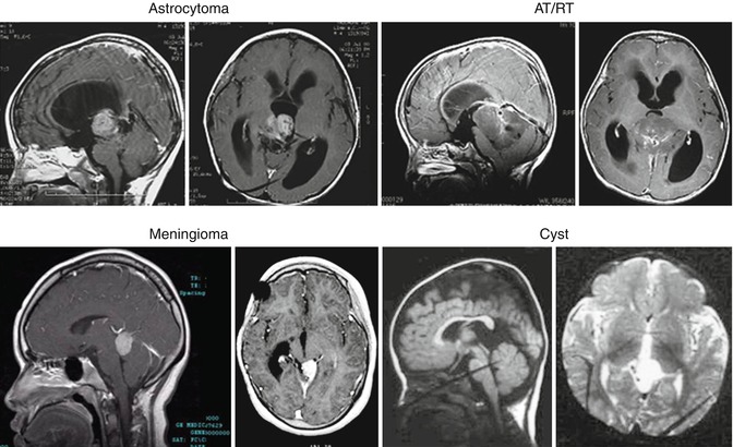
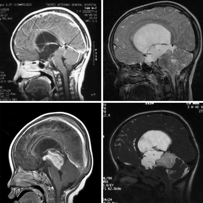

Fig. 14.3
MR scans with examples of other tumor types and cysts in the pineal/juxtapineal region

Fig. 14.4
MR scans with examples of Juxtapineal tumors – midbrain tumors
3.
Get Clinical Tree app for offline access

In case of midbrain tumors, we propose endoscopic third ventriculostomy combined with biopsy in selected cases. In some cases, an endoscopic removal of the tumor can be achieved [9–11] (Figs. 14.5, 14.6, and 14.7).
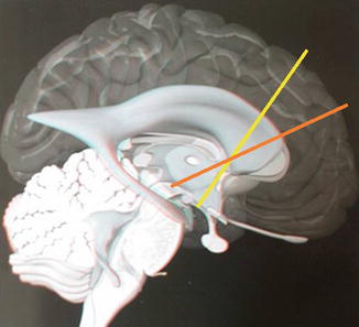
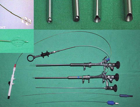

Fig. 14.5
Trajectories of coronal burr hole for ETV (yellow line) and behind hairline burr hole for ETB (orange line) (Drawings modified from Martin C. Hirsch, Thomas Kramer)

Fig. 14.6


Endoscopic instruments. The STORZ DECQ endoscope is used, with the 0° and 30° rod lenses. At the upper left the Integra neuroballoon and urological basket perforator (KARL STORZ GmbH & Co., Tuttlingen, Germany; Integra, Plainsboro, NJ, USA)
Stay updated, free articles. Join our Telegram channel

Full access? Get Clinical Tree


