Fig. 16.1
Diagrams showing the possible endoscopic approaches that can be employed in the removal of cervical, thoracic, and lumbar herniated discs. (a) Anterior, to remove the herniated part of the disc. (b) Posterior, to decompress the spinal root canal as well, in the presence of a lateral osteophyte. (c) Lateral, to remove extraforaminal disc herniation
16.3.2 Epiduroscopy
The usefulness and effectiveness of this technique is a matter of discussion. It is most commonly applied in post-laminectomy peridural fibrosis [5, 14–17]. Via the hiatus sacralis and with the use of intraoperative radiology, a 0.9–2.5-mm 0° semiflexible or flexible neuroendoscope is introduced through a sheath (Fig. 16.2). Adhesiolysis is performed with a balloon catheter and cauterization. No more than 120 ml of irrigation is recommended. Our series includes 26 patients with confirmed peridural fibrosis on epiduroscopy despite negative MRI scans. Targeted adhesiolysis was performed in half of the patients, whereas the other half underwent nontargeted adhesiolysis. Epidural adhesiolysis was followed by the injection of steroids (triamcinolone) and local anesthetic (lidocaine) during epiduroscopy. In one patient, the procedure was repeated 6 months later, and an epidural catheter was left in place for 3 days postoperatively.
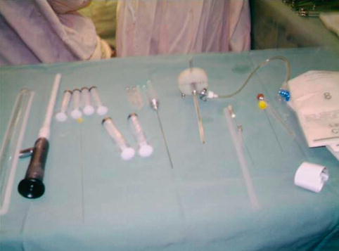
Fig. 16.2
Semiflexible and flexible spinal 0.9-mm 0° neuroendoscopy equipment used for epiduroscopy
After treatment, sciatic pain improved in all patients; however, low back pain was persistent, a finding that is concordant to previous literature reports. Based on the findings during epiduroscopy, 83.3 % of all patients with persistent pain after back surgery had severe (grade 3 or 4) epidural fibrosis, while 91.0 % had significant (grade 2, 3, or 4) fibrosis. In patients who had undergone more extensive surgery, severe fibrosis was present in 91.1 % and significant fibrosis in 95.6 %. Using MRI, epidural fibrosis was diagnosed only in 16.1 % of these patients. All patients with severe epidural fibrosis had a filling defect on epidurography. Concordant pain was present in 84.3 % of patients and was dependent on the severity of fibrosis. Epiduroscopy demonstrates that the prevalence of severe epidural fibrosis after back surgery syndrome is substantially higher than is generally reported in MRI evaluations. Severe epidural fibrosis is the underlying pathology in most patients with failed back surgery syndrome. Epidural adhesiolysis followed by the injection of steroids and local anesthetic during epiduroscopy alleviates pain and functional disability and reduces dysfunction of A-β and A-δ fibers in patients with chronic sciatica. Complication rate was about 2–7 % and included bleeding, visual loss and retinal hemorrhages, root injury, and infection. There are still some problems to be solved in the epiduroscope development, but its dramatic effect in pain reduction has been recognized, and it is considered as an attractive diagnostic and therapeutic tool.
16.3.3 Spinaloscopy
The lumbar and lower thoracic subarachnoid space of 26 human autopsy subjects was studied using rigid endoscopy by Blomberg in 1994 [1] with the aim to detect fibrous structures. This procedure is known as spinaloscopy. Fibrous attachments were found between nerve roots and/or nerve roots and the arachnoid membrane at least at one spinal level in 16 subjects. The appearance and density of the structures varied and caused restriction of nerve root mobility in nine subjects. In three of them, the impeded mobility prevented the nerve root from yielding to the contact and pressure exerted either by the tip of the endoscope or by a spinal needle introduced into the subarachnoid space. In another three subjects, a distinct membranous structure was identified in the posterior midline of the subarachnoid space in the lower thoracic and upper lumbar regions. These findings may be possibly associated with the variation in the extent of subarachnoid block and to the development of isolated nerve root trauma in connection with this procedure. The learning curve for spinal endoscopy is steep, and the procedure should not be attempted alone by a novice surgeon. Nevertheless, with training and experience, the spine surgeon can achieve better outcomes and reduced morbidity with spinal endoscopy, and the operating times are comparable to open procedures. As technology evolves and more experience is obtained, neuroendoscopy will likely achieve a more central role in spine surgery. In our clinical series we used flexible and semiflexible 0.9-mm 0° neuroendoscope, in a minimal approach (small laminectomy) or percutaneous procedure in lumbosacral and dorsal lesion like tumor biopsy. We closed with fibrin glue. Different kinds of pathologies were approached by this technique.
Our clinical series consists of 30 cases within a 9-year period (2002–2010). We used a flexible and semiflexible 0.9-mm 0° neuroendoscope either through a minimal surgical approach (small laminectomy) or as a percutaneous procedure in the management of dorsal lumbosacral lesions. Fibrin glue was utilized during closure. Overall, there was no associated intra- or postoperative mortality or morbidity. Infection rate was 1.6 %. Mean follow-up period was 48 months. Our total list of patients includes diverse pathologies:
13 arachnoid cysts, of which 8 are subdural and 5 are extradural. All of the arachnoid cysts were seen in pediatric patients (age range 2–14 years) and 50 % in patients with MMC and secondary tethered cord. Only 2 patients demonstrated radiological improvement. Clinical and/or EMG improvement after endoscopy for arachnoid cyst varied from 53.5 % in patients with previous MMC to 87.4 % in patients without MMC.
6 spinal tumors, of which 3 are less than 2 cm, 1 extended dorsal tumor which was biopsied, 1 meningioma, and 1 non-Hodgkin lymphoma. There are no previous reports of gross total resections of spinal tumors endoscopically. Published papers mention partial resections and biopsies.
14 patients with syringomyelia (7 posttraumatic, 2 in the cervical region, 5 dorsolumbar). Success rate was 82 % in the management of posttraumatic syringomyelia. Sometimes a septated syringomyelia is treated with endoscopic fenestrations in order to achieve free communication of fluid between the cavities and subsequent collapse with clinical improvement. However, despite the fenestrations, very often the cavity remains unchanged in size. In a second operation under endoscopic view, a small tube can be inserted into the cystic cavity to drain the fluid. Its efficacy is a matter of discussion and controversy.
16.4 The Future: Stem Cell Implants Through Neuroendoscopy (Restorative Neurosurgery)
Experimental and clinical research [7, 9, 10, 20] has shown the plasticity of the multipotent adult progenitor cell (MAPC) in various conditions of tissue damage. It was observed that in some neuromuscular disorders, clinical improvement of the pathological condition can be reached after the transplantation of MAPCs.
Amyotrophic lateral sclerosis (ALS) is a degenerative disorder characterized by loss of upper and lower motor neurons resulting in progressive muscle weakness, respiratory insufficiency, and ultimately death. Up to now, there are no effective treatments to reverse the natural course of the disease that is fatal. Considering the devastating effects of the ALS, we have designed a trial in which stem cells were implanted in the spinal cord and in the CSF of patients with ALS through a minimally invasive neuroendoscopic procedure. We also utilized stem cell implants in 7 patients with traumatic spinal cord injury and 1 patient with Alzheimer’s disease.
16.5 Our Experience in Patients with Amyotrophic Lateral Sclerosis (ALS)
16.5.1 Materials and Methods
Twenty-four patients with ALS met the inclusion criteria for this study. Spirometry with a forced expiratory volume (FEV) and forced vital capacity (FVC) ratio not inferior to 60 % was required. All patients were measured with an ALS function scale to set an individual initial score. Based on this scale, all our patients with ALS were graded from a minimum of 0 to a maximum of 40 points. A minimum score of 17 points was a prerequisite for inclusion in the study.
Related posts:
 Current Status and Future Developments of Neuroendoscopic Management of Pituitary Tumours and Craniopharyngiomas
 Endoscopy-Assisted Craniosynostosis Surgery
 Endoscopic Treatment of Hypothalamic Hamartomas
 Neuroendoscopic Treatment of Colloid Cysts
 The Application of Neuroendoscopic Techniques in Improving Altered CSF Physiology
 Neuroendoscopic Management of Intraparenchymal Lesions
Stay updated, free articles. Join our Telegram channel

Full access? Get Clinical Tree


