Chapter 8 Neurologic Presentation of Spinal Tumors
INTRODUCTION
One common feature of spinal tumors is their insidious course and the frequent delay between the onset of symptoms and the diagnosis of the neoplasm. In many cases, the delay can be measured in months or even years. A major reason for the delay in diagnosis is the myriad of symptoms with which these patients present, and the overlap of these symptoms with common conditions such as degenerative spine disease. Adding to the challenge of diagnosis are a number of case reports in the literature describing spinal neoplasms with unusual clinical presentations, such as severe ear pain or subarachnoid hemorrhage.1,2 As would be expected, the most common presenting neurologic signs and symptoms of spinal tumors are dependent on their location. One way to classify the location is with respect to the dura and the spinal cord. Spinal tumors can be located in the intramedullary, intradural extramedullary, or extradural space.
EXTRADURAL TUMORS
CRANIOCERVICAL JUNCTION
Perhaps the most interesting and initially perplexing neurologic signs and symptoms of spinal neoplasms arise from tumors in the craniocervical junction. Initially, it may be very difficult to localize the tumor based on the symptoms. About 49–66% of patients will report neck and/or suboccipital pain as primary symptoms at the time of initial presentation.3,4 By the time the diagnosis is made, up to 75% will have suboccipital or neck pain, usually referable to the C2 dermatome. Dysesthesias are the second most common initial complaints, occurring in 39–54% of patients, and up to 95% of patients may complain of dysesthesias by the time of diagnosis.3 In decreasing order of frequency, the dysesthesias are found in the hands, arms, and lower extremities with up to 10.5% complaining of sensory disturbance in the face.3
Tumors compressing on the lower medulla and upper cervical spinal cord and thus affecting the descending trigeminal nucleus partly explain this finding. Cold dysesthesia of the lower limb has actually been described as a symptom pathognomonic for high cervical tumors.5,6 However, this is seen in fewer than 7% of patients. Weakness is the first symptom apparent to the patient in as few as 5.3%, but it is noted at some point by 40–46% in the upper extremities and 29–42% in the lower extremities. The sensory abnormalities and weakness lead to gait disturbance in roughly half of the patients by the time of diagnosis. Bladder dysfunction is present in about 22–33% by the time of diagnosis.
Even though up to 20% of patients may actually have a normal neurologic examination at the time of initial clinical presentation,4 most patients have at least some signs of a deficit. Tetraplegia or tetraparesis can actually be found in 15–25% of patients with a craniocervical region tumor. Others may present with a monoparesis/monoplegia of the upper extremity (13–17%), hemiparesis/hemiplegia (14–15%), or paraparesis/paraplegia of the upper extremities (13–20%). The pattern of motor deficit development is quite fascinating. An alternating, rotational pattern of weakness is known to be a hallmark of craniocervical junction tumors. Typically, the weakness will begin in one upper extremity. The patient then develops weakness in the ipsilateral lower extremity. If enough time passes, the weakness may then also involve the contralateral lower extremity, and finally the contralateral upper extremity. Of course there are patients who do not display this pattern, but its presence should certainly alert the physician to the possibility of a tumor in this region. The most likely explanation for this unique finding is the decussation of the medullary corticospinal tracts near the craniocervical junction.7 The upper extremity motor fibers are carried medially in the corticospinal tract before the decussation. Just past the decussation, the fibers destined for upper extremities are the most lateral, with the lower extremity fibers being medially located. Therefore, a slightly lateral tumor anterior to the spinal cord will have mass effect, first on axons to the ipsilateral upper extremity motor fibers, followed by the ipsilateral lower extremity, contralateral lower extremity, and finally the contralateral upper extremity.
Sensory findings also can be quite variable in these patients. Some loss of pain and temperature is found in 37–57%.3,4 Other sensory modalities affected include touch (22–30%), proprioception (27–38%), and hypalgesia of C2 (18–23%). A “cape distribution” of sensory loss can be found in 7–15%, and there is a dissociated sensory loss in 23–25%. As with motor findings, the sensory deficit is usually greater in the upper extremities compared with the lower extremities.
Other neurologic findings on examination are found in Table 8-1, which uses data from Yasuoka et al and Meyer et al.3,4
Table 8-1 Neurologic Findings with Extradural Tumors at the Craniocervical Junction*
| Neurologic Sign | Frequency (%) |
|---|---|
| Atrophy | |
| Hands | 15–17 |
| Arms | 7–9 |
| Legs | 1–4 |
| Hyperreflexia | 71–83 |
| Babinski sign | 57–58 |
| Brown–Séquard | 23–29 |
| Gait disturbance | 40–47 |
| Nystagmus | 13–25 |
| Horner syndrome | 4–6 |
| Cranial nerve palsy | |
| V | 6 |
| VII | 1 |
| VIII | 1 |
| IX | 4–7 |
| X | 2–7 |
| XI | 28–32 |
| XII | 4–8 |
* Data from Yasuoka et al3 and Meyer et al.4
CERVICAL AND THORACIC
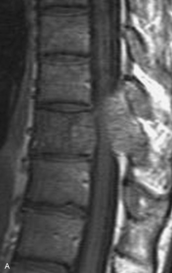
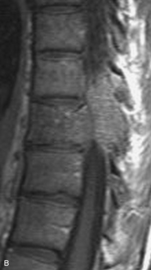
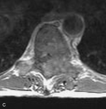
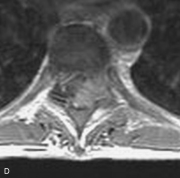
Spinal tumors in the cervical and thoracic regions may vary in their clinical presentation from absence of symptoms to complete spinal cord compression. Estimates of patients presenting with neurologic deficits range from 20–90%.8,9 Approximately 42% will have at least a serious neurologic deficit, such as paraplegia or complete loss of sphincter tone, by the time a diagnosis is made (Fig. 8-1).8 Metastatic lesions to the spine are the most common spinal tumors and are therefore reported on the most extensively in the literature. In retrospective studies of cancer patients, approximately 5–20% had evidence of spinal metastatic disease, yet fewer than 2% showed neurologic signs of spinal cord compression.10,11 As with craniocervical junction extradural tumors, pain is the most common initial symptom and has been reported as the presenting symptom in 61–96% of patients.8,9,12–14 Other symptoms at the time of diagnosis include weakness (69–76%), autonomic dysfunction (45–57%), sensory loss (51–55%), ataxia (3–5%), and even herpes zoster (2%).12,13,15 Actual findings on examination are tenderness (63%), weakness (77–97%), autonomic dysfunction (40–45%), sensory loss (66–90%), and ataxia (7%).13,16 The specific motor and sensory deficits depend on the location of the tumor within the cervical and thoracic spine (Fig. 8-2).
Stay updated, free articles. Join our Telegram channel

Full access? Get Clinical Tree


