‘Where’
It is often the case that the neurologist or neurosurgeon, when confronted with a patient, will first ask themselves, where is the lesion?, meaning where is the predominant focus of pathology within the neurological system.
The lesion may be at a single focus within the nervous system, such as entrapment of the median nerve at the wrist in the case of carpal tunnel syndrome or entrapment of an L5 nerve root in a patient with foot drop. Alternatively, the lesion may be multifocal, as in multiple sclerosis (MS) where demyelinating plaques may be found throughout the brain and spinal cord, or it may be diffuse or generalised, as in the case of encephalopathic patient with a reduced level of consciousness secondary to a systemic infection or metabolic derangement.
In some instances, gross localisation is very straightforward, for example, in a patient with traumatic brain injury post a road traffic accident, it is relatively implicit to focus localisation within the brain. In case of a patient complaining of weak legs (see Chapter 9), the problem could however variably be within the brain, brainstem, spinal cord (central nervous system (CNS)) or within the anterior horn cell, nerve root, lumbar sacral plexus, peripheral nerves or neuromuscular junction (peripheral nervous system (PNS)) or be isolated to skeletal muscle. In the latter scenario, the neurological examination is pivotal to localising the problem where, for example, upper motor neurone (UMN) signs will point to a problem in the central nervous system and lower motor neurone (LMN) signs will point to a problem in the peripheral nervous system. The distribution of signs will also point towards the potential site of the lesion; therefore, hemisphere lesions usually lead to contralateral weakness affecting face, arms and legs, variably depending on the exact site of the lesion, whereas a cord lesion usually produces bilateral weakness with possible evidence of LMN signs at the level of the lesion and UMN signs below the level of the lesion with a sensory level. Patients may present with typical syndromes (Table 3.1) observed with pathology in different parts of the CNS, thereby further helping in localisation (also refer to Chapter 35).
Table 3.1 Key areas within CNS with description of clinical syndromes related to pathology.
| Relevant areas within CNS | Clinical syndromes related to pathology |
| Frontal lobe | Dominant frontal lobe only (Broca’s area): Broca’s or motor or expressive or non-fluent dysphasia with intact comprehension of written and spoken language but reduced verbal output with difficulty in putting words together and difficulty with repetition |
| Primary motor cortex: Contralateral hemiparesis or hemiplegia | |
| Premotor (anterior to primary motor cortex) and supplementary motor cortex: Apraxia (i.e. performance of movements is impaired despite normal strength) as this area plays a role in contralateral motor programming | |
| Prefrontal area: Frontal lobe syndrome (problems with behaviour and cognition; intelligence and memory; apathy, including lack of motivation and abulia, i.e. lack of initiative or awareness) usually resulting from a large unilateral or bilateral lesions | |
| Parietal lobe | Primary somatosensory cortex: Problem with perceiving somatic sensation on contralateral side of body |
| Superior parietal lobule (usually with dominant hemisphere lesions): Usually result in ideomotor apraxia (inability to carry out tasks on command despite absence of motor deficit or weakness) or constructional apraxia (inability to copy a diagram) or astereognosia (inability to recognise an object without looking at it, for example a coin in the palm of a patient with eyes closed) | |
| Inferior parietal lobule (angular and supramarginal gyrus) in right hemisphere: Contralateral neglect (asomatognosia) despite intact somatic sensation, i.e. patients neglect the left side of body including dressing or looking after it; classically these patients when asked to draw a clock face only draw numbers on the right side. With left parietal lobe lesion, contralateral neglect is uncommon | |
| Inferior parietal lobule in left or dominant hemisphere: Gerstmann’s syndrome (finger agnosia, i.e. inability to recognise finger by name; agraphia without alexia, i.e. cannot write but can read; left–right disorientation and acalculia, i.e. inability to perform simple arithmetic calculations); with angular gyrus lesions (alexia, i.e. inability to read or comprehend written text and agraphia, i.e. inability to write); supramarginal gyrus and posterior part of superior temporal gyrus in dominant lobe (Wernicke’s or fluent or sensory or receptive aphasia characterised by fluent speech without meaning) | |
| Occipital lobe | Contralateral homonymous haemianopia with macular sparing results with lesions of primary visual cortex, usually due to occlusion of a branch of posterior cerebral artery (for field defects associated with other sites, see Chapter 7) |
| Cerebellum | Unilateral cerebellar hemisphere lesions: Ipsilateral gait ataxia (impaired heel to toe test and broad-based gait), problem with coordination (intention tremor on finger to nose, i.e. increase in tremor amplitude as target is approached; dysdiadochokinesia on repeated movements and impaired heel to shin test) |
| Cerebellar vermis: Truncal ataxia | |
| Refer to Chapter 2 and Figure 2.1 therein. | |
‘What’
Having decided where the lesion lies, the next question to consider is what is the problem? Here we are thinking particularly of the aetiological group to which the potential diagnosis belongs. This is typically based upon the temporal sequence of events largely elicited by obtaining the history of the presenting complaint (Table 3.2).
Table 3.2 Potential causative groupings along with brief temporal and other clues associated with a particular aetiological group.
| Aetiology | Characteristics/clues |
| Traumatic | Preceding history of trauma. Symptoms are typically acute but may, as in the case of chronic subdural haematoma, evolve over time |
| Vascular | Typically indicated by abrupt nature of onset, reaching maximum severity from outset |
| Infective | Typically a history evolving over days to weeks, although may evolve over months or be chronic. Supported by systemic markers of infection like raised white cell count or CRP/ESR |
| Metabolic | Typically symptoms evolve over days to weeks, although may be episodic or chronic |
| Malignant | Often symptoms evolve over weeks to months and may be due to direct tumour effects or as a paraneoplastic phenomena |
| Degenerative | Typically degenerative diseases evolve over months to years |
| Genetic | May be suggested by family history and typically symptoms evolve over months to years |
| Iatrogenic | This may be acute, subacute or chronic but clues are from the past medical history and history of medication exposure |
| Idiopathic | A causative category ascribed by a process of exclusion |
| Psychogenic | Be wary of ascribing a psychogenic cause, although very important to identify if this is the explanation |
‘Why’
Having decided where the potential lesion lies and also to which causative group the diagnosis may belong, the next step is to consider why might this disease or entity have occurred?. For example, the patient presenting with acute onset of right arm weakness and language disturbance is likely to have an abnormality localising to the left frontal lobe with the sudden onset suggesting a vascular cause. The question then arises regarding the reason for the occurrence of the vascular event. With a preceding history of trauma, an extradural haemorrhage may be the explanation, whereas a past history of smoking, diabetes and hypertension might point more towards an ischaemic stroke. This gathering of information regarding such associated factors should be covered in the history of the presenting complaint and other components such as the past medical history, the medication list, family history and social history.
The Neurological History
Many of the questions pertinent to be asked when considering patient complaining of specific neurological symptoms are covered in Parts II and III of this book dealing with presenting complaints and specific disease entities, which may be referred to when confronted with specific neurological complaints. Below are some additional points that should be included in the medical student or junior doctor clerking when obtaining a neurological history.
In addition to detailing the patient’s name, age, occupation and marital status, in a neurological history it is also common to record patient’s handedness. Handedness suggests the dominant (language functioning) hemisphere of the brain, where nearly all right-handed people are left hemisphere dominant whereas only approximately 70% of left-handed people are left hemisphere dominant. Further understanding of patient’s handedness will increase understanding of impairment the patient may be experiencing due to their deficit. For example, a patient with right body predominant Parkinson’s disease who is right-handed will typically have greater impairment in fine finger function than a patient with left body predominant Parkinson’s disease who is right-handed.
Complaint and History of the Presenting Complaint (see Part II)
Complaints may concern problems with senses, including sight, smell, hearing and taste. Other complaints may include blackouts, dizziness, headache, numbness, paraesthesia (pins and needles) and weakness in arms or legs. These are covered in detail in Part II. Disturbances with cognition, speech, swallowing and bowel or bladder function may also be reported. Psychiatric symptoms may also be present. A full history of presenting complaint is warranted, including mode of onset, provoking or triggering causes and progression of symptoms, for example gradual (with a slowly growing tumour) or rapid (e.g. in a vascular event like stroke). Also record the associated factors, for example in the case of a migraine headache, the associated factors of photophobia and nausea.
Past Medical History
Events from the past medical history are often useful in understanding why patients may have developed a particular disease. Check regarding meningitis, foreign travel and previous history of CNS trauma (minor or major, for example with reference to extradural or subdural haematoma). Past medical history may also help localise the lesion and point to the causative aetiology, for example the patient with a recent history of tooth abscess and dental extraction in preceding weeks who then develops focal onset of seizures affecting the right hand may have a brain abscess affecting the left motor strip.
Medication History
Neurological symptoms as a side effect of medication are extremely common. A detailed list of the medications (including anticonvulsants, antidepressants or antipsychotics), the dose and frequency of administration, as well as documentation of previous medication exposures is extremely important, for example a patient presenting with numbness of both feet evolving over months who has had chemotherapy in the preceding year may have a peripheral sensory axonal neuropathy secondary to chemotherapy. Isoniazid similarly can also cause peripheral neuropathy. Compliance with medications and any relevant side effects should be checked, for example this can be particularly important in patients with epilepsy as ongoing epileptic seizures may be due to non-compliance or due to taking anticonvulsant medication at an inadequate dose.
Family History
Conditions with a genetic basis are relatively common in neurological practice (see Part III). It is therefore important to obtain a detailed family history. Furthermore, understanding the patient’s fears about their symptoms or illness relies upon understanding of their prior exposure to and experiences of particular illnesses in their family.
Social History
Obtain history regarding smoking and drinking habits. This is relevant, for example peripheral neuropathy can occur in a patient with excessive alcohol drinking. Also detail exposure to illicit drugs and recent foreign or local travel. This may be relevant, for example in a patient with a lower motor neurone facial palsy who in preceding weeks suffered a tick bite while walking in a forest and now has neurological Lyme disease (neuroborreliosis).
Functional History
Understanding the level of disability or handicap a patient experiences due to an impairment (e.g. of arm function) is important in patients with long-term neurological conditions. One can work through a list of activities of daily living. Alternatively, one can work from head to toe asking the patient about specific functions, i.e. visual impairment, speech and swallowing impairment, upper limb impairment, impairment of bladder, bowel and sexual function and lower limb impairments, understanding along the way the consequences of these upon patient’s occupation or on performance of activities of daily living and the impact on family/carers.
Systems Review
Always review other body systems by means of direct questioning at the end of the history to pick up additional associated features of a systemic disturbance which may highlight the cause of a specific neurological symptom. As an example, a patient with nocturnal breathlessness on lying flat may have neuromuscular and ventilatory impairment, which when considered with a history of weight loss, wasting and lack of sensory disturbance will point towards motor neurone disease.
Neurological Examination
The typical clerking style of neurological examination performed by medical students and junior doctors includes the neurological examination of the head and neck (cranial nerve (CN) examination) as well as the upper and lower limbs. Additional routines of examination are also used to specifically test the cerebellar system, gait (see Chapter 11) and extrapyramidal system, domains of cognition or lobar functions of the brain (Table 3.1).
Suggested Order of Examination
General examination as a screening test including general inspection; mental status examination (not fully and formally undertaken on every neurological patient, however, may be important as neurological and psychiatric symptoms may coexist in illnesses like dementia), including checking patient’s appearance, behaviour, mood and for presence of delusions or hallucinations among others; higher mental function and speech; cranial nerves; upper and then lower limbs (inspection, tone and power); reflexes; sensory exam and finally coordination and gait.
Higher Mental Function and Speech
Check patient’s conscious level (using Glasgow Coma Scale as detailed in Appendix 2); orientation in time, place and person and finally perform a cognitive screening using abbreviated mental test score or mini-mental state examination if there is suspicion of cognitive impairment. Higher mental function may be impaired in various lobar syndromes, as detailed in Table 3.1. Note that ‘apraxia’ refers to the inability to perform an action despite an intact motor and sensory function while ‘agnosia’ refers to impaired perception despite the presence of an intact sensory function during neurological examination.
Check speech (usually assessed during obtaining history) for dysphasia syndromes (Table 3.1), including conduction dysphasia seen with lesions of the arcuate fasciculus (fibre tract connecting Broca’s and Wernicke’s area) and characterised mainly by impaired repetition; dysarthria implying difficulty with articulation by asking the patient to say, for example, ‘British Constitution’ (types include cerebellar characterised by slurred (drunk) and scanning speech, spastic as seen in pseudo-bulbar palsy in motor neurone disease and characterised by ‘Donald Duck’-type voice, monotonous speech in extrapyramidal disease and bulbar speech in lower motor neurone syndromes like facial nerve palsy and characterised by nasal quality to speech) and dysphonia (impaired speech volume due to problem with respiratory muscles, for example myasthenia gravis).
Cranial Nerves
For a detailed paper on the topic of ‘cranial nerve examination’, refer to Asghar and Abhinav (2010). Brief notes on examination of CN are as follows (Table 3.3):
- I (Olfactory): Smell is not routinely tested unless patients complain of problem with smell; if needed, check ability of each nostril to distinguish different types of smell, including camphor, peppermint and others.
- II (Optic): Each eye should be checked separately. Check acuity (Snellen chart), visual fields (to confrontation) and pupils (size, shape, reactivity to light, including direct and consensual response and accommodation) and perform a detailed opthalmoscopic examination inspecting the optic disc for papilloedema or atrophy.
- III (Oculomotor), IV (trochlear), and VI (abducens): Check extraocular movements for full range of motion and evidence of diplopia, nystagmus, ptosis or internuclear opthalmoplegia (INO) (Chapter 18). Nystagmus refers to involuntary eye oscillations and can be normal at extreme lateral gaze. Direction of the fast phase should be noted; nystagmus can occur in the context of cerebellar or vestibular lesions. INO, for example, due to multiple sclerosis results due to a lesion in the medial longitudinal fasciculus and refers to failure of adduction of the ipsilateral eye with nystagmus in contralateral eye on abduction.
- V (Trigeminal): Check facial sensation (pinprick and light touch) in all three divisions (ophthalmic—V1, maxillary—V2 and mandibular—V3), corneal reflex (CN V1—afferent and CN VII—efferent) and motor function (CN V3) by asking patient to open their mouth and clench in order to feel masseter and temporalis (muscles of mastication).
- VII (Facial): Ask patient to raise eyebrows or look at the ceiling, puff up their cheeks or whistle and screw their eyes shut. Taste can also be checked in the anterior two-thirds of the tongue. Distinction between UMN type versus LMN type of facial nerve weaknesses is important (Table 3.3). Bell’s palsy with its clinical features (as listed in Table 3.3) is a diagnosis of exclusion and typically has an abrupt onset with features of a unilateral LMN facial nerve palsy. Bell’s phenomenon (inability to close eyes with rolling up of eye on attempted closure) may be seen. Short course of steroids (Prednisolone) may be tried with artificial tears and eye patch to protect the cornea.
- VIII (Vestibulocochlear): Whisper a number while blocking the contralateral ear and ask patient to repeat the number. If there is unilateral hearing disturbance, carry out Weber’s and Rinne’s tests to distinguish between conductive and sensorineural hearing loss.
- IX (Glossopharyngeal) and X (vagus): Ask patient to say ‘Ahh’ and look at the uvula for central elevation. Gag reflex (CN IX—afferent and CN X—efferent) may be checked.
- XI (Accessory): Ask patient to shrug their shoulders (trapezius) or turn head to one side against resistance (sternocleidomastoid).
- XII (Hypoglossal): Check for fasciculation and wasting with tongue inside mouth (LMN lesion). Ask the patient to move it from side to side checking for any slowing of the tongue movement (UMN lesion).
Upper and Lower Limbs
Typically start with inspection and then examine tone and power. Tendon reflexes, sensation and finally coordination and gait are also tested.
Table 3.3 Summary of signs and symptoms in cranial nerve lesions with their associated aetiology.
| Cranial nerve | Signs or symptoms of a lesion | Cause or lesion |
| I (Olfactory) | Change in sense of smell | Nasal obstruction |
| Polyps or foreign bodies | ||
| Viral infections | ||
| Unable to identify common substances | Neurological causes | |
| Head injury | ||
| Nasofrontal tumours | ||
| Parkinson’s disease | ||
| Alzheimer’s disease | ||
| II (Optic) (see Chapter 7) | Monocular blindness | Lesions of the eye |
| Cataracts | ||
| Intraocular haemorrhage | ||
| Retinal detachments | ||
| Diseases of the optic nerve | ||
| Multiple sclerosis | ||
| Tumours | ||
| Bitemporal haemianopia | Compression of optic chiasm | |
| Pituitary tumour | ||
| Homonymous haemianopia | Lesions of the optic tract | |
| Vascular lesions | ||
| Neoplasm | ||
| Optic radiation | ||
| Lesions of the occipital lobe | ||
| Visual inattention | Parietal lobe lesions | |
| Reduced visual fields | Glaucoma | |
| Chronic papilloedema | ||
| Marcus Gunn Pupil (relative afferent pupillary defect) observed during the swinging flashlight test [patient’s pupils constrict less (therefore appearing to dilate) when the light swings from the pupil of the unaffected eye to the pupil of the affected eye] | Damaged optic nerve pathway—indicating a decreased pupillary response to light in the affected eye (this detects less light than the functioning pathway) | |
| Constricted pupil | Horner’s syndrome | |
| Opiate overdose | ||
| Brainstem stroke | ||
| Nystagmus | Physiological | |
| Congenital | ||
| Visual impairment (difficulty in fixing gaze) | ||
| Vestibular disease | ||
| Cerebellar disease | ||
| Papilloedema | Increased intracranial pressure | |
| Tumour | ||
| Abscess | ||
| Encephalitis | ||
| III (Oculomotor) | Divergent squint and diplopia (eyes down and out) | Paralysis of extraocular muscles (superior rectus, inferior rectus and medial rectus) |
| Ptosis | Paralysis of levator palpebrae | |
| Horner’s syndrome | ||
| Myasthenia gravis | ||
| Dilated pupil | Paralysis of sphincter pupillae | |
| Tumour | ||
| Aneurysm | ||
| Brainstem stroke | ||
| IV (Trochlear) | Eye elevation and outward rotation and diplopia (on looking down) | Paralysis of superior oblique muscle |
| Tumour | ||
| Aneurysm | ||
| V (Trigeminal) | Localised pain and vesicular eruption | Herpes zoster infection |
| Anaesthesia and dissociated sensory loss | Syringobulbia | |
| Brisk jaw jerk | Bilateral upper motor neuron lesion | |
| Loss of corneal reflex, paralysed muscles of mastication (deviation of jaw towards side of the lesion with unilateral lesion) and loss of facial sensation | CN V palsy | |
| Neoplasm | ||
| Infection | ||
| VI (Abducens) | Convergent squint and horizontal diplopia (with all movements excluding adduction) | Paralysis of lateral rectus muscle |
| Tumour | ||
| Aneurysm | ||
| VII (Facial) | Unilateral complete facial paralysis, hyperacusis, altered taste and impaired corneal reflex on affected side | Bell’s palsy |
| Unilateral lower facial palsy only (as forehead or frontalis has bilateral UMN innervations, so relative sparing of forehead is observed with unilateral UMN lesion) | UMN lesion | |
| Stroke | ||
| Tumour | ||
| MS | ||
| Unilateral entire facial palsy (observed with LMN lesion where complete ipsilateral palsy with droopy mouth and loss of eyebrow lines and nasolabial folds is noted) | LMN lesion | |
| Stroke and tumour (acoustic neuroma) | ||
| Ramsay Hunt syndrome | ||
| Lyme disease, TB and HIV | ||
| Diabetes and sarcoidosis | ||
| VIII (Auditory) | Conductive deafness | Ear disease |
| Otitis externa or otitis media | ||
| Paget’s disease | ||
| Perforated ear drum | ||
| Sensorineural deafness | Congenital | |
| Acquired | ||
| Presbycusis (ageing) | ||
| Noise induced | ||
| Ototoxicity (drugs) | ||
| Attacks of dizziness and deafness | Acoustic neuroma (benign tumour). As it expands, it may compress adjacent CN V–VII | |
| IX (Glossopharyngeal) | Altered sensation to palate and pharynx | CN IX palsy |
| Base of skull tumour | ||
| Stroke or trauma | ||
| X (Vagus) | Weak cough or dysphonia | Lesion of the recurrent laryngeal branch |
| Asymmetrical soft palate (with unilateral weakness palatal deviation towards the normal side) and loss of gag reflex | CN X palsy | |
| Base of skull tumour | ||
| Stroke or trauma | ||
| XI (Accessory) | Loss of power to sternocleidomastoid (SCM) or trapezius muscles | CN XI palsy |
| Tumour | ||
| Stroke | ||
| Trauma | ||
| XII (Hypoglossal) | Tongue deviation (towards side of lesion) or weakness | Lower motor neuron lesion |
| Tongue fasciculation | Motor neurone disease | |
| Adapted from Asghar and Abhinav (2011). | ||
Specifically inspect for muscle wasting or atrophy (in LMN syndromes) and fasciculations (e.g. in motor neurone disease) and observe the posture (e.g. stooped posture of Parkinson’s) and check for presence of any abnormal movements, for example tremor (see Chapter 20). Check for pyramidal drift (pronation and drifting down of outstretched arms) occurring for example with UMN lesions in corticospinal tract.
Tone
Testing tone can help determine whether the lesion is within the central or peripheral nervous system. Tone may be increased, decreased (hypotonia) or normal and is best assessed at wrist, elbow, hip and knee. If increased, it can be either in spastic or rigid fashion. Lesions affecting the corticospinal tract (UMN) produce a spastic increase in tone, best elicited by a rapid flexion extension movement of the elbow where a so-called spastic catch or a sudden increase in tone may be felt. In contrast, a rigid increase (increased through a whole range of movement) in tone typically localises to problems within the basal ganglia (extrapyramidal system, for example Parkinson’s disease) and is best elicited either by a slow flexion extension movement at the elbow, knees or ankles or by a slow circling movement at the wrist. Rigidity may be described as ‘lead pipe’ and when combined with breaks or tremors as being ‘cogwheeling’. Rigidity may be heightened by asking the patient to perform movement of the contralateral limb (synkinesis).
Power Testing
Power testing involves assessing power in a given muscle group around each joint. For example at the elbow, power is assessed for elbow flexion and extension. Power testing is documented using the Medical Research Council (MRC) grading scale shown in Table 3.4. Always compare with the strength on the contralateral side.
Table 3.4 Muscle strength grading (MRC scale)
| Grade | Muscle strength |
| 0 | No contraction |
| 1 | Flicker of contraction |
| 2 | Movement with elimination of gravity |
| 3 | Active movement against gravity |
| 4 | Active movement against resistance (4− slight, 4 moderate and 4+ strong) |
| 5 | Normal power |
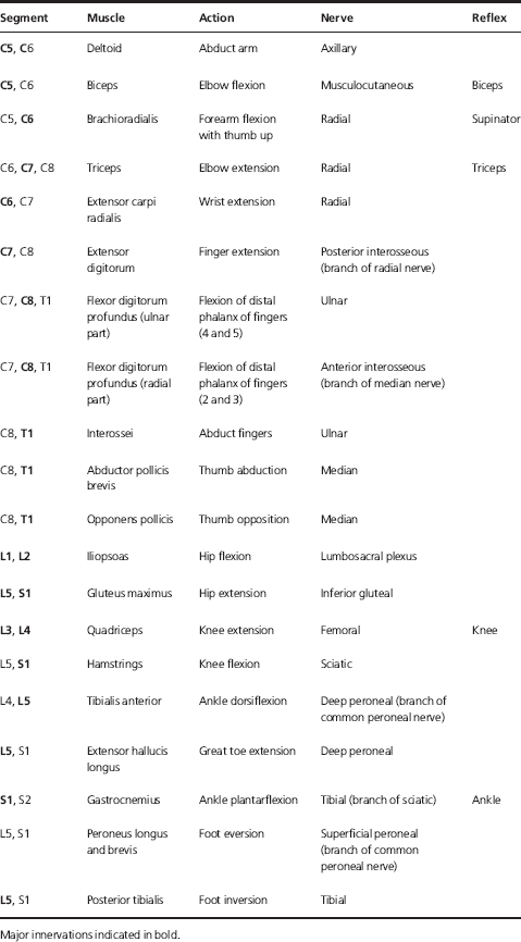
It is important to be aware of the muscle groups being tested along with innervating nerve roots and named nerves (Table 3.5).
Table 3.5 Important muscle groups and spinal nerve roots with reflexes for upper and lower limbs.
Reflexes
Reflex (segmental levels in Table 3.5) testing requires practice and reflexes may be absent, decreased, normal, increased and finally increased with clonus (contraction of muscle when stretched and tested for at the ankle and quantified in terms of number of beats). Increased reflexes are associated with a lesion in the UMN pathway (corticospinal tract) whereas decreased or absent reflexes occur with a problem in the peripheral nervous system (LMN pathway) with a loss of a reflex or reflexes being due to failure of either the afferent (sensory) or the efferent (motor) arm of the reflex arc. The plantar response is assessed using stimulus on lateral sole with an upgoing or extensor plantar or Babinski sign (associated with UMN lesions within the corticospinal tract) being defined as extension of big toe with fanning out or spreading out of other toes. Normal response is flexion of the toes.
Note on UMN versus LMN lesions
- LMN lesions lead to hypotonia, hyporeflexia or areflexia; fasciculations with atrophy of the affected muscles therefore causing a flaccid paralysis on the same side and at the level of the lesion.
- UMN lesions lead to hypertonicity, hyperreflexia with or without clonus, Babinski sign, loss of abdominal and cremasteric reflex and disuse atrophy. UMN lesions always lead to spastic weakness which is below the level of the lesion and could be ipsilateral (if lesion affects corticospinal tract in spinal cord) or contralateral (if lesion is between the cerebral cortex and medulla above the pyramidal decussation).
Sensory
Different modalities of sensation are conveyed by different pathways (see Figure 2.3) and each should be assessed. Always teach the test first prior to carrying it out.
Dorsal Column (fine touch, joint position and vibration sense on ipsilateral side)
Vibration sense is assessed with 128 Hz tuning fork; start with sternum to familiarise patient with the vibration sense and then apply to bony prominences (including base of big toe, medial malleolus, knee and elbow) starting distally. If sensation is normal distally, there is no need to carry out tests more proximally.
Test for joint position sense is taught with patient’s eyes open followed by movement of the joint with patient’s eyes closed and asking them regarding the direction of movement of the joint, i.e. up or down.
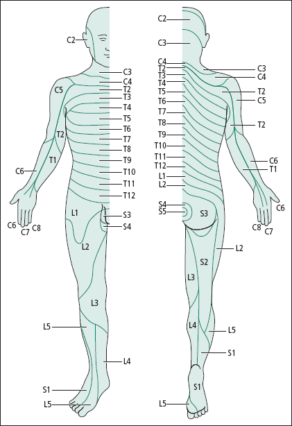
Light touch is tested using cotton wool or fingertip in all dermatomes (Figure 3.1), including those in the affected ones.
< div class='tao-gold-member'>