Fig. 1
Representatives of carbachol-induced neuronal oscillations at temperature of 27–35 ∘C. Perfusion of 30 μM carbachol induced the bursts of the oscillation. The left trace reveals successive bursts of neuronal oscillation with the regular burst duration (white arrow) and IBI (black arrow). The right trace expands the asterisk portion of the left trace
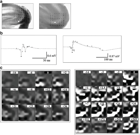
3.2 Different Spatio-Temporal Patterns of CITO and PIED
CSD analysis was performed using the field potentials recorded from 64 electrodes of MED64. During the generation of CITO, a cluster of a pair consisted of a source and a sink made a line, and propagated from dentate gyrus to CA1 along the stratum pyramidale, a pyramidal cell layer in CA3 (Fig. 2). The cluster diminished after the propagation. Then the source covered the entire cell layer. On the other hand, in the PIED, a larger cluster of a sink and source pair was induced suddenly, it propagated faster than CITO, and it lasted for a longer time (Fig. 2).


