Neuropathology


Neuropathology


Peter Falkai

Bernhard Bogerts


Introduction

The traditional domains of neuropathology are well-defined organic brain diseases with an obvious pathology, such as tumours, infections, vascular diseases, trauma, or toxic and hypoxemic changes, as well as degenerative brain diseases (e.g. Alzheimer’s disease, Parkinson’s disease, and Huntington’s chorea). Neuropathological investigations of these brain disorders have been rewarding, because patients with any of these conditions can be expected to have gross morphological or more or less specific neurohistological anomalies related to the clinical symptoms of the disorders. Moreover, the type of brain pathology of these well-defined disease entities is quite homogenous. For example, it is highly unlikely that a patient with Parkinson’s disease would not exhibit morphological changes and Lewy bodies
in the nigrostriatal system, just as much a person with Huntington’s chorea would have a normal striatum, or a patient with Pick’s or Alzheimer’s disease would have no changes in the cerebral cortex.

In contrast, the history of the neuropathology of psychiatric disorders outside primary degenerative diseases is much more controversial, because no such obvious and homogenous types of brain pathology (as seen in neurological disorders) have yet been detected for the major psychiatric illnesses such as schizophrenia, affective disorders, substance-related disorders, or personality disorders.

The scope of this chapter is to summarize the neuropathological findings in schizophrenia, affective disorders, and alcoholism. Tables 2.3.5.1, 2.3.5.2, 2.3.5.3, and 2.3.5.4 highlight the significant findings. It goes beyond the scope of this chapter to review the large body of literature on the dementias, including specifically Alzheimer’s disease. Concerning this matter, the reader is referred to several comprehensive reviews (e.g. Jellinger and Bancher 1998).(1)


Table 2.3.5.1 Gross morphometric findings in schizophrenia


Region/parameter


Finding


General


Brain length


(↓)


Brain weight


Ventricular area/volume


Cortex thickness


Temporal lobe


Lobar area/volume


Hippocampal area/volume


Parahippocampal area/volume


Parahippocampal cortical thickness


Amygdala area/volume


Sylvian fissure length, planum temporal volume


Sulcogyral pattern


Abnormal


Frontal, parietal, and occipital lobes


Cingulate cortical thickness


Insula area/volume


Corpus callosum thickness


(↑)


Internal capsula area/volume


Basal ganglia


Globus pallidum area/volume


(↓)


Nucleus accumbens area/volume


Cautdate-putamen area/volume


Thalamus


Mediodorsal nucleus area/volume


(↓)


Whole and various nuclei area/volume


Cerebellum


Anterior vermis area


Brainstem


Substantia nigra volume


Locus coeruleus volume


Periventricular grey volume


In comparison with controls: ↓, reduced; ↑, increased; —, no difference; ( ), finding not or only partially replicated.


Adapted from Arnold and Trojanowski.(3)


Schizophrenia and other psychotic disorders


Studies between 1898 and 1975

In 1898, Alois Alzheimer (1898)(2) described subtle changes in the neocortex of patients with schizophrenia. Subsequently to Alzheimer, Southard reported cortical atrophy in schizophrenia and mentioned association areas of the cerebral cortex to be most affected in this disorder.(4) Vogt and Vogt and their coworkers reported cellular alterations in the cortex, thalamus, and basal ganglia of schizophrenics.(5) These considerable efforts on the part of many well-known neuroanatomists and neuropathologists to prove schizophrenia to be a primary brain disorder ended in inconsistent and unsubstantiated findings.(6) To a large extent, these inconsistencies can be attributed to a variety of methodological inadequacies including diagnostic uncertainties, inadequate control samples, flawed tissue-handling procedures, variable choice of brain regions for neuropathological studies, limitations in the sensitivity and specificity of classical histological stains, as well as lack of quantitative methods to delineate and analyse subtle brain abnormalities.(7)


Table 2.3.5.2 Neuronal morphometric findings in schizophrenia


Region/parameter


Finding


Temporal lobe


Superior temporal gyrus (Tpt) neurone density


Hippocampal neurone density


(↓)


Hippocampal neurone size


(↓)


Entorhinal cortex neurone density


(↓)


Entorhinal cortex neurone size


Amygdala neurone density (basolateral n.)


Frontal lobe


Prefrontal cortex pyramidal neurone density


Prefrontal cortex interneurone density


(↓)


Prefrontal cortex neurone size


Cingulate (anterior) pyramidal neurone density


Cingulate interneurone density


Cingulate neurone size


Motor cortex neurone density


(↓)


Motor cortex neurone size


Basal ganglia


Globus pallidus neurone counts


Nucleus accumbens neurone counts


Nucleus basalis of Meynert neurone counts


Thalamus


Mediodorsal nucleus neurone counts


(↓)


Cerebellum


Purkinje cell density


Brainstem


Substantia nigra neurone density


Substantia nigra neurone size


Locus coeruleus neurone density


Locus coeruleus neurone size


Pedunculopontine nucleus neurone density


In comparison with controls: ↓, reduced; ↑, increased; —, no difference; ( ), finding not or only partially replicated.


Adapted from Arnold and Trojanowski.(3)


Neuropathological findings in schizophrenia since 1975

Advances in the last 30 years have produced more reliable psychiatric diagnostic criteria, improved structural and functional neuroimaging techniques, a large array of highly sensitive and specific molecular probes and labeling procedures, suitable for use in neuropathological studies, and computer-assisted image analysis methodologies. For these and other reasons, there has been a resurgence of interest in the neurobiological substrates of schizophrenia, and contemporary neuropathological studies have enumerated many findings in the brains of patients with schizophrenia (for reviews see(7, 8, 9)). Finally, the recent description of the first risk genes like Neuregulin-1 or Dysbindin has provided this field with reliable research targets.(8, 9) To identify the role of these genes for the pathophysiology of schizophrenia their expression pattern in human brain tissue has to be established in the near future.


(a) Diagnostic neuropathology

Stevens (1982)(10) surveyed the brains of 28 schizophrenic patients for gross and microscopic abnormalities using standard diagnostic stains. She discovered no abnormalities in temporal (including the amygdalohippocampal region), frontal, or parietal lobes or in the thalamus, but detected assorted abnormalities in other regions, including neuronal loss or infarction in the globus pallidus in five patients, increased cerebellar white matter gliosis in five patients, excessive Purkinje cell loss in 13 cases, and, most notably, increased fibrillary gliosis in periventricular, periaqueductal, and basal forebrain regions bilaterally.

In another prospectively accrued series,(10) she found that of 56 schizophrenics five were afflicted with other distinct neurological illnesses (multiple sclerosis, Friedreich’s ataxia, epilepsy, stroke) and three had been treated with prefrontal leukotomies. The remaining 48 showed no differences to controls in the frequency of large- or small-vessel cerebrovascular disease, senile plaques, or neurofibrillary degeneration. However, there was an ‘increased incidence of unexpected pathology in the schizophrenic group compared with the control group’. Of these 48 schizophrenics, 21 exhibited some degree of focal pathology compared to 12 of 56 controls, but these abnormalities were diverse in nature and location. Holzer staining suggested a significant increase in fibrous gliosis in the cortex, white matter, and periventricular structures, but generally for those brains showing other focal pathology. After removal of these cases, the ‘adjusted’ group showed no evidence of increased gliosis.


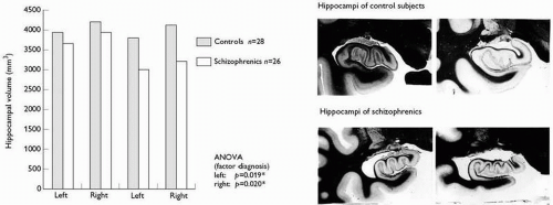
Fig. 2.3.5.1 Left side: hippocampal volumes in schizophrenic patients and controls; Right side: lippocampal atrophy macroscopically seen in about one-third of patients with schizophrenia (upper row) compared to control subjects (lower row) (from Bogerts 1990).

In a series of 101 elderly schizophrenics,(12) Golier et al. found only 10 with definite or probable Alzheimer’s disease by modern neuropathological diagnostic criteria, 29 with senile plaques, 15 with vascular lesions, two with Parkinson’s disease, three with unspecified tumour, and five with ‘other’ findings.

Another review concluded extensive neuropathological investigations due to lack of any evidence of neurodegeneration or neural injury beyond what typically is observed in brains of individuals without neuropsychiatric illness.(8)


(b) Morphometric studies


Macroscopic findings (Table 2.3.5.1)

Several planimetric postmortem studies of the entire cortex have been performed, some reporting significant reduction of cortical volume (12 per cent) and central grey matter (6 per cent), and others reporting no difference in volumes of cortex, white matter and whole hemispheres between schizophrenics and controls. Others that measured general brain parameters have shown reduced brain length, brain weight, and increased ventricular area/volume.

Since the publication of the first report of reduced tissue volume in temporolimbic structures of schizophrenics,(13, 14) numerous quantitative or qualitative anatomical postmortem studies on limbic structures of schizophrenics have been conducted. The majority of these studies substantiated subtle structural changes (15-20 per cent mean volume reduction) in at least one of the investigated areas, whereas only a few yielded entirely negative results. The findings comprise reduced volumes or cross-sectional areas of the hippocampus, amygdala, parahippocampal gyrus, which were later corroborated by morphometric magnetic resonance imaging (MRI) studies. Figure 2.3.5.1 demonstrates the subtle bilateral volume reduction of the hippocampus in schizophrenics and furthermore visualizes the kind of hypoplastic appearance of the anterior hippocampus, which can be seen in about one third of the patients
(lower row of the photographs). Other findings in limbic brain regions are left temporal horn enlargement, white-matter reductions in parahippocampal gyrus or hippocampus, and an increased incidence of a cavum septi pellucidi.

Unchanged volumes of the striatum and external pallidum but a subtle volume decrease in the internal pallidal segment were found in brains from the preneuroleptic era. Pallidal volume reduction was due to a reduction in the catatonic subgroup.(15) These initial findings have to be pursued, as longitudinal MRI studies suggest that enlargement of basal ganglia can be seen in schizophrenia as a consequence of treatment with classical neuroleptics, which can be reversed by the use of atypical substances.(16)

After initially finding no volumetric changes in the thalamic nuclei, subsequently the area/volume of the mediodorsal nucleus and anteroventral thalamic nucleus were found to be decreased.(17)

Changes in area measurements of the corpus callosum were described in some studies. The findings, however, are inconsistent; there are reports of increased as well as of decreased midline areas. More consistent are reports of shape abnormalities, in that the sex difference in anterior and posterior callosal thickness in normal controls seems to be reversed in schizophrenics and the mean curvature in the corpus callosum is bent upwards.(18)

Findings of decreased volume of the substantia nigra and the periventricular grey matter as well as no volumetric change in the locus coeruleus await replication.


Microscopic findings (Table 2.3.5.2)

There are a number of studies of neurone number, density, and size in schizophrenia. As summarized in Table 2.3.5.2, the majority of these have focused on the ventromedial temporal and frontal lobes.

In the lateral prefrontal cortex, an increase in neurone density has been reported inconsistently, which may relate to the observed decrease in neurone size (with decreased dendritic arborization and a decreased neuropil compartment).(20) In the anterior cingulate could be observed decreased pyramidal and local circuit neurone density accompanied by increased vertical axon density and altered dopaminergic innervation. These findings have been interpreted as representing disturbed connections in the anterior cingulate.

Within the ventromedial temporal lobe, reduced cell numbers or cell size and abnormal cell arrangements in the hippocampus or entorhinal cortex were described. However, some groups could not confirm cellular disarray in the hippocampus(21) just as little find significant volume and cell number reductions in the hippocampus and entorhinal cortex.(22)

Original studies demonstrating decreased neuronal counts in the mediodorsal and anteriorventral nucleus of the thalamus have been partially supported by subsequent investigations.(17)

The lateral (nigrostriatal) and medial (mesolimbic) parts of the mesencephalic dopaminergic systems have been evaluated and the size of the nerve cell bodies found to be significantly reduced in the medial part by 16 per cent, while the cell numbers were unchanged. The reduced cell size of the medial, mesolimbic neurones were taken to indicate dopaminergic underactivity. Two qualitative reports on degenerative changes in cholinergic cells in the basal nucleus of Meynert of schizophrenics have been published; more recent quantitative studies found normal cell numbers in the basal nucleus of schizophrenics. Volume measurements and cell counts in the noradrenergic locus coeruleus revealed a trend for decreased locus coeruleus volume without loss of neurones, indicating a reduction of neuropil in schizophrenics. These results appear comparable to those described in the substantia nigra, as mentioned above. Investigating the brainstem reticular formation revealed a twofold increased number of the cholinergic neurones of the pedunculopontine nucleus and the dorsal tegmental nucleus as well as a reduced cell size in the locus coeruleus.(23,24) However, these results are not undisputed as newer studies using state of the art stereology demonstrate opposite findings.(25)


Schizophrenia as a disorder of brain maturation

There is evidence from clinical research implicating aberrant neurodevelopmental processes in the pathophysiology of schizophrenia,(26) but there is also a growing literature suggestive of progressive deterioration in the disease for a substantial proportion of patients.(27) It should be noted that abnormal neurodevelopmental processes are not mutually exclusive of neurodegenerative mechanisms in the pathogenesis of complex neuropsychiatric disorders. Indeed, while some genetic disorders are mainly developmental (e.g. fragile X syndrome) and others mainly neurodegenerative (e.g. Huntington’s disease), some have both developmental and degenerative pathologies (e.g. Down syndrome). Based on the neuropathological literature of the last 30 years some suggestions can be made concerning the pathophysiology of schizophrenia.


(c) Gliosis

Glial cells, mainly astrocytes (Figs. 2.3.5.2 and 2.3.5.3), show changes in response to almost every type of injury or disease in the central nervous system. Therefore, in typical degenerative brain disorders such as Alzheimer’s disease or Huntington’s chorea increased glial cell densities are found. Most studies using glial cell counts, neuron-to-glial ratios and glial cell nuclear volumes found no difference between schizophrenics and controls in temporolimbic structures, the thalamus, and cingulate cortex. In our own large-scale study we counted the number of astrocytes in several key regions such as the area surrounding the temporal horn and found no evidence for astrogliosis in schizophrenia (Fig. 2.3.5.3).(28) Although the question of fibrous gliosis (i.e. increase in glial cell fibres) remains more controversial, the well-controlled study by Bruton et al. (1990)(11) also rejects fibrous gliosis in schizophrenia.
Therefore, it seems unlikely that the majority of schizophrenic patients show a considerable degree of astrogliosis. There is, on the contrary, some evidence demonstrating reduced macroglial densities in major depression and schizophrenia.(29) In this respect, specifically the oligodendroglia demonstrates qualitative and quantitative changes in schizophrenia(30) which is an interesting view of the riskgene Neuregulin-1 regulating myelin thickness via these cells. Some recent studies found evidence for the activation of microglia in the cortex of patients with schizophrenia.(31, 32, 33) As microglia respond to neuronal injury within 24 to 48 hours, studies are needed to link psychopathology with these markers. The examination of apoptic processes constitutes another interesting line of research supporting atypical degeneration with schizophrenia. Some recent studies demonstrate low-grade apoptotic processes in circumscribed brain regions in schizophrenia(34, 35) which is in line with other degenerative disorders showing similar features.(36)

Only gold members can continue reading. Log In or Register to continue

Stay updated, free articles. Join our Telegram channel

Sep 9, 2016 | Posted by in PSYCHIATRY | Comments Off on Neuropathology

Full access? Get Clinical Tree

Get Clinical Tree app for offline access