Nonviral Infectious Diseases of the Nervous System


Nonviral Infectious Diseases of the Nervous System


Karen L. Roos


An acquired immunodeficiency occurs with aging that increases an individual’s susceptibility to infection. The older adult is at risk for disseminated disease that, in a younger person, may remain localized to the primary site of infection. The older adult is at risk for reactivation of latent infection in the central nervous system (CNS) that has remained latent for years because of intact cell-mediated immunity. The increased risk for shingles with aging is a classic example of this. The older adult may also have chronic illness (diabetes mellitus, cardiopulmonary disease, or chronic renal insufficiency) or cancer or be treated with immunosuppressive therapy or corticosteroid therapy, all of which contribute to impaired cell-mediated immunity. For these reasons, the older adult is at risk for CNS infections that are classically associated with an immunocompromised state.

This chapter reviews the nonviral infectious diseases of the nervous system, which include bacterial, fungal, and tuberculous meningitis and brain abscess. A concise discussion of the infectious etiologies of stroke and the infectious etiologies of dementia is provided. An understanding of the acquired immunodeficiency that occurs with aging is needed in order to prevent the reactivation of latent CNS infections and the acquisition of opportunistic CNS infections in the older adult. Until then, a high degree of suspicion for the possibility of these infectious diseases in the older adult should be maintained.


MENINGITIS


BACTERIAL MENINGITIS

Bacterial meningitis is an acute purulent infection in the subarachnoid space that is associated with an inflammatory reaction in the brain parenchyma and cerebral blood vessels that causes decreased consciousness, seizure activity, raised intracranial pressure (ICP), and stroke.


Epidemiology in Older Adults

The most common etiologic organisms of bacterial meningitis in older adults are Streptococcus pneumoniae, Listeria monocytogenes, and gram-negative bacilli (Escherichia coli, Klebsiella species, Pseudomonas aeruginosa, and Enterobacter species). Streptococcus agalactiae is a leading cause of bacterial meningitis and sepsis in neonates and is increasingly seen in older adults with underlying diseases. Haemophilus influenzae type b, once the most common causative organism of bacterial meningitis in children, is now rarely seen in children but remains a causative organism of bacterial meningitis in older adults, especially those with chronic lung disease, those who have had a splenectomy, and patients who are immunocompromised. The most common causative organisms of bacterial meningitis in patients who have had a neurosurgical procedure are gram-negative bacilli and staphylococci.


Clinical Presentation

The classic triad of symptoms of meningitis is fever, headache, and stiff neck. An altered level of consciousness ranging from lethargy to stupor or coma and seizure activity can accompany or follow the initial symptoms. The combination of fever, headache, stiff neck, and an altered level of consciousness is highly suggestive of bacterial meningitis. Nuchal rigidity is the pathognomonic sign of meningeal irritation and is present when the neck resists passive flexion. Neck stiffness is sometimes difficult to interpret in the older adult. In this age group, resistance to passive movement of the neck may be due to meningeal infection or inflammation, cervical spondylosis, parkinsonism, or paratonic rigidity. When neck stiffness is due to meningitis, the neck resists flexion but can usually be passively rotated from side to side. When neck stiffness is due to cervical spondylosis, parkinsonism, or paratonic rigidity, any passive movement of the neck (lateral rotation, extension, or flexion) meets with resistance. The possibility that nuchal rigidity is due to meningeal infection or inflammation is supported by a positive Brudzinski and/or Kernig sign. Brudzinski sign is elicited with the patient in the supine position and is positive when passive flexion of the neck results in spontaneous flexion of the hips and knees. Kernig sign is elicited with the patient in the supine position, and the thigh is flexed on the
abdomen with the knee flexed. Passive extension of the leg is limited by pain when meningeal irritation is present.

Seizures occur as part of the initial presentation of bacterial meningitis or during the course of the illness in up to 40% of patients. The majority of seizures have a focal onset, suggesting that focal arterial ischemia or infarction is a major cause of seizure activity in bacterial meningitis. Focal seizures may also be due to cortical venous thrombosis with hemorrhage or focal edema. Generalized seizure activity and status epilepticus are due to fever, anoxia from decreased cerebral perfusion, spread from a focal onset to a generalized tonic-clonic convulsion, or, less commonly, toxicity from antimicrobial agents.

Raised ICP is an expected complication of bacterial meningitis and is the major cause of obtundation and coma in this disease. Signs of increased ICP include an altered or deteriorating level of consciousness, papilledema, dilated poorly reactive pupils, sixth-nerve palsies, decerebrate posturing, and the Cushing reflex (bradycardia, hypertension, and irregular respirations).


Diagnosis

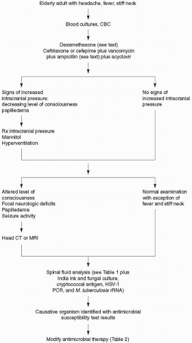
When the clinical presentation is suggestive of bacterial meningitis, Gram’s stain and blood cultures should be immediately obtained, and empiric antimicrobial therapy should be initiated without delay (Fig. 26-1). The diagnosis of bacterial meningitis is made by examination of the cerebrospinal fluid (CSF) (Table 26-1). As stated earlier, raised ICP is an expected complication of bacterial meningitis and is the major cause of obtundation and coma in this disease. The role of lumbar puncture as a causative factor for cerebral herniation in patients with acute bacterial meningitis has been debated for years and remains unresolved. The risk of cerebral herniation from acute bacterial meningitis independent of lumbar puncture is approximately 6% to 8%. When the possibility of increased ICP exists because of a decreased level of consciousness, lumbar puncture should either be delayed (but empiric antimicrobial therapy should be initiated) until the increased ICP can be treated or performed with a 22-gauge needle 30 to 60 minutes after mannitol 1 g/kg has been administered intravenously. The patient can also be intubated and hyperventilated, in addition to being treated with mannitol, to decrease ICP prior to lumbar puncture. CSF should be obtained for cell count (1.0 mL), glucose and protein concentrations (1.0 mL), Gram’s stain and bacterial culture (1.0 mL), latex agglutination test (0.5 mL), and/or polymerase chain reaction (PCR) for bacterial nucleic acid. CSF should also be examined for viral DNA because herpes simplex virus encephalitis is the leading disease in the differential diagnosis of bacterial meningitis.

The classic CSF abnormalities in bacterial meningitis are as follows: (a) increased opening pressure; (b) a pleocytosis of polymorphonuclear leukocytes (10 to 10,000 cells/mm3); (c) decreased glucose concentration (<45 mg/dL and/or CSF:serum glucose ratio of <0.31); and (d) an increased protein concentration. CSF bacterial cultures are positive in >80% of patients, and CSF Gram’s stain demonstrates organisms in >60%.

Opening pressure should be measured with the patient in the lateral recumbent position. The normal opening pressure for adults is <180 mm H2O. In obese adults, however, the normal opening pressure is <250 mm H2O. In adults, in uninfected CSF, the normal white blood cell (WBC) count ranges from 0 to 5 mononuclear cells (lymphocytes and monocytes)/mm3. In normal uninfected CSF in the adult, there should be no polymorphonuclear leukocytes; however, a rare polymorphonuclear leukocyte can be found in concentrated CSF specimens. If the total WBC count is <5 cells/mm3, the presence of a single polymorphonuclear leukocyte may be considered normal. The latex particle agglutination (LA) test for the detection of bacterial antigens of S. pneumoniae, Neisseria meningitidis, H. influenzae type b, S. agalactiae, and E. coli K1 strains in the CSF can be used to make a rapid diagnosis of bacterial meningitis and to make a diagnosis of bacterial meningitis in those patients who have been pretreated with antibiotics in whom Gram’s stain and CSF culture are negative. The LA test has a rapid turnaround time, usually less than a few hours, but a negative LA test does not rule out bacterial meningitis. A broad-range PCR can detect small amounts of viable and nonviable organisms in CSF. When the broad-range PCR is positive, a PCR that uses specific bacterial primers to detect the nucleic acid of S. pneumoniae, N. meningitidis, E. coli, L. monocytogenes, H. influenzae, or S. agalactiae should be done based on the clinical suspicion of the meningeal pathogen. However, the turnaround time for PCR may be several days. The Limulus amebocyte lysate assay is a rapid diagnostic test for the detection of gram-negative endotoxin in CSF and thus for making a diagnosis of gram-negative meningitis.

All patients with bacterial meningitis should have neuroimaging performed, either before or after lumbar puncture. Indications for performing neuroimaging prior to lumbar puncture are an altered level of consciousness, papilledema, a focal neurologic deficit, focal or generalized seizure activity, or an immunocompromised state. Magnetic resonance imaging (MRI) is preferred over computed tomography (CT) because of its superiority in demonstrating areas of cerebral edema and ischemia.


Figure 26-1. Algorithm for management of older adult with headache, fever, and stiff neck.


Treatment

Antimicrobial therapy should be initiated immediately in every older adult with fever, headache, and stiff neck. There is a heightened sense of urgency in initiating antimicrobial therapy when these symptoms are accompanied by an altered level of consciousness. The choice of antimicrobial therapy should be based on the predisposing conditions for bacterial meningitis. There are a
number of predisposing conditions that increase the risk of pneumococcal meningitis in the older adult, the most important of which is pneumococcal pneumonia. Due to the emergence of penicillin- and cephalosporin-resistant Streptococcus pneumoniae, empiric therapy of community-acquired bacterial meningitis in the older adult should include a third- or fourth-generation cephalosporin, vancomycin, and acyclovir. Acyclovir is added to the initial empiric regimen because herpes simplex virus encephalitis is the leading disease in the differential diagnosis. Ampicillin should be added to the empiric regimen for coverage of L. monocytogenes in older adults. In hospital-acquired meningitis and, particularly, meningitis following neurosurgical procedures, staphylococci and gram-negative organisms, including P. aeruginosa, are the most common etiologic organisms. In these patients, empiric therapy should include a combination of vancomycin and ceftazidime, cefepime, or meropenem. If a third-generation cephalosporin is chosen for empiric therapy of meningitis in neurosurgical patients and in neutropenic patients in whom P. aeruginosa is a possible meningeal pathogen, ceftazidime should be substituted for ceftriaxone or cefotaxime because ceftazidime is the only third-generation cephalosporin with sufficient activity against P. aeruginosa in the CNS. Once the organism has been identified by Gram’s stain or by bacterial culture of CSF and the results of antimicrobial susceptibility tests are known, antimicrobial therapy can be modified accordingly (Table 26-2). Some strains of pneumococci are sensitive to penicillin, but in clinical practice, few physicians use penicillin to treat pneumococcal meningitis. A CSF isolate of S. pneumoniae is considered to be susceptible to penicillin with a minimal inhibitory concentration (MIC) <0.06 µg/mL, to have intermediate resistance when the MIC is 0.1 to 1.0 µg/mL, and to be highly resistant when the MIC is >1.0 µg/mL. Isolates of S. pneumoniae that have cephalosporin MICs ≤0.5 µg/mL are considered sensitive to the cephalosporins (cefotaxime, ceftriaxone, cefepime). Those with MICs equal to 1 µg/mL are considered to have intermediate resistance, and those with MICs ≥2 µg/mL are considered resistant (11). For meningitis due to pneumococci with cefotaxime or ceftriaxone MICs of 0.5 µg/mL or less, treatment with cefotaxime or ceftriaxone is usually adequate. If the MICs are ≥1 µg/mL, vancomycin is the antibiotic of choice. Patients with S. pneumoniae meningitis should have a repeat lumbar puncture performed 24 to 36 hours after the initiation of antimicrobial therapy to document sterilization of the CSF unless findings on the neurologic examination are worrisome for a risk of herniation. Use of intraventricular vancomycin should be considered when intravenous vancomycin fails to sterilize the CSF after 24 to 36 hours of therapy. The intraventricular route of administration is preferred over the intrathecal route because adequate concentrations of vancomycin in the cerebral ventricles are not always achieved with intrathecal administration. Intrathecal administration of vancomycin is safe and is not associated with a risk of seizure activity. Cefepime is a broad-spectrum fourth-generation cephalosporin that is increasingly seen on the medication charts of hospitalized patients. Cefepime has in vitro activity similar to that of cefotaxime or ceftriaxone against S. pneumoniae and greater activity against Enterobacter species and P. aeruginosa. The dose of cefepime is 3 g intravenously every 8 hours in adults. In clinical trials, cefepime has been demonstrated to be equivalent to cefotaxime in the treatment of pneumococcal meningitis, but its efficacy in bacterial meningitis caused by penicillin- and cephalosporin-resistant pneumococcal organisms, Enterobacter species, and P. aeruginosa has not been established. A 2-week course of intravenous antimicrobial therapy is recommended for pneumococcal meningitis.


Table 26-1. Cerebrospinal Fluid (CSF) Analysis in Bacterial Meningitis


Opening pressure


>180 mm H2O


White blood cells


>10 to <10,000/mm3 – neutrophils predominate


Red blood cells


Absent unless traumatic tap


Glucose


<40 mg/dL


CSF:serum glucose ratio


<0.40


Protein


>50 mg/dL


Gram’s stain


Positive in 70% to 90% of untreated cases


Culture


Positive in 80% of cases


PCR


Detects bacterial nucleic acid


Latex agglutination


Specific for antigens of S. pneumoniae, N. meningitidis, E. coli, Hib and group B streptococcus


Limulus amebocyte lysate assay


Positive in gram-negative meningitis


Table 26-2. Antimicrobial Therapy of Bacterial Meningitis


Organism


Antibiotic


Total Daily Adult Dose (dosing interval)


Streptococcus pneumoniae


Sensitive to penicillin


Penicillin G


24 million U/day (every 4 hours)


Relatively resistant to penicillin


Ceftriaxone or cefotaxime or cefepime


4 g/day (every 12 hours)


12 g/day (every 4 hours)


6 g/day (every 8 hours)


Resistant to penicillin


Vancomycin plus cefepime, ceftriaxone, or cefotaxime ± intraventricular vancomycin


30-45 mg/kg/day (every 8 hours)


Gram-negative bacilli (except P. aeruginosa)


Ceftriaxone or cefotaxime or cefepime


20 mg/day


Pseudomonas aeruginosa


Ceftazidime or meropenem


8 g/day (every 8 hours)


6 g/day (every 8 hours)


Staphylococci


Methicillin-sensitive


Nafcillin


9-12 g/day (every 4 hours)


Methicillin-resistant


Vancomycin


30-45 mg/kg/day (every 8 hours)


Listeria monocytogenes


Ampicillin ± gentamicin


12 g/day (every 4 hours)


6 mg/kg/day (every 8 hours)


Haemophilus influenzae


Ceftriaxone or cefotaxime


4 g/day (every 12 hours)


12 g/day (every 4 hours)


Streptococcus agalactiae


Ampicillin or penicillin G


12 g/day (every 4 hours)


20-24 million U/day (every 4 hours)


All antibiotics are administered intravenously. Doses indicated are for patients with normal renal function.


The third-generation cephalosporins—cefotaxime, ceftriaxone, and ceftazidime—are equally efficacious for the treatment of gram-negative bacillary meningitis, with the exception of meningitis due to P. aeruginosa. Ceftazidime or meropenem is recommended for meningitis due to this organism. A 3-week course of intravenous antimicrobial therapy is recommended for meningitis due to gram-negative bacilli. Meningitis due to S. aureus or coagulase-negative staphylococci is treated with nafcillin or oxacillin. Vancomycin is the drug of choice for methicillin-resistant staphylococci. The CSF should be monitored during therapy, and if the spinal fluid continues to yield viable organisms after 48 hours of intravenous therapy, then intraventricular vancomycin 20 mg once daily can be added.

A 3- to 4-week course of ampicillin is recommended for L. monocytogenes meningitis. Gentamicin should be added to ampicillin in critically ill patients because the combination of ampicillin and gentamicin has greater bactericidal activity than ampicillin alone in experimental models of meningitis (15).

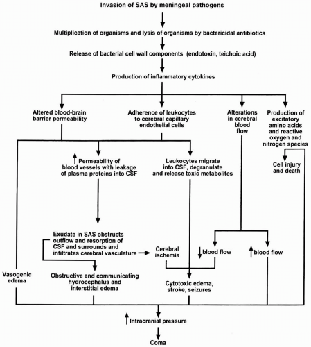
Adjunctive TherapyThe neurologic complications of bacterial meningitis (raised ICP, seizure activity, stroke) continue long after the CSF has been sterilized by antimicrobial therapy (20). The pathophysiology of the neurologic complications of bacterial meningitis is shown in Figure 26-2. The release of bacterial cell wall components by the multiplication of bacteria and the lysis of bacteria by bactericidal antibiotics leads to the production of the inflammatory cytokines, interleukin-1 (IL-1) and tumor necrosis factor (TNF), in the subarachnoid space, which initiates a cascade of events that ultimately leads to increased ICP, coma, stroke, seizures, and obstructive and communicating hydrocephalus. Dexamethasone has a beneficial effect by inhibiting the synthesis of IL-1 and TNF at the level of mRNA and by decreasing CSF outflow resistance and stabilizing the blood-brain barrier. IL-1 and TNF are produced by microglia, astrocytes, monocytes, and macrophages. The rationale for giving dexamethasone before antibiotic therapy is that dexamethasone inhibits the production of TNF if administered to macrophages and microglia before they are activated by bacterial cell wall components. A meta-analysis of randomized, concurrently controlled trials of dexamethasone therapy in childhood bacterial meningitis
published from 1988 to 1996 confirmed benefit for H. influenzae type b meningitis if dexamethasone was begun with or before intravenous antibiotics and suggested benefit for pneumococcal meningitis in children (10). In the European Dexamethasone in Adulthood Bacterial Meningitis Study of 301 adults with bacterial meningitis, 157 patients were randomized to receive dexamethasone and 144 patients were assigned to placebo 15 to 20 minutes before the first dose of an antimicrobial agent. There were 108 cases of pneumococcal meningitis. Within 14 days, five (9%) of 58 patients with pneumococcal meningitis died in the dexamethasone group and 13 (26%) of 50 patients in the placebo group died (2). In addition to reducing mortality, dexamethasone was associated with a reduction in the number of patients who had an unfavorable outcome. Present recommendations are to initiate dexamethasone therapy (10 mg) before or with the first dose of antibiotics and continue dexamethasone, regardless of the bacteria causing meningitis, in a dose of 10 mg every 6 hours for 4 days (23).


Figure 26-2. The neurologic complications of bacterial meningitis are the result of this inflammatory cascade. ((c) Roos KL, Tyler KL. Meningitis, encephalitis, brain abscess, and empyema. In: Kasper DL, Braunwald E, Fauci AS, et al., eds. Harrison’s principles of internal medicine. 16th ed. New York: McGraw Hill; 2005:2471-2490.)


FUNGAL MENINGITIS

Aging is associated with immune dysfunction, especially in cell-mediated immunity (25). This places older adults at risk for fungal meningitis. There are a number of fungi that can cause meningitis, the most common of which are Cryptococcus neoformans, Histoplasma capsulatum, and Coccidioides immitis. Blastomyces dermatitidis, Candida species, and Sporothrix schenckii may also be the causative organisms of meningitis, as may the rare fungi Paracoccidioides brasiliensis, Chaetomium atrobrunneum, Cladophialophora bantiana, Pseudallescheria boydii, and Schizophyllum commune.

Only gold members can continue reading. Log In or Register to continue

Stay updated, free articles. Join our Telegram channel

Jul 14, 2016 | Posted by in NEUROLOGY | Comments Off on Nonviral Infectious Diseases of the Nervous System

Full access? Get Clinical Tree

Get Clinical Tree app for offline access