and Aditya G. Shivane1
(1)
Cellular and Anatomical Pathology Level 4, Derriford Hospital, Plymouth, UK
Abstract
An understanding of the pathology of human nervous system requires a good basic knowledge of the normal cellular components and their architecture. This chapter starts with the discussion of normal cellular constituents of the nervous system, their morphology and some of their salient functions. The general architecture of central and peripheral nervous tissues is then described. The nervous tissue and their various components are studied using traditional tinctorial stains. Immunohistochemical preparations which target specific cellular antigens are now being increasingly used for both diagnostic and research purposes.
Keywords
CellsHistologyNervous systemStainsImmunohistochemistryThe human nervous system can be broadly divided into two parts- the central and peripheral nervous system. The central nervous system (CNS) includes the brain and spinal cord. Both brain and spinal cord are surrounded by tough coverings called the meninges and are encased in a protective bony structure, the skull and the vertebral column respectively. The peripheral nervous system (PNS) includes the nerves (cranial, spinal and peripheral nerves), sensory ganglia (dorsal root ganglion) and autonomic ganglia (sympathetic and parasympathetic ganglia).
1.1 Cells of the Nervous System
The cells which make up the nervous system are the neurons and other supporting cells (glial cells) which include astrocytes, oligodendrocytes, Schwann cells, ependymal cells and microglia.
1.1.1 Neurons
A neuron is the basic functional unit of the nervous system. It is primarily responsible for collecting information, processing and then generating response. During development, they are derived from the neural tube and eventually migrate and populate different regions of the nervous system. A neuron is a post-mitotic cell and therefore cannot be replaced when damaged. It is also a highly metabolically active cell and requires continuous supply of nutrition for normal functioning. In an adult brain, neural stem cells have been identified within the subventricular zone of lateral ventricles, in the dentate gyrus of hippocampus and in the olfactory bulb.
The basic structure of a neuron include a cell body or perikaryon, many short processes or dendrites which receive information from other neurons and a single long process called ‘axon’ which transmits signals to other neurons. The dendrites and axons are collectively referred to as neurites. The cell body appears large in some types of neurons and contains a large nucleus with a prominent nucleolus. The cytoplasm of the neuron also contain granular dark staining material rich in rough endoplasmic reticulum termed ‘Nissl substance’ which is one of the important distinguishing feature on microscopy (Figs. 1.1a and 1.2; Box 1.1). The region where axon begins is called an ‘axon hillock’ from where action potentials are generated.
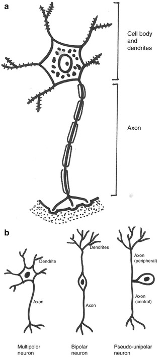
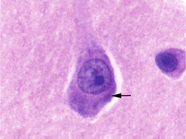
Fig. 1.1
(a) Diagram showing different parts of a neuron and, (b) the basic morphological subtypes of neurons
Fig. 1.2
A cortical pyramidal neuron showing a large nucleus, prominent nucleolus and purplish granules (Nissl substance) in the cytoplasm (arrow). H&E stain
The neurons come in various shapes and sizes. Neurons in some locations such as dentate gyrus and cerebellum appear small and rounded with no visible cytoplasm and are referred to as granular neurons. Neurons can be either multipolar (many dendrites, single axon. e.g. motor neurons), bipolar (single dendrite, single axon. e.g. sensory neurons in retina) or unipolar/pseudo-unipolar (single process which divides into central and peripheral axons, no dendrites. e.g. sensory neurons in dorsal root ganglia). The majority of neurons within the nervous system are multipolar (Fig. 1.1b).
The cell bodies of neurons make up the bulk of grey matter and deep nuclei of the brain and spinal cord. Their axons run as bundles within the white matter. Occasional neurons can be seen within the white matter, especially in the temporal lobe. This should not be mistaken for a neuronal migration abnormality.
Box 1.1 How to Identify a Neuron
Large cell and cell body
Large nucleus
Single prominent nucleolus
Nissl substance (purplish granules on H&E)
1.1.2 Astrocytes
Astrocytes are the most numerous of the glial cells and give structural and metabolic support to a neuron. Astrocytes are derived from radial glial cells during embryonic development. Astrocytes appear to have several complex roles in healthy tissue, some of which include- development of grey and white matter, regulation of blood flow, maintaining biochemical homeostasis, synapse function, and CNS metabolism [1]. Astrocytes have star-shaped cytoplasmic processes which can be identified with a special stain such as phosphotungstic acid haematoxylin (PTAH) (Fig. 1.3 and Box 1.2). These processes abut on capillaries, neuron, axon, dendrites and pia mater (the innermost layer of meninges). The cytoplasmic processes contain characteristic filaments termed ‘glial fibrillary acidic protein’ or GFAP which can be demonstrated using immunohistochemistry. Astrocytes can be of two morphological subtypes- (1) fibrous or fibrillary astrocytes which have long processes rich in GFAP-positive filaments and are present in the white matter and, (2) protoplasmic astrocytes with short processes and few GFAP-positive filaments and mainly confined to the grey matter.
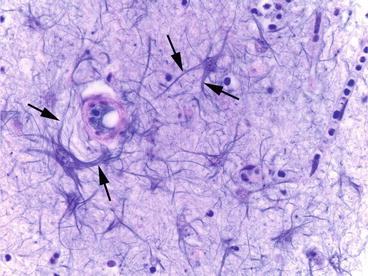
Fig. 1.3
Fibrous or fibrillary astrocytes with stellate cytoplasmic processes (arrows). PTAH stain
The cell processes of astrocytes form the background fibrillary meshwork called ‘neuropil’ (seen as pink background on the standard H&E stain).
Box 1.2 How to Identify an Astrocyte
Medium-size cell
Round or oval nucleus
Indistinct nucleolus
‘Stellate’ cell processes (seen with special stains such as PTAH; appear as pink background on H&E stain)
1.1.3 Oligodendrocytes
Oligodendrocytes are the myelin producing cells within the CNS. Oligodendrocytes are presumed to be derived from a common progenitor cell which also gives rise to neurons and depend on various regulatory factors for their differentiation and migration. The process of myelination is complex and relies on neuronal and axonal signals [2]. Each oligodendrocyte forms myelin segments on multiple axons. They are mainly present within the white matter along bundles of axons which they myelinate. Within the grey matter they are often seen around the cell body of neurons as ‘satellite cells’ (Fig. 1.4a). Oligodendrocyte, as the name implies, has fewer cytoplasmic processes compared to that of an astrocyte. These cell processes are not clearly visible on tissue sections. Therefore, these cells appear as naked dark round nuclei with ‘perinuclear halo’ or ‘fried egg’ appearance (Fig. 1.4b, Box 1.3). This cytoplasmic clearing is not evident in intraoperative frozen sections or tissue which is rapidly fixed in formalin and is considered an artefact of delayed fixation. However, this is a very helpful feature in recognising a cell as being oligodendroglial in origin within a glial neoplasm.
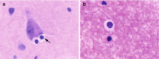
Fig. 1.4
(a) Oligodendrocytes (arrow) clustered around a neuron (satellite cells) and, (b) a typical oligodendrocyte with round nucleus and perinuclear halo in white matter. H&E stain
Box 1.3 How to Identify an Oligodendrocyte
Small to medium-size cell
Dark round nucleus
Cytoplasmic clearing or ‘perinuclear halo’ or ‘fried egg’ appearance
1.1.4 Schwann Cells
Schwann cells perform the function of electrically insulating axons of the peripheral nervous system. Unlike an oligodendrocyte, each Schwann cell myelinates only one axon. The cell processes of a Schwann cell wrap around an axon in multiple layers forming the myelin sheath (Fig. 1.5a). Large diameter axons are always myelinated whereas the small diameter axons can be myelinated or unmyelinated. Myelinated nerve fibers conduct electrical signals faster than unmyelinated fibers. Schwann cells also surround unmyelinated axons (Remak cell), with their cytoplasm wrapping around and isolating each axon from its neighbours (Fig. 1.5b). A Schwann cell also performs an important role in nerve regeneration after injury.
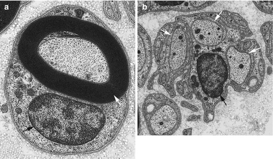
Fig. 1.5


(a) Ultrastructure of a peripheral nerve showing a large myelinated axon (white arrow) surrounded by a Schwann cell (black arrow) and, (b) Schwann cell (black arrow) wrapping three unmyelinated axons (white arrows) also referred to as a ‘Remak cell’
Related posts:


Stay updated, free articles. Join our Telegram channel
Full access? Get Clinical Tree
