Fig. 35.1
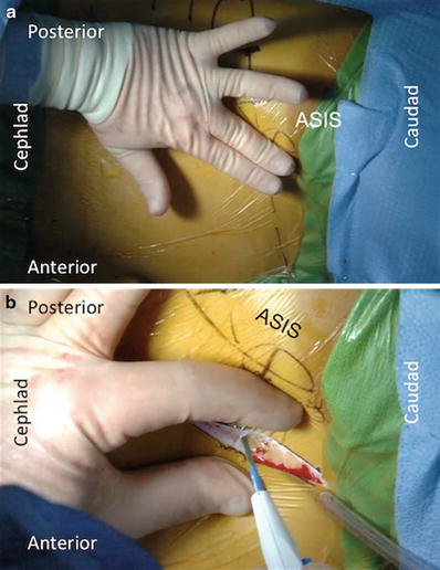
This image illustrates skin marking that needs to be performed prior to incision to ensure adequate exposure to the L5–S1 disc space
See Fig. 35.1—This image illustrates skin marking that needs to be performed prior to incision to ensure adequate exposure to the L5–S1 disc space.
2.
Approximately 1–2 finger breaths (depending upon size of the patient) from the ASIS and pelvis, between the two lines, the incision is made from postero-rostral to antero-caudal medial as depicted in the figure drawing. There is great flexibility in this incision as it can be extensile to higher levels of the spine and distally to the symphysis pubis as needed (see Fig. 35.2a, b).


Fig. 35.2
(a, b) An illustration of the recommended skin incision
See Fig. 35.2a, b—An illustration of the recommended skin incision.
3.
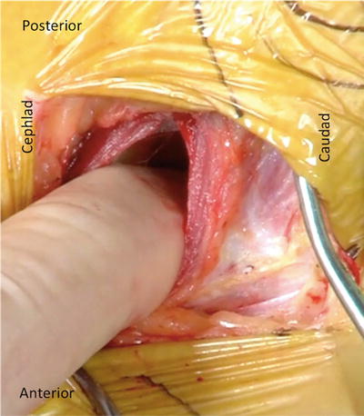
Depending upon the variability in each patient, the surgeon will either encounter the external oblique muscle or the aponeurosis and fascia. This is opened to expose the muscle. Using a blunt finger, just as described in O25, retroperitoneal dissection and exposure is done. Using a two-finger technique facilitates a wider exposure for the retractors. The ureter is more likely to be visualized in this exposure and dissection therefore a wide rostral to caudal development of the retroperitoneal plane is worthwhile and protective of the ureter, thereby maintaining its attachment to the posterior peritoneum while mobilizing anteriorly (see Fig. 35.3).


Fig. 35.3
Blunt finger dissection into the retroperitoneal space
See Fig. 35.3—Blunt finger dissection into the retroperitoneal space.
4.
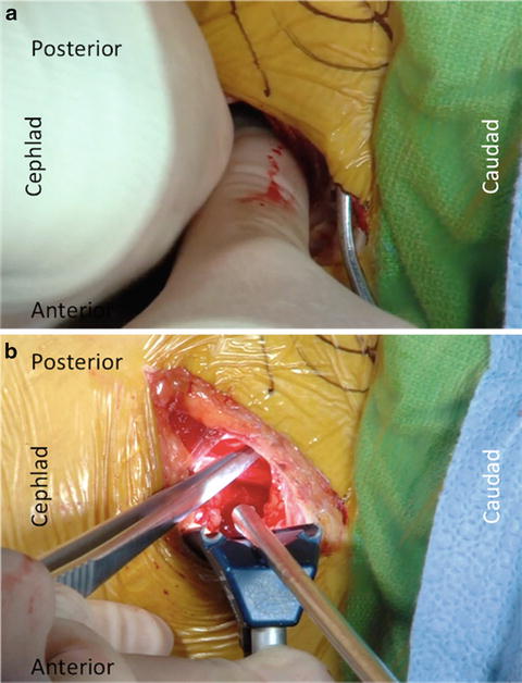
As the two-finger dissection progresses down the pelvis and across the psoas, it is noted that the psoas is many times lifting off the spine to leave the pelvis. The dissection continues anteriorly from the pelvis while searching for the very palpable common iliac artery pulse. The common iliac vein is medial to the artery. Continue the gentle finger dissection past the pulsating common iliac artery with appreciation that the vein is encountered just medial to the artery on the sacral promontory and L5/S1 disc space (see Fig. 35.4a, b).


Fig. 35.4
(a) Two-finger dissection into the retroperitoneal disc space. (b) The common iliac artery after exposure
See Fig. 35.4a—Two-finger dissection into the retroperitoneal disc space.
Figure 35.4b—The common iliac artery after exposure.
5.
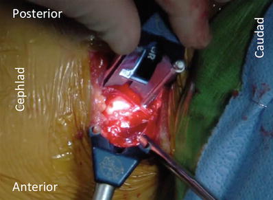
The lighted retractors are then placed sequentially down onto the anterior spine and the adventitial layers that are on the anterior disc and sacrum are encountered (see Fig. 35.5).


Fig. 35.5
The exposed anterior spine of the L5–S1 disc space
See Fig. 35.5—The exposed anterior spine of the L5–S1 disc space.
6.
This layer may have elements of the superior hypogastric plexus and sympathetic chain within. This thin adventitial layer adheres and tethers the common iliac vein to the annulus and must be mobilized before posterior retraction of the vein is done. It is generally accepted that gentle blunt dissection and avoidance of bovie usage of this layer with kitners may mitigate against injury to the superior hypogastric plexus and venous structures.
7.


Because the O51 approach accesses the L5/S1 disc in the classic supine ALIF interval below the bifurcation, the iliolumbar vein does not need to be identified nor ligated since posterior retraction of the left common iliac vein and artery laterally does not cause stretch and potential avulsion. Anterior retraction at L4/L5 in ALIF, when the major vessels are mobilized to access the disc space directly anteriorly, may cause injury to the iliolumbar vein and it must be identified before retraction. In the O51 approach, the disc space is accessed in a similar fashion as traditional ALIF and at L45, because the graft is inserted obliquely, no mobilization of the vessels is required.
Stay updated, free articles. Join our Telegram channel

Full access? Get Clinical Tree


