Obstructive Sleep Apnea Hypopnea Syndromes: Definitions, Epidemiology, Diagnosis, and Consequences


Obstructive Sleep Apnea Hypopnea Syndromes: Definitions, Epidemiology, Diagnosis, and Consequences


Richard B. Berry


HISTORY AND DEFINITIONS

The obstructive sleep apnea hypopnea syndrome (OSAHS) was recognized as a significant health problem only over the last three decades of the 20th century. In 1956, Burwell (1) used the term Pickwickian syndrome to describe individuals with obesity, hypersomnolence, hypercapnia, cor pulmonale, and erythrocytosis. We now know that Pickwickian patients represent the “tip of the iceberg” of the larger group of patients with sleep-disordered breathing (SDB). Subsequently, the connection between nocturnal respiratory events and daytime sleepiness was recognized. Guilleminault and coworkers (2) described the obstructive sleep apnea syndrome as patients with daytime sleepiness and obstructive apneas on polysomnography (PSG). By convention, in adults, an apnea is a cessation of airflow at the nose and mouth for 10 seconds or longer. An obstructive apnea is characterized by absent airflow despite persistent respiratory effort (Fig. 18-1). An apnea index (AI) of ≥5 was considered abnormal (3). Obstructive apneas are secondary to airway closure at a supraglottic location that reverses at apnea termination often associated with a brief awakening (arousal) (4). A fall in arterial oxygen saturation (Sao2) of varying severity follows the event. An arterial oxygen desaturation is usually defined as a fall in the Sao2 of 4% or more. Central apneas are defined as absent airflow for 10 seconds or longer associated with an absence of respiratory effort. Mixed apneas are those apneas consisting of an initial central apnea portion followed by an obstructive portion (Fig. 18-2). Respiratory effort is commonly detected by the movement of bands around the chest and abdomen in response to breathing efforts. Esophageal pressure monitoring detects changes in pleural pressure during inspiration and is a more sensitive method to detect inspiratory effort. The nadir in negative pressure swings or the size of the deflections can be used to quantify the amount of effort (5). The deflections in chest and abdominal bands during obstructive apnea may or may not increase prior to apnea termination. In nonrapid eye movement (NREM) sleep, the esophageal pressure deflections routinely increase during the terminal portion
of obstructive apnea, reflecting an increase in inspiratory effort during the apnea (Fig. 18-3).


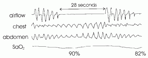
FIGURE 18-1 An obstructive apnea is characterized by absent airflow and persistent respiratory effort (movement of chest and abdominal bands). An arterial oxygen desaturation to 82% follows this 28-second apnea. The drop in the Sao2 to 90% is from a preceding apnea. In this case, the chest and abdominal tracing deflections progressively increase during the event. This is not always seen.

It was soon realized that the episodes of severe airway narrowing (partial obstruction) resulting in reduction of airflow or tidal volume (hypopneas) are also important (Fig. 18-4) (6). Patients primarily with hypopneas had the same symptoms, arousals, and arterial oxygen desaturation as did patients with obstructive apneas (7). Hence, the term OSAHS has been used to be more inclusive. However, many clinicians still use the term obstructive sleep apnea (OSA) to refer to the apnea and hypopnea syndrome. Of note, patients with the OSAHS will have variable proportions of obstructive apneas and hypopneas. For example, they may have apneas during rapid eye movement (REM) sleep or in the supine position and hypopneas during NREM sleep or in the lateral position. Variable amounts of mixed and central apneas may also be present.

The apnea-hypopnea index (AHI) is the number of apneas and hypopneas per hour of sleep. The AHI has been used to quantify the severity of SDB. An AHI of ≥5 per hour in the presence of symptoms is said to define OSAHS. However, there has been considerable controversy about the definition of hypopnea and the frequency of events that is considered abnormal. As will be discussed later, the AHI value for a given patient can vary tremendously depending on the definition of hypopnea and the technology used to monitor airflow (8,9). It has also been recognized that the correlation between the AHI and measures of impairment, such as subjective or objective sleepiness, although significant, is low (correlation coefficient in the range of 0.4-0.5) (10). However, to date, efforts to find better indexes of impairment on the basis of polysomnographic findings, such as the arousal index, have not substantially improved the correlation with symptoms. The wide variability in symptoms is likely due, in part, to different individual susceptibility to sleep fragmentation or other factors contributing to symptoms of daytime sleepiness, such as medications or reduced sleep time. Two patients with the same AHI can have vastly
different degrees of arterial oxygen desaturation. This could be relevant for many of the cardiovascular consequences of sleep apnea.


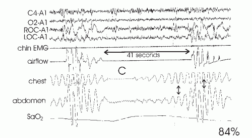
FIGURE 18-2 This tracing shows a 41-second mixed apnea. The initial portion (C) is a central apnea characterized by absent ventilatory effort. The small deflections in the abdomen tracing are from cardiac pulsations. After the central portion, an obstructive portion is noted. In this case, there is a progressive increase in chest and abdominal movements prior to apnea termination. In addition, there is paradoxical motion of the chest and abdomen tracings (small arrow), which vanishes after apnea termination. The apnea is followed by an arterial oxygen desaturation to 84%. In this case, an arousal is not seen in the central (C4-A1) or occipital (O2-A1) EEG tracings, although there is sudden movement in the eye tracings (ROC-A1 and LOC-A1). Here ROC and LOC are the right and left outer canthus electrodes, and A1 is the left mastoid electrode.


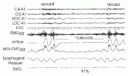
FIGURE 18-3 This tracing shows the end of one obstructive apnea followed by a 16-second obstructive apnea during NREM sleep. Arousal is noted at the termination of both events. The esophageal pressure swings increase toward the end of the event, consistent with a progressive increase in inspiratory effort. The moving time average MTA-EMGgg of the genioglossus EMG (EMGgg) was measured using a mouthpiece electrode. At the start of the apnea, the genioglossus activity falls; at apnea termination, there is a large increase in the EMGgg coincident with arousal and opening of the airway.


Hypopnea Controversy

It is not possible to discuss the hypopnea controversy without briefly discussing the methods of airflow monitoring. Many early sleep studies used thermal-sensitive devices (thermocouples and thermistors) to detect airflow. These are fairly accurate in detecting apnea, but the signal is not proportional to the flow rate (5,11). Thus, they tend to be less sensitive at detecting hypopneas than the systems that accurately measure airflow, such as pneumotachograph-mask systems (5,11). The latter are somewhat uncomfortable and confined to research studies. Early definitions of hypopnea required a reduction in airflow coupled with a 4% or greater drop in the Sao2 (6). This certainly meant that the reduction in airflow had physiologic significance for the patient. However, subsequently it became clear that it is possible for patients to have episodes of increased inspiratory effort sufficient to induce arousal from sleep without a drop in the Sao2 (12,13). Guilleminault and coworkers (12) described a group of patients who were sleepy, but traditional airflow monitoring with thermal-sensing devices showed an AHI of <5 per hour and most had minimal arterial oxygen desaturation. Monitoring of esophageal pressure in these patients demonstrated increased respiratory effort and subsequent arousal. In the original description, a respiratory arousal index (RAI) of >10 per hour was required to diagnose these patients as having the “upper-airway resistance syndrome.” Respiratory arousals were defined as arousals after a period of abnormally negative esophageal pressure. The upper-airway resistance events were associated with minimal changes in flow detected by thermal devices but mildly reduced airflow and flattening (airflow limitation) by pneumotachograph monitoring. Thus, esophageal pressure was needed to demonstrate the events in the absence of an accurate measurement of airflow. Studies of experimental sleep disturbance in normal subjects also demonstrated that repetitive brief awakenings (arousals)
could induce daytime sleepiness in the absence of arterial oxygen desaturation (14,15 and 16). These developments suggested that the sleep-disturbing consequences of respiratory events were as clinically important as the respiratory event-associated arterial oxygen desaturation.


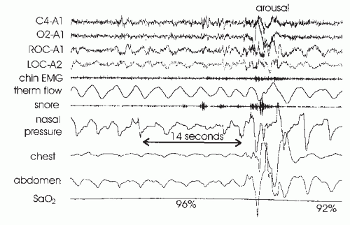
FIGURE 18-4 Obstructive hypopnea with arousal at event termination. Note that the decrease in airflow by nasal pressure is greater than by thermistor (thermal flow). Also note the flattening of the nasal pressure signal during the event. Obstructive hypopnea may be associated with paradoxical motion of the chest and abdomen band tracings, but this is not seen in this tracing. The reduction in airflow is associated with a 4% arterial oxygen desaturation and would therefore meet the hypopnea ciriteria based on oxygen desaturation. After arousal, the flattening in the nasal pressure signals resolves and the negative pressure swings decrease.

Despite the ability of esophageal pressure monitoring to detect subtle episodes of increased respiratory effort, the technique was never widely used in clinical sleep medicine. Instead, nasal pressure monitoring has gained popularity, being both well tolerated and more accurate than thermal devices at detecting changes in airflow during sleep (5,17). In addition, nasal pressure monitoring can often detect many of the subtle respiratory arousal events detected by esophageal pressure monitoring. A sensitive pressure transducer is connected to a nasal cannula inserted in the nose. The pressure drop across the nasal inlet is measured and is proportional to the square of the nasal flow (5,17,18). During obstructive hypopnea, the nasal pressure signal demonstrates a reduction in magnitude and the shape of the inspiratory portion shows flattening (flow plateau) during flow limitation (airway narrowing/increased upper-airway resistance) (see Fig. 18-4). Patients monitored with nasal pressure will often have many more events, defined as a reduction in flow, than detected by a thermal device (17). Arousal associated with minimal changes in thermistor flow will often be seen to follow a more obvious reduction in the nasal pressure signal with flattening (see Fig. 18-4).

Respiratory inductance plethysmography (RIP) provides another alternative to thermal devices for detecting respiratory events. The signal from bands around the chest and abdomen is added to give a RIPsum signal. The fluctuations in the RIPsum give an estimate of tidal volume when the device is calibrated (19,20 and 21). The individual chest and abdomen signals give an estimate of the surface area they encircle (inductance of a coil is proportional to the area it encloses). During hypopnea, the chest and abdominal band fluctuations and RIPsum decrease (Fig. 18-5). Either nasal pressure or RIP will detect many instances of obvious reductions in airflow (tidal volume) in the absence of a 4% arterial oxygen desaturation. Should such events be classified as hypopneas?


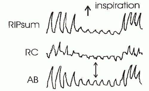
FIGURE 18-5 This tracing shows an obstructive hypopnea detected by RIP. Fluctuations in the RIPsum are estimates of tidal volume. The rib cage (RC) and abdominal (AB) band tracing are decreased during the hypopnea and also show paradoxical motion.

A task force of the American Academy of Sleep Medicine (AASM) published guidelines for monitoring respiration during sleep and graded the accuracy of monitoring technology on the basis of the literature. The task force defined a hypopnea (Chicago criteria) on the basis of a 50% reduction in airflow or any discernable reduction in flow associated with either an arousal or a 3% arterial oxygen desaturation (22) (Table 18-1). Although this definition of hypopnea is inclusive, there are no large population studies using these definitions to determine a frequency of events associated with cardiovascular risk or to define normal limits. The AASM task force also published criteria for estimation of severity of OSAHS on the basis of the AHI. Values of 5 to <15, 15 to 30, and >30 per hour were termed mild, moderate, and severe, respectively. These are somewhat arbitrary but are widely used.

The task force also defined a respiratory effort-related arousal (RERA) as an event lasting 10 seconds or longer with a “pattern of progressively more negative esophageal pressure, terminated by a sudden change in pressure to a less-negative level and an arousal.” The event may be associated with a drop in airflow but does not qualify as a hypopnea (Fig. 18-6). In actual clinical practice, most sleep centers detect RERAs on the basis of the nasal pressure signal rather than esophageal pressure. A flowlimitation RERA would be one associated with flattening in the airflow signal >10 seconds followed by an arousal and an abrupt reversal in flow to a round shape (that does not qualify as a hypopnea). Although one may have airflow limitation without an increase in respiratory effort and vice versa, most episodes of airflow limitation (increased upper-airway resistance) during sleep are associated with increased esophageal pressure deflections. One study showed that the RERA indexes determined by nasal pressure and esophageal pressure monitoring were similar (23).

A consensus statement for the indications for continuous positive airway pressure (CPAP) treatment from another group suggested that a respiratory-disturbance index (RDI) be defined as the AHI + the RERA index. The RERA index
is defined as the number of RERAs per hour of sleep. The RDI could then be used to assess the severity of SDB (24). The RAI can be defined as the number of arousals per hour of sleep associated with apnea, hypopnea, or a RERA.


TABLE 18-1 HYPOPNEA DEFINITIONSAa


AIRFLOW


OTHER CRITERIA


AASM task force


50% reduction


None


“Chicago criteria”


Discernable but <50% reduction


Arousal or ≥3% desaturation required


AASM CPRC


30% or greater reduction


4% or greater drop in the Sao2


aAirflow reduction must be 10 seconds or greater in duration.


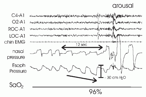
FIGURE 18-6 An RERA. The esophageal pressure shows a progressive increase in negative pressure followed by an arousal and reduction in the pressure swings. No desaturation was noted. There is also a reduction in flow by nasal pressure with a flattening in the inspiratory shape. After arousal, the flattening is abruptly reversed. Of note, this event would qualify as a hypopnea if an associated desaturation was not required (reduction in flow + arousal).

Subsequent to the publication of the above definitions, the Clinical Practice Review Committee (CPRC) of the AASM advocated another definition of hypopnea on the basis of a 30% reduction in airflow of 10 seconds or longer and a 4% or greater desaturation (drop in the Sao2) (25). The presence or absence of arousal was not considered. The CPRC provided several reasons for their choice of hypopnea definition. First, the scoring of hypopnea based on airflow and desaturation has good intra-and interscoring reliability, whereas the scoring of arousals does not (26,27). Second, the Sleep Heart Health study, using this definition of hypopnea, was able to show that even mild elevations of the AHI (≥5 per hour) are associated with an increased risk of cardiovascular disease (28). Of note, the Centers for Medicare & Medicaid Services has adopted the CPRC definition for hypopnea to determine the qualification for CPAP reimbursement. However, the CPRC hypopnea definition does not recognize the sleep-disturbing effects of reductions in airflow associated with arousal but less than a 4% desaturation. For example, the event in Fig. 18-6 would not be considered a hypopnea if a 4% or greater arterial oxygen desaturation is required. It would qualify as a RERA (or flowlimitation RERA if esophageal pressure was not monitored). Some sleep centers report an AHI based on a hypopnea definition requiring a 4% desaturation and an RDI = AHI + RERA index. In summary, a reduction in airflow (nasal pressure) with flattening that is followed by an arousal, but less than a 4% arterial oxygen desaturation, could be classified as either a hypopnea or a flow-limitation RERA, depending on the definition of hypopnea that is used. Clearly, the hypopnea controversy is not over. However, if one reads a sleep study report or a published sleep medicine investigation, it is important to know both the definition of hypopnea and the technology used to detect airflow.

The new requirements for an event to be classified as a hypopnea are as follows. A hypopnea should be scored only if all of the following criteria are present.


  • The nasal pressure signal excursions (or those of the alternative hypopnea sensor) drop by >30% of baseline.


  • The event duration is at least 10 seconds.


  • There is a >4% oxygen desaturation from pre-event baseline.


  • At least 90% of the event’s duration must meet the amplitude reduction of criteria for hypopnea.

Alternatively, a hypopnea can also be scored if all of the following criteria are present.


  • The nasal pressure signal excursions (or those of the alternative hypopnea sensor) drop by >50% of baseline


  • The duration of the event is at least 10 seconds.


  • There is a >3% oxygen desaturation from pre-event baseline or the event is associated with arousal.


  • At least 90% of the event’s duration must meet the amplitude reduction of criteria for hypopnea.


VARIANTS OF THE OBSTRUCTIVE SLEEP APNEA HYPOPNEA SYNDROME


Upper-airway Resistance Syndrome

As noted earlier, Guilleminault and coworkers identified a group of patients who exhibited subjective and objective (Multiple Sleep Latency Test [MSLT]) daytime sleepiness but did not have an AHI of >5 per hour (thermal devices measured airflow). The group was defined by having an RAI of >10 per hour using esophageal pressure monitoring (12). The events were not associated with desaturation or a change in thermal device-detected airflow. The symptom of sleepiness responded to CPAP treatment. The mean arousal index of the group was 33 per hour (range
16-52), and the mean maximally negative esophageal pressure nadir was -37 cm H2O. There has been controversy as to whether the upper-airway resistance syndrome is a distinct entity or simply a milder form of OSAHS (29,30). Individuals without daytime sleepiness may have a RERA index of >10 per hour, although the mean RERA for a group of persons without symptoms is usually <10 per hour (31). Of note, the mean total arousal index in a group of normal persons using AASM criteria was 21 per hour in one study (32). The 95% confidence limit of normal for the arousal index was very wide due to high arousal rates in older patients. It is possible that respiratory arousals cause more potent sleep disruption than “spontaneous arousals.” However, this has never been experimentally addressed. In any case, there is some overlap in the arousal index between groups of normal subjects and patients with upper-airway resistance syndrome/mild OSAHS. If one uses nasal pressure monitoring for airflow and a definition of hypopnea that utilizes arousal, as well as a drop in the Sao2, most “upper-airway resistance” patients will have an AHI of >5 per hour and, hence, be classified as having the OSAHS. Alternatively, an RDI = AHI + RERA index will be >5 per hour if hypopneas are required to be associated with a 4% or greater desaturation.


Obesity Hypoventilation Syndrome

Most patients with the OSAHS do not have daytime hypoventilation. Obese patients with daytime hypoventilation, not secondary to lung disease, are said to have the obesity hypoventilation syndrome (OHS). These patients were previously referred to as “Pickwickian patients” (1). Patients with the OHS are a heterogeneous group. The etiology includes upper-airway obstruction, decreased respiratory system compliance from obesity, and intrinsic or acquired abnormalities in ventilatory drive. Most OHS patients will have a high AHI and severe arterial oxygen desaturation (33,34 and 35). A few will exhibit worsening hypoventilation and severe arterial oxygen desaturation during sleep, without many discrete apneas or hypopneas. A recent study characterized the patients on the basis of their response to treatment (35). Some OHS patients could be adequately treated with CPAP alone. Opening the upper airway with CPAP during sleep restored adequate oxygenation. Others still had hypoventilation despite the absence of apnea or hypopnea. Some patients with persistent airflow limitation responded to higher levels of CPAP (decreasing the upper-airway resistance). Presumably, they could not compensate for a high upperairway resistance even if apnea and hypopnea were not present. Another group of patients required either nasal bilevel pressure-support ventilation or mechanical ventilation with or without oxygen. This group was felt likely to have abnormal ventilatory drive or very decreased respiratory compliance due to massive obesity. Despite the fact that the term OHS describes a very diverse group, a better terminology for the group of patients is not currently available. The sleep hypoventilation syndrome is an alternate term used by a task force of the AASM to include all forms of abnormal sleep-induced hypoventilation (22).

OHS patients may present with acute respiratory failure (36,37). The treatment of choice is positive airway pressure (usually bilevel positive airway pressure and oxygen). Very severe patients may require temporary endotracheal intubation and mechanical ventilation. For stable chronic OHS patients, treatment with positive airway pressure may reduce the daytime PCO2 as well as apnea and hypopnea and nocturnal desaturation (33,34 and 35). Berthon-Jones and Sullivan (38) showed that chronic CPAP treatment of OSA patients with daytime hypoventilation resulted in a leftward shift in the ventilatory response to CO2 (ventilation plotted vs. PCO2) during the day without a change in slope. The PCO2 set point is lowered, and there is higher ventilation at any given PCO2. Medroxyprogesterone, a respiratory stimulant, may improve the daytime PCO2 but does not reduce the AHI (39).


Overlap Syndrome

Patients with the OSAHS and chronic obstructive pulmonary disease (COPD) may have daytime hypoventilation and severe nocturnal oxygen desaturation. Of note, patients with COPD alone rarely retain CO2 until the forced expiratory volume in 1 second (FEV1) is below 1 L or 40% of predicted values. However, patients with the OSAHS and mild-to-moderate COPD may retain CO2 (40,41). Patients with the overlap syndrome tend to have particularly severe arterial oxygen desaturation at night. They are often assumed to simply have COPD and are treated with nocturnal oxygen alone. This may incompletely reverse the nocturnal hypoxemia and worsen the CO2 retention during sleep (42). The long-term outcome of patients with overlap syndrome may worsen if upperairway obstruction is not addressed (43). Proper treatment usually requires CPAP or bilevel positive airway pressure and supplemental oxygen, if needed (44). The daytime PCO2 may improve in some patients with adequate treatment of upper-airway obstruction during sleep.


Pathogenesis of Upper-airway Obstruction

A detailed discussion of the pathogenesis of upper-airway obstruction is beyond the scope of this chapter. The reader is referred to several excellent reviews on this topic (45,46). Multiple factors determine upper-airway patency during sleep (Table 18-2). Different factors may be more or less important in a given individual. Patients with the OSAHS tend to have small upper airways either secondary to bony or soft-tissue alterations (47). In general, a short and posteriorly placed mandible, a long dependent palate, a large tongue, nasal obstruction, and thick lateral pharyngeal walls all predispose to upper-airway collapse during sleep. Patients with OSAHS tend to have a different shape of the upper airway with the narrowest dimension laterally versus anterior-posterior in normal persons (47,48). Dynamic
imaging of the upper airway during wakefulness shows the smallest diameter at end expiration. Studies of the upper airway during general anesthesia (passive properties) have shown the upper airway of OSAHS patients to be narrower and more collapsible (49). When the Starling resistor model is applied to the upper airway, one can define a critical closing pressure (Pcrit) during sleep such that lower intraluminal pressures are associated with airway closure (50). In normal persons, Pcrit is negative, whereas in OSAHS patients, it is positive. That is, a positive intraluminal pressure is required to keep the airway open during sleep.


TABLE 18-2 FACTORS DETERMINING UPPER-AIRWAY PATENCY


OPEN UPPER AIRWAY


CLOSED UPPER AIRWAY


Active upper-airway muscle activity


Decreased upper-airway muscle activity


Active negative pressure reflexes


Decreased pressure reflexes (sleep)


Increased lung volume


Decreased lung volume


Less negative intraluminal pressure


More negative intraluminal pressure


Less positive extraluminal pressure


More positive extraluminal pressure


Stable ventilatory drive


Fluctuating ventilatory drive


Larger, stiffer upper airway


Smaller, more compliant upper airway


Lateral decubitus posture


Supine posture


During wakefulness, upper-airway muscle activity maintains an open upper airway even if the airway is anatomically narrow. Some upper-airway muscles, such as the genioglossus (tongue protruder) and palatoglossus, show increases of activity with inspiration (phasic activity), while others such as the tensor veli palatini (a muscle of the palate) show tonic (constant) activity (51). At the onset of NREM sleep, the activity of upper-airway muscles decreases (51,52 and 53) and upper-airway resistance increases (54) (Fig. 18-7). With stable sleep, the activity of the genioglossus may actually return to waking or higher than wakefulness levels. While chemostimulation from hypoxia and hypercapnia stimulates respiratory muscle activity during sleep, simultaneous increases in genioglossus activity appears to be related, in large part, to stimulation of upperairway mechanoreceptors by negative pressure (55,56). Upper-airway reflexes triggered by negative pressure also help maintain upper-airway patency during wakefulness. The sudden application of negative pressure elicits a reflex increase in the genioglossus (57) and palatal muscle activity (58). This reflex is diminished during sleep (58,59 and 60).


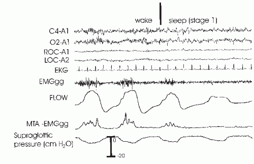
FIGURE 18-7 This tracing shows the onset of sleep with the coincident fall in genioglossus muscle activity and a fall in airflow with evidence of flow-limitation flattening. As the supraglottic pressure is similar but flow is lower, this means that upper-airway resistance has increased.

Patients with OSAHS tend to have higher than normal basal genioglossus activity (61,62) and a greater genioglossus response to negative airway pressure (63). The higher activity and response to negative pressure are believed to be a compensation for an intrinsically narrowed airway. In contrast, the response of the palate muscles to negative pressure may be impaired in OSAHS patients (64). Despite evidence for higher upper-airway muscle activity during wakefulness, the upper airways of OSAHS patients are still more collapsible than those of normal persons (65). At sleep onset, some studies have suggested that OSAHS patients have a greater than normal fall in upperairway muscle activity (66). In any case, the upper-airway activity is not sufficient to maintain an open upper airway. In Figure 18-3, a fall in genioglossus activity as the patient returns to sleep is associated with an obstructive apnea. Posture also has important effects on airway patency (67,68), and some patients with OSAHS have apnea or hypopnea only in the supine position (postural OSAHS).

During upper-airway obstruction, phasic genioglossus muscle activity increases proportionally to esophageal pressure deflections (ventilatory drive) (see Fig. 18-3) (4,69). At apnea termination, both genioglossus and palatal muscle activities are preferentially augmented (4,69,70)
and the upper airway opens. While it was once assumed that increasing genioglossus activity during obstructive apnea or hypopnea was driven by hypoxia and hypercapnia, a study of the effect of upper-airway local anesthesia suggests that mechanoreceptor stimulation (from increasingly negative pressure below the site of airway closure) is responsible for a large proportion of the augmentation (69). Traditionally a concept of a balance between negative inspiratory pressure tending to collapse the airway and upper-airway muscle dilating forces was assumed to determine the state of the airway. However, more recently the concept of passive collapse at sleep onset has gained favor. In fact, at sleep onset, the ventilatory drive decreases and supraglottic pressure actually may initially decrease (less negative) in some patients (although resistance increases). Upper-airway closure has also been documented during central apnea where there is no inspiration or negative collapsing forces (71). Therefore, suction pressure during obstruction may help keep the airway closed but is not necessary for the onset of airway occlusion.

Upper-airway volume also has a dependence on lung volume, with decreasing airway size as lung volume decreases (72,73). The lung volume dependence may be greater in patients with OSAHS (72). The lung volume dependence of the upper airway may be mediated via passive distending forces due to a downward tension on upper-airway structures during inspiration (tracheal tug) (74). Another way of thinking of the tracheal tug is a decrease in extramural pressure surrounding the airway (46). Any fall in end-expiratory volume or tidal volume would then reduce upper-airway size. Functional residual capacity (FRC) is known to decrease during sleep (75), and this would tend to predispose to airway closure. Morrell and coworkers (76) demonstrated a progressive fall in end-expiratory retropalatal cross-sectional area as well as end-expiratory lung volume in the breaths leading up to obstructive apnea.

Ventilatory instability generated by arousal and hyperventilation postapnea may also predispose to subsequent upper-airway closure and help perpetuate repetitive cycles of respiration and apnea. If the patient falls asleep rapidly after arousal, the arterial PCO2 may be near or below the apneic threshold, the level of PCO2 below which ventilation is no longer triggered during sleep (77). If the PCO2 falls below the apneic threshold, a central apnea may occur, followed by an obstructive apnea when ventilatory drive returns (mixed apnea) (78). Alternatively, a hypopnea or obstructive apnea may occur as ventilatory drive and upper-airway muscle activity fall (79). Of note, obstructive apnea may occur before the nadir in inspiratory effort is reached. If one monitors esophageal pressure in patients with OSAHS, the nadir in deflections in some patients can occur two or three breaths into the apnea (Fig. 18-8) (5,13).


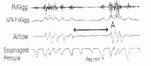
FIGURE 18-8 An obstructive apnea is shown with the raw genioglossus EMG (EMGgg) and the moving time average of the genioglossus EMG (MTA-EMGgg) as well as airflow and esophageal pressure. Note that the genioglossus EMG fall at airflow limitation (flattening) is noted on the last breath before apnea. At arousal (A), there is a large increase in genioglossus EMG. Note also that both EMGgg and the esophageal pressure deflections fall at apnea onset. In this case, the nadir in esophageal deflections (per minute) occurs on the second breath of apnea.

During REM sleep, ventilation is irregular, even in normal persons with the greatest irregularity during periods of phasic eye movements (80). As periods of REM sleep are longest and the REM density (number of eye movements per time) is the highest during the early morning hours, it is not surprising that this is the time of the greatest changes in ventilation during sleep. Although the diaphragm is not affected by the generalized muscle hypotonia of REM sleep, periodic decrements in diaphragmatic activity are associated with periods of reduced tidal volume. Upper-airway muscles are also affected during REM sleep. In normal persons, during REM sleep, genioglossus tonic activity is reduced but phasic activity can still be detected if intramuscular electromyography (EMG) electrodes are used (Fig. 18-9). During bursts of eye movements, both diaphragmatic and genioglossus phasic activity is often decreased (81). REM sleep without phasic eye movements is called “tonic REM” and with eye movements “phasic REM.” The latter is associated with more breathing irregularity. Many patients with OSAHS have a much higher AHI during REM sleep or have events only during REM (REM-related OSAHS). However, two studies have found no greater collapsibility of the upper airway during REM than NREM sleep (67,68). This variance with clinical experience could be due to either the nonhomogeneous nature of REM or the fact that drops in diaphragm activity are also important for inducing apnea and hypopnea.


Mechanisms of Apnea Termination and Arousal

Obstructive apnea or hypopnea termination is believed to depend on arousal mechanisms. During obstructive apnea in NREM sleep, upper-airway muscle activity increases, as does inspiratory effort. Airway opening
does not occur until there is a preferential increase in upper-airway muscle activity (4). This is often associated with signs of cortical arousal, although the electroencephalography (EEG) changes may not meet AASM criteria (16), for example, the sudden onset of delta activity A recent study suggested that arousal can be detected at the termination of the majority of respiratory events if frontal electroencephalographs, as well as central EEG, is monitored (82). If cortical arousal does not occur, there is still believed to be a “state” change in the brainstem—socalled subcortical or autonomic arousals. The term autonomic arousal is used because an abrupt change in heart rate or blood pressure can be detected at apnea termination, even if cortical arousal is absent. While hypercapnia and hypoxia drive the increase in respiratory effort, the level of effort rather than individual values of hypoxia or hypercapnia seems to trigger arousal (13,83). Thus, the level of effort is an index of the combined arousal stimulus. Studies have suggested that information from upperairway mechanoreceptors may contribute to the arousal stimulus (84,85). In NREM sleep, arousal appears to occur when inspiratory effort reaches an “arousal threshold.” Normal subjects tend to arouse during mask occlusion when suction pressure reaches 20- to 40-cm H2O. In contrast, many patients with OSAHS arouse only after pressure reaches -60 to -80 cm H2O (13). The increased arousal threshold in OSAHS patients is probably due, in part, to chronic sleep deprivation or hypoxemia. Withdrawal of CPAP for even three nights has been shown to increase the arousal threshold in OSAHS. However, chronic CPAP treatment does not restore the arousal threshold to normal (86). Patients with OSAHS could have an intrinsically increased respiratory arousal threshold. Alternatively there could be damage to mechanoreceptors from years of snoring or to chemoreceptors from repetitive nightly stimulation. A study of respiratoryrelated evoked potentials (RREP) in patients with mild OSAHS suggested that there is a sleep-specific blunted cortical response to inspiratory occlusion (87). At least in these milder patients, there was no evidence of impaired mechanoreceptor function as the respiratory-related evoked potential was normal during wakefulness. One study found that the prolongation in event duration that occurs overnight in patients with OSAHS is secondary to a blunting of the cortical response as the level of inspiratory effort at apnea termination increased during the night (88). Another study found that the within-night variation in the arousal threshold followed the cycles of NREM sleep (89) with a higher arousal threshold associated with higher EEG delta power (deeper sleep).


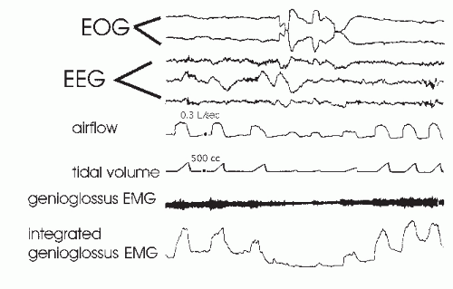
FIGURE 18-9 This tracing shows the fall in integrated genioglossus EMG activity and tidal volume during a burst of eye movements during REM sleep in a normal individual. The tracing illustrates that REM sleep is not homogeneous with respect to the effects on ventilation or upper-airway muscle control. (From Wiegand L, Zwillich CW, Wiegand D, et al. Changes in upper-airway muscle activation and ventilation during phasic REM sleep in normal men. J Appl Physiol. 1991;71:488-497, with permission.)

Only gold members can continue reading. Log In or Register to continue

Stay updated, free articles. Join our Telegram channel

Jul 14, 2016 | Posted by in PSYCHIATRY | Comments Off on Obstructive Sleep Apnea Hypopnea Syndromes: Definitions, Epidemiology, Diagnosis, and Consequences

Full access? Get Clinical Tree

Get Clinical Tree app for offline access