♦ Preoperative
Imaging
- Magnetic resonance imaging to assess need for neural element decompression (transoral odontoidectomy, enlargement of foramen magnum, C1 or subaxial laminectomies)
- Computed tomography with thin-cut reconstructions for bone depth and screw lengths, relationship of vertebral artery to C2 pedicle
Preoperative Care
- Patients with traumatic occipitocervical dislocation should be placed in halo.
- Patients with basilar invagination are frequently admitted before surgery and placed in traction to determine if odontoidectomy needed.
Equipment
- Occipital screw set and connector to secure rods to cervical construct, standard lateral mass system of choice, or
- Threaded Steinmann pin, double Songer cable (DePuy Spine Inc., Raynham, MA) for sublaminar wires, single Songer cable for occiput, BendMeister Stein-mann pin bender (Sofamor Danek, Memphis, TN)
- Halo adapter for Mayfield head holder and halo vest removal tools
Operating Room Set-up
- Somatosensory and motor evoked potential monitoring (optional)
- Fluoroscopy
Positioning
- Regular bed with rolls with head in Mayfield holder or Jackson table (Mizuho OSI) and foam or horseshoe headrest with traction
- If in halo, turn prone in halo vest, fix head to Mayfield with halo adapter, then remove the posterior part of the vest and posts.
- Head must be in neutral position; confirm with lateral fluoroscopy.
♦ Intraoperative
Exposure
- Subperiosteal exposure from inion to each lamina and lateral mass
- Decompression of neural elements if necessary
Screw-Rod Technique
- Place C1 lateral mass, C2 pedicle or pars screws, subaxial lateral mass screws as indicated
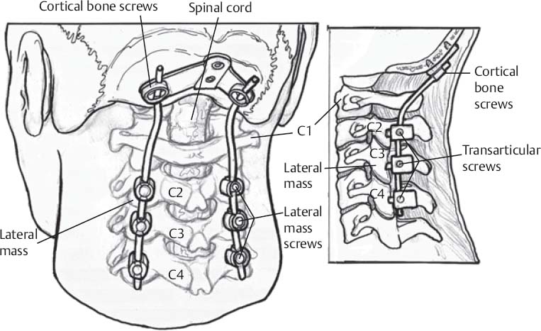
Placement of Occipital Screws (Fig. 91.1)
- Determine where the rods and connector will sit on the occipital bone.
- Mark the entry points for the screws in the occipital bone through the holes in the connector.
- Drill the pilot holes with Midas Rex AM-8 bit, then use a hand drill to the predetermined depth (usually 8 to 12 mm).
- Place appropriate sized screw.
Secure the Rods to the Screw Heads
- Torque-limited final tightener
- Consider cross-link for multiple subaxial levels.
Bone Graft
- Decorticate lateral masses, C1–C2 facet joint, and occiput.
- Lay cancellous iliac crest autograft or autograft over lateral masses and occiput, pack into facet joints
< div class='tao-gold-member'>
Only gold members can continue reading. Log In or Register to continue
Stay updated, free articles. Join our Telegram channel

Full access? Get Clinical Tree


