24 | Odontoid Fracture Nonunion |
 | Case Presentation |
History and Physical Examination
A healthy 26-year-old woman presented with long-standing neck pain and base-of-skull headaches. Her history was notable for a motor vehicle accident 10 years prior. She was told at that time she had a fracture in her neck and was treated with a cervical collar for 10 weeks. Eight years later she developed intermittent neck pain. It worsened over the most recent 2 years. Her current circumstance is one of consistent, often severe, lancinating pain, worse with movement of her neck, especially with flexion and extension. She also reported intermittent, radiating pain to her right shoulder.
On physical exam, the patient was a healthful young woman who weighed 125 pounds on a 5-foot, 5-in. frame. She had limited neck range of motion with flexion, extension, and lateral rotation due to pain. Motor, sensory, and reflex testing in the upper and lower extremities were normal.
Radiological Findings
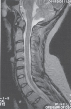
The patient presented with a contemporary magnetic resonance imaging (MRI) scan of the cervical spine and flexion and extension lateral cervical spine radiographs. These imaging studies revealed an old type II odontoid fracture with nonunion. The dynamic radiographs revealed an unstable fracture with 5 mm of posterior displacement of C1 on C2 with neck extension. MRI revealed a fibrous nonunion without evidence of acute injury (Fig. 24–1). There was no associated pannus or mass. The cervical spinal cord was normal.
Diagnosis
The patient was given a diagnosis of odontoid type II fracture nonunion with instability, and operative treatment was recommended.
 | Background |
Prior to discussion of the surgical treatment options for odontoid fracture nonunion, it is necessary to discuss the pathology and natural history of odontoid fractures and to review factors that may predispose patients with acute traumatic odontoid fractures to nonunion.
Odontoid fractures represent between 10 and 15% of cervical spine fractures and 60% of all acute traumatic axis fractures.1 The most common mechanism of injury is forcible flexion with anterior displacement of C1 on C2. Odontoid fractures are classified under the system of Anderson and D’Alonzo.2

Figure 24–1 T2-weighted magnetic resonance imaging study showing evidence of an old type II odontoid fracture with fibrous nonunion. There is no evidence of acute injury.
Type I odontoid fractures result from avulsion of the tip of the dens above the transverse ligament. They are very rare and, although considered to be stable fracture injuries, they are typically due to distraction injury and may be an indication of atlanto-occipital instability. In the presence of a type I odontoid fracture, an atlanto-occipital dislocation injury must be considered and carefully ruled out. Type II odontoid fractures are by far the most common of the odontoid fractures and consist of a fracture across the base of the dens. Type II fractures also include a further subtype, IIA, which consists of a comminuted base-of-dens fracture associated with free bone fragments at the fracture site. These rare and unstable fracture injuries are thought to account for roughly 5% of type II fractures.3 Type III odontoid fractures are, in essence, fractures of the dens that extend into the body of the axis.
Initial treatment options for odontoid fractures vary from cervical collar immobilization to operative reduction with internal fixation and fusion. Rates of nonunion of odontoid fractures vary depending on the type of dens fracture. Type I fractures in isolation are very rare; therefore, analysis of optimal treatment and nonunion rates is difficult. Available data, however, suggest that type I fractures will heal with immobilization alone in close to 100% of cases.2,4,5 The optimal treatment of type II dens fractures is more controversial. Several investigators have tried to accurately predict rates of nonunion and the factors associated with failure of the dens fracture to heal. Widely different rates of nonunion for type II dens fractures (5 to 60%) have been reported with external immobilization alone.6–9 Data from some of the better designed studies of odontoid fractures suggest that the rate of nonunion for type II odontoid fractures is roughly 40% for patients treated in a cervical collar and between 10 and 30% for those treated with halo vest immobilization.6–10 Two important trends are noted in the literature on this subject. Both patient age and the degree of initial fracture displacement appear to have a large effect on rates of fusion using immobilization alone.7,11–15
Class II medical evidence suggests that patients who have a type II dens fracture injury and are 60 years of age or older have a 21-fold higher incidence of failure of healing with an external immobilization device. Type II odontoid fractures in patients 60 years of age or older should be considered for early surgery for internal fixation and fusion.14
Type III odontoid fractures have an excellent fusion success rate with external immobilization alone. Successful fusion is achieved in over 90% of patients if maintained in external immobilization for 8 to 14 weeks.10,13 The halo vest immobilization device appears to be the most successful orthosis for type II and type III odontoid fracture injuries.16
 | Methods of Surgical Management |
The management of odontoid type II and III fracture nonunion is internal fixation and fusion. As in our example case, patients present with neck pain from C1-2 instability. Rarely physicians will see patients with myelopathy secondary to high cervical spinal instability with cord compression.16 Patients with evidence of C1-2 instability due to odontoid nonunion should be treated with operative internal fixation and fusion. Observation and radiographic follow-up may be acceptable in older patients who are at a high risk for surgical complications, or the rare patient who is asymptomatic without evidence of C1-2 instability on dynamic lateral cervical spinal radiographs.
Once the decision to proceed with surgical treatment has been made, the surgeon must decide the best operative approach for individual patient pathology. Procedures for internal fixation of odontoid fractures fall into two main categories: anterior and posterior procedures.
Anterior odontoid screw fixation of an unstable dens to the C2 body is well established as an effective treatment for acute odontoid type II and III fractures. However, the usefulness of this technique for older fractures or in the treatment of nonunion is highly suspect. Apfelbaum et al compared fusion rates for anterior odontoid screw fixation for both acute and remote fractures.17 They found that, although fusion rates for odontoid screw fixation approached 90% in the treatment of acute dens fracture injuries, odontoid fractures 18 months and older had a fusion rate of only 28%. The older or less acute the dens fracture, the more likely that the vascularity of the fracture edges has been lost and that the gap in the fracture line has been bridged by fibrinous granulation material. The chance for ossification and therefore long-term union to occur across this gap is remote even with subsequent, solid odontoid screw fixation.
Posterior approaches offer the best option for successful treatment of odontoid fracture nonunion. It is important to remember that the goal of treatment is to address and eliminate instability at the C1-2 level. Therefore, although the fracture site itself is located anteriorly at the C2-dens junction, fusion of the C1-2 articular surfaces, facets, and/or the posterior arch elements will still achieve the desired goal of atlantoaxial stabilization.
Posterior operative techniques for C1-2 fusion have long been employed with good results. The treatment of type II and type III odontoid fractures with posterior instrumentation and fusion is associated with reported fusion rates of 87% and 100%, respectively.4,5,12,18,19 Maiman and Larson reported that the fusion rate at the fracture site itself was only 35%, but approached 100% at the posterior operative fusion site.20 This illustrates why the posterior approach is superior for cases of odontoid fracture nonunion. The surgeon does not have to rely on healing across a fracture site with little hope of bony fusion to achieve the goal of C1-2 stabilization with fusion.
Historically, posterior C1-2 internal fixation and fusion was accomplished using a wire/cable construct with interspinous or intralaminar fusion followed by immobilization in a rigid orthosis. Variations of this procedure consist of sublaminar C1-2 wires or cables, sublaminar C1 cables around the spinous process of C2, or laminar clamps over C1 and C2 with autologous bone graft placed under the lamina of C1 and superior to the spinous process and lamina of C2. Effective stabilization procedures and more recent innovations in instrumentation and techniques have led to biomechanically superior C1-2 internal fixation procedures. The use of C1-2 transarticular fixation with intralaminar C1-2 fusion, or, more recently, C1 lateral mass screws secured by rods to pedicle screws in the axis, have proven to be effective in the treatment of odontoid fracture in both the acute and remote settings.21,22 Although associated with higher C1-2 fusion rates without the need for rigid external immobilization, these newer techniques are technically more challenging and surgical morbidity and mortality rates are higher, reportedly between 2 and 4%.23 Major complications include vertebral artery injury, stroke, and new neurological deficits.
 | Authors’ Preferred Method of Surgical Management |
In our example case, we chose to offer the patient C1-2 transarticular screw fixation. We chose the posterior approach because of its proven effectiveness in the treatment of remote dens fractures. In our opinion, the two best methods of posterior C1-2 internal fixation and fusion currently available are transarticular fixation and C1-2 fusion using lateral mass screws at C1 and pedicle screws at C2. Either of these procedures would have been appropriate in this case. We chose the transarticular approach based on the rigid fixation it provides; our familiarity, experience, and comfort with the techniques; and the fact that our patient’s C1-2 subluxation would reduce to anatomical alignment.
In addition to the preoperative MRI and plain films, we obtained a computed tomographic (CT) scan through the upper cervical spine. This was to ascertain the location of the transverse foramen of C1 and at C2, and the course of the vertebral arteries. Even in the best of situations, transarticular screws pass very close to the C1 and C2 transverse foramen, and any aberrance in the location of the foramen or course of the vertebral artery must be known in advance. If transarticular screw fixation is deemed too risky due to an aberrant or unusually large vertebral artery or an unusually medial C1 or C2 transverse foramen, or if the C1-2 subluxation cannot be completely reduced, we then opt to proceed with C1 lateral mass screws coupled to a rigid rod to pars screws in C2.
In this case, the patient’s anatomy was favorable (Fig. 24–2) and we proceeded with atlantoaxial transarticular screw fixation. This was combined with a sublaminar C1 to C2 spinous process cable, intralaminar fusion construct utilizing autologous iliac crest bone graft substrate. In this case, due to the patient’s young age and nonsmoking status, fusion adjuncts such as rh-bone morphogenetic protein or demineralized bone matrix were not used. In geriatric patients or smokers, however, they may enhance fusion success rates.
Pearls
We position the patient prone on the operating room table on chest rolls and affix the patient’s head to the operating room table with Mayfield pins. We accomplish a military, chin-tucked position, avoiding extremes of flexion or extension of the neck, Intraoperative fluoroscopy helps to confirm position and alignment of C1 and C2.
We expose the dorsal arches of C1 and C2 and identify the C2-3 facet capsule. We dissect around the arch of C2 to identify the medial portion of the pars bilaterally. This is accomplished easily with little epidural bleeding.
The insertion site for transarticular screws is marked, 2 mm superior and 3 mm lateral to the medial portion of the C2-3 facet capsule. A small shallow drill hole is made bilaterally to break the cortex. A threaded K-wire is then drilled through this entry site, up the C2 pars across the C1-2 facet capsule, angled medially 5 to 7 degrees and superiorly toward the cephalad portion of the anterior arch of the atlas.
Anatomical realignment of C1 and C2 is crucial to accomplish internal fixation and fusion in this way and to avoid vertebral artery injury. Occasionally we will pass a braided cable sublaminar at C1 to pull the atlas dorsal, and/or will push down on C2 to force the axis more ventral into perfect anatomical position when drilling or inserting transarticular screws.
The K-wire is left in the appropriate position and a drill is used to overdrill the pilot wire across the C1-2 articular surfaces. A threaded cannulated screw of measured length is passed over the K-wire to appropriate depth and achieving dorsal cortical purchase. This is accomplished bilaterally.
Fusion at C1-2 is accomplished by decortication of the dorsal aspect of the C1-2 facet capsule and by placing autologous iliac crest in the intralaminar space between inferior C1 and superior C2. Great care must be used to decorticate the dorsal and inferior surfaces of the arch of C1 and the superior surfaces of the arch and spinous process of C2, and to fashion the autologous graft to fit precisely between. It must be strapped or secured in place by C1-2 sublaminar braided cables, with care to avoid ventral displacement of the bone fusion substrate toward the thecal sac and underlying spinal cord.


