Fracture Type
Fracture type and the amount of displacement are important factors in selecting a patient for surgery.
Type II and “shallow” or rostral type III fractures according to the classification of Anderson and D’Alonzo are an indication for odontoid screw fixation [4]. Apfelbaum et al. have suggested a subclassification of type II fractures in three types according to the orientation of the fracture line in one of their series. According to this subclassification horizontal as well as oblique antero-superiorly oriented fracture lines on the sagittal plane predict higher fusion rates than oblique antero-inferiorly oriented fracture lines. This subdivision has however not been reported in series by other authors and given that the overall fusion rate in the oblique antero-inferior group is still 75 % this information is probably not indispensable, but can be of help in difficult decisions [11].
In addition to the above criteria the axis needs to be carefully examined for additional fractures or fracture lines extending into the body, as this could have negative repercussions on screw purchase. The same goes for the fractured dens fragment. Type IIA fractures (comminution of the dens) might not warrant sufficient screw purchase, which is particularly important at the cortical tip of the dens that needs to be engaged by the screw [8–12].
As far as the amount of displacement is concerned the literature shows that values over 4–6 mm seem to represent a threshold beyond which the risk of non-fusion gets significantly and probably inacceptably high with conservative treatment [13–15].
Finally, an MRI scan of the area to check for integrity of the transverse ligament is indicated even though not indispensable, if not available, in our opinion due to the relatively low association of ligamentous rupture in association with an odontoid fracture. However, due to the diffuse availability of the imaging technique and its routine use in spinal trauma in many centres, particular attention to the state of the transverse ligament should be paid. In case of a clear rupture this would represent a contraindication to odontoid screw fixation.
Timing of Surgery
The general rule “The earlier you fuse, the better” surely also applies to this type of procedure. Evidence from the literature seems to suggest that there are no differences in fusion rates during the first 6 months, whilst after 18 months fusion rates drop clearly. This information is based on a series published by Apfelbaum et al. and has initially led to a division of the patients in “early” and “late”. The fact that there is essentially no information on patients who had a fracture between 6 and 18 months is due to the lack of such patients in that study and to our knowledge no other study has examined this thereafter [10–12, 16–18].
Knowing, however, that it apparently does not make a difference whether a patient undergoes fusion immediately or after 6 months is surely valuable information, as it gives ample time to first try conservative treatment in those patients where deemed appropriate and still have the time to offer surgery should that treatment fail.
Age
Even though again a matter of controversy in the literature with a vast amount of related articles, there is a certain prevalence towards the opinion that age influences the outcome of odontoid screw fixation [11–29]. The younger the patients, the more likely it is that fusion may be obtained by external immobilization . Sherk et al have reported a series of 35 children (under 7 years), in whom only one failed to fuse after halo immobilization [27]. The more age advances, however, the more the fusion rate seems to correlate inversely and Lennarson et al. have even shown in one of their studies that the nonunion rate in patients over 50 years is 21 times higher than in those that are younger [18].
Even if one tends to not believe in the inverse relationship between age and fusion rate in conservative management , the important message surely seems to be that older patients do at least as well as younger patients with surgery and this can be an important piece of information when tailoring a specific treatment plan. In older patients the compliance with external immobilization like a Halo Jacket and the resulting overall movement restriction, can have significant negative repercussions on outcome and the patients general health.
We therefore tend to favour surgery in older patients, that is patients over age 50, taking only unfitness for general anesthaesia or severe osteopenia as contraindications.
A third age group we consider in selecting treatment is the geriatric population with patients over 75–80 years of age. In this age group pseudoarthrosis rate has been described to be as high as 85 % [30].
Recent papers based on the AOSpine North America Geriatric Odontoid Fracture Study showed an increase in mortality of non surgically treated patients at one year but non significant difference between the surgical and conservative groups after that. As the patients in this study were not randomized, the difference in the first year likely reflects the poorer general conditions of those that were not operated and could be the result of a selection bias. Other weak points of this study are that neurological status in the patients groups was not accounted for. Furthermore, a group of patients out of the same study that was followed after conservative treatment showed a “union rate”, whether fibrous or bony, of as high as 75 %. The remaining 25 % eventually developed “non union” following which around 2/3 underwent surgery. It is however not clear how this non union reflected itself clinically, in particular with regards to the neurological status. The only conclusion that is drawn is that operated patients demonstrated a significant benefit on the Neck Disability Index (NDI ) and the SF-36 version 2 Bodily Pain dimension. Again, this difference could reflect the difference in the general conditions of the two groups and thus be a selection bias [30, 31].
In our eyes this study failed to show convincing elements to favour surgery in the geriatric population and if anything showed an increased incidence of dysphagia and permanent feeding tube placements in the surgical group.
In addition there are papers that describe series of patients, even though small, followed after conservative treatment who did not develop any complications, as well as our own experience which seems to reproduce these findings, and this makes us withhold from surgery in the geriatric population , that is over 75–80 of age, unless the patient is in particularly good general conditions and most of all still very active [32, 33].
We tend to treat these patients with a cervical collar choosing the type, either rigid or soft, and the duration of treatment based on the level of activity of the patient, as well as on the range of movement of the fracture on dynamic x-rays.
Surgical Technique
The following is a description of the operative technique used in odontoid screw fixation outlining the most significant general steps that need to be followed. Various manufacturers offer different systems to carry out this operation and some specific steps might thus differ according to the specific system. It is not our intent to specifically describe the use of any of these systems but outline the important points that are necessary for a smooth and successful performance of the operation.
Patient Positioning and Setup
The patient is under general anesthaesia with endoscopic endotracheal intubation . This can be accomplished either via the nose or the mouth making sure that a non armoured tube is used. The patient is then positioned neutrally supine on the operating table. The head can either be secured in a three pin fixation device, rest on a horseshoe or directly on the operating table with a towel roll under the neck. What is essentially important is that the head remains immobile throughout the procedure.
Once the patient is thus positioned, two C-arm image intensifiers are brought in and centred on the C1-C2 complex, with one obtaining a lateral view and the other one obtaining an anteroposterior view (Fig 17.1).
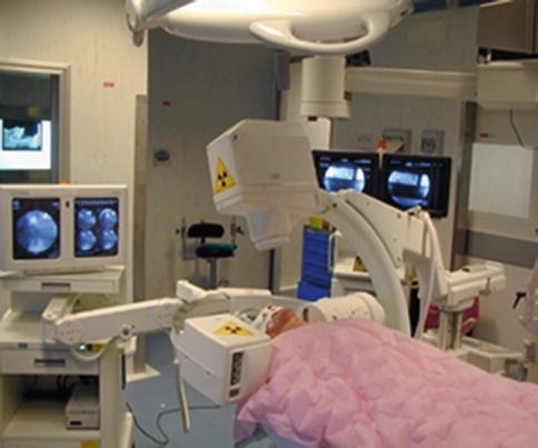
Fig. 17.1
Intraoperative photograph showing patient positioning for operation. Note the biplanar flouroscopy left in place throughout the entire procedure
In case of dislocated fractures, the patients head is now gently manipulated under lateral fluoroscopy in an attempt to reduce the fracture as much as possible, and the head is finally secured in the desired position. In repositioning the fracture direct manipulation by pressure on the pharynx through the mouth can be of help. The two C-arms will remain in position during the entire procedure, as frequent imaging is crucial during the various steps. In order to obtain a good anteroposterior view it is often helpful to obtain open mouth views by inserting a radiolucent mouth opener or bite-blocks in the patient’s mouth.
Once all these steps are accomplished, the C-arms are both in a position to obtain good views, the patients head in the correct position to obtain as much fracture reduction as possible and the head well secured, the patient and the equipment are draped in the usual fashion.
Related posts:


Stay updated, free articles. Join our Telegram channel

Full access? Get Clinical Tree


