Fig. 1
(a) Organotypic cortex-striatum-substantia nigra pars compacta culture. The white circles indicate the electrodes for which representative LFP traces are shown in (b). (b) Example of LFP activity for four cortical and striatal electrodes. Red dots indicate significant negative LFP peaks
Local Field Potential Analysis
Local field potential (LFP) activity was recorded at (or down-sampled to) 1 kHz, and subsequently band-pass filtered at 1–50 Hz. Negative LFP (nLFP) deflections were detected by finding the minimum value of the LFP signal that crossed a threshold of z = −4.5 standard deviations (SDs) (Fig. 1b). Spatiotemporal clusters were detected as in previous work [2].
Computational Model
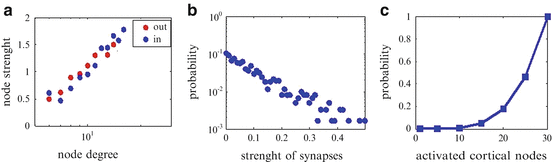
Our abstract model of the cortical network was designed in order to reproduce some of the main statistics observed in experimental data [10]. First, each of the N nodes was assigned a number that determined the out-degree of each of them. The average number of connectivity was chosen to be 10 [10], and afterwards we applied the preferential attachment rule. Each node was attached to other nodes in proportion to the out-degrees of those nodes in order to get the node degree linearly related to the average node strength for both in- and out-degrees (Fig. 2a). Transmission probabilities pij (from node j to node i) were picked from an exponential distribution (Fig. 2b). There were no self-connections and no more than one connection between attached pairs of nodes. The weights were then scaled (p ij ′) such that the branching parameter for the entire network was equal to 1. The probability that node i fired at time t + 1 was:
where J(t) was a set of nodes that fired at time t. Node i actually fired at time t + 1 only if piJ>ϵ, where ϵ was a random number from a uniform distribution on [0, 1]. Then, each of the N = 30 nodes in the striatum was randomly connected with a certain number (Nk = 4 here) of nodes in the cortex. A node in striatum always fired if the full pattern in cortex assigned to that node was present; otherwise it fired with a very low probability (Fig. 2c). In a first approach, we focus on the contribution of the corticostriatal pathway to explain the experimental results while ignoring intrastriatal inhibition. Our future work will explore the role of the intrastriatal GABAergic network.
(1)
Fig. 2
(a) Node degree is linearly related to the average node strength for both in and out degrees. (b) Plot of link weight probability. (c) Probability of activation for striatal nodes depending on the cortical activity
3 Results
We recorded spontaneous LFP activity in organotypic cortex-substantia nigra cultures simultaneously from 31 electrodes in the cortex to 28 electrodes in the striatum [12, 13]. Negative LFPs were detected by applying a negative threshold to each electrode. Analyses of the cluster size distributions in the striatum pointed to a more negative exponent than the one obtained for the cortex [2, 13] (Fig. 3). The steeper slope of the striatal cluster size distributions indicates reduced spatiotemporal correlations.
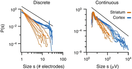
Fig. 3
Experimentally measured discrete (left panel) and continuous (right panel) cluster size distributions for cortex and striatum. Black line indicates a power law with α = −1.5 for comparison
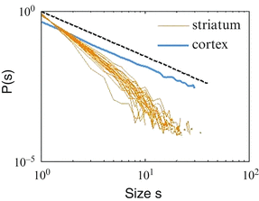
In order to investigate how the observed striatal dynamics could be explained by the corticostriatal connectivity, we developed an abstract computational model (see section “Method”). We stimulated population events by randomly choosing and triggering a single node (this procedure was repeated 30,000 times for each trial) in the cortex, while simultaneously monitoring the resulting activity in the striatum. When we gave to each striatal node the probability of activation presented in Fig. 2c, we got a steeper slope of the striatal cluster size distribution, similar to experimental results (Fig. 4).
 Integrated Neuropsychiatric Assessment System: A Generic Platform for Cognitive Neurodynamics Research
 Dynamic Temporal-Topological Structure of Brain Network Within ADHD
 Activity Patterns in Cortical Dynamics and the Illusion of Localized Representations
 State Functions Characterizing the Acquisition of New Motor and Cognitive Skills
 Interactions of Two Individual Arm Robots Using Independent Chaos in Recurrent Neural Networks
 as Bifurcations Shaped Through Sequential Learning


Related posts:
Integrated Neuropsychiatric Assessment System: A Generic Platform for Cognitive Neurodynamics Research
Dynamic Temporal-Topological Structure of Brain Network Within ADHD
Activity Patterns in Cortical Dynamics and the Illusion of Localized Representations
State Functions Characterizing the Acquisition of New Motor and Cognitive Skills
Interactions of Two Individual Arm Robots Using Independent Chaos in Recurrent Neural Networks
Stay updated, free articles. Join our Telegram channel
Full access? Get Clinical Tree
