Comparison of Positional Therapy to CPAP Therapy
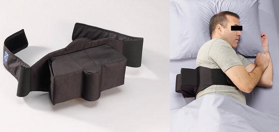
As of this point in time, there have only been three prospective studies that have compared positional therapy to CPAP therapy in patients with positional OSA (Table 1) [36–38]. Jokic et al. [36] in a randomized crossover study of 13 patients with mild-to-moderate OSA (AHI 17 ± 8 events/h) compared positional therapy using their soft ball in a backpack device to CPAP therapy after 2 weeks of using each treatment modality. Positional OSA was defined as an AHI during supine sleep that was two or more times the AHI during sleep in the lateral position. In addition, the AHI in the lateral position had to be <15 event/h, during a minimum duration of 1 h of sleep in the lateral position and the inclusion of at least 1 rapid eye movement (REM) period. In a cross-over designed study, Skinner et al. [37] compared their TASB to CPAP therapy in 22 patients with mild to moderately severe positional OSA (AHI 22.7 ± 12 events/h) after utilizing each treatment modality for 1 month. Positional OSA was defined as an AHI in the supine position that was greater or equal to twice the AHI in other positions. Permut et al. [38] compared the Zzoma Positional Device to CPAP therapy in a crossover designed study after 1 night of use in 38 patients with mild-to-moderate positional OSA (AHI of 13 ± 5 events/h). Positional OSA was defined on the baseline study as an overall apnea–hypopnea index (AHI) of ≥5 events/h with symptoms of excessive daytime sleepiness or an AHI of ≥15 events/h, with a 50 % decrease in the AHI in the non-supine position as compared to the supine position. Additionally, the AHI had to fall to <5 events/h in the non-supine position, and the patient must have slept in the lateral position for a minimum of 1 h during the baseline study.
Table 1
Comparison of positional therapy to CPAP therapy
Study | N | Length of study | Baseline AHI (events/h) | Effects on AHI | Effects on nocturnal oxygenation | Effects on sleep quality |
|---|---|---|---|---|---|---|
Jokic et al. [36] | 13 | 2 weeks | 18 ± 5 | CPAP and PD decreased AHI—but lower with CPAP | Lowest SaO2 lower with PD | No difference in SE and TST |
Skinner et al. [37] | 22 | 1 month | 23 ± 12 | 72 % with PD and 89 % with CPAP had an AHI < 10 events/h | Both PD and CPAP increased the mean SaO2 | No difference in SE and TST |
Permut et al. [38] | 38 | 1 night | 13 ± 5 | 92 % with PD and 97 % with CPAP normalized AHI to <5 events/h | No change in mean SaO2 with PD and increase with CPAP | No difference in SE but TST lower with CPAP |
Sleep-Disordered Breathing
Many would consider the AHI as the most important parameter to be evaluated in regard to assessing the effectiveness of treatment in patient with OSA. That is also the case in regard to studies that have assessed patients with known positional OSA, including those that have compared positional therapy to CPAP therapy. Jokic et al. [36] noted that although 2 weeks of treatment with both their positional device and CPAP decreased the AHI (from 18 to 10 and 3 events/h, respectively), the decrease with CPAP was statistically more significant and was associated with a normalization of the AHI (<5 events/h). Only 3 of the 13 patients (23 %) slept supine during their study night. Compliance as it relates to use of the positional device and CPAP was not assessed during the study. Skinner et al. [37] noted that both the TASB and CPAP decreased the AHI, from 23 to 12 and 5 events/h, respectively, with a significant difference noted between the two forms of therapy. Using a definition of successful treatment as an AHI of <10 events/h, treatment success was noted in 13/18 subjects using the TASB and 16/18 subjects using CPAP therapy. Supine sleep was significantly decreased but not completely eliminated with the TASB, with 6 % of the total sleep time spent in the supine position. Adherence with the TASB was based on a self-recorded diary, with a significantly higher adherence rate reported for TASB as compared to CPAP therapy. Permut et al. [38] noted that when compared to baseline, both the Zzoma Positional Device and CPAP therapy (mean 10 ± 3 cm H2O) significantly decreased the AHI, from 11 (9–15, 6–26) events/h to 2 (1–4, 0–8) and 0 (0–2, 0–7) events/h, respectively (p < 0.001), with a difference between the two treatments (p < 0.001). In addition, the Zzoma Positional Device was equivalent to CPAP (92 % vs. 97 %, respectively [p = 0.16]) at normalizing the AHI. The Zzoma Positional Device eliminated supine sleep in 37 of the 38 patients, with only a mean of 1 ± 4 % of total sleep time spent supine. However, the study was only an acute single night intervention, and more long-term results are being examined.
Nocturnal Oxygenation
Jokic et al. [36] noted no difference in mean SaO2 between their positional device and CPAP therapy. However, the lowest SaO2 during the night was lower with positional therapy as compared to CPAP. Skinner et al. [37] noted a clinically insignificant difference in mean SaO2 during the night between CPAP and the TASB. In comparison, Permut et al. [38] noted the mean SaO2 during the night was unchanged compared to baseline with the use of the Zzoma Positional Device, but was increased with CPAP therapy. In addition, there was an increase in the lowest SaO2 during the night with both the Zzoma Positional Device and CPAP therapy, with no difference between the two treatment modalities. The percent of total sleep time with a SaO2 < 90 % was significantly decreased compared to baseline with the Zzoma Positional Device and CPAP therapy.
Sleep Quality
Jokic et al. [36] demonstrated no difference in sleep quality between their positional device and CPAP therapy, as measured by total sleep time and sleep efficiency. In addition, the arousal index and sleep architecture were not different between the two treatment modalities. Similar results were noted by Skinner et al. [37] when comparing the TASB to CPAP therapy, with no change in the total sleep time with either form of therapy as compared to baseline. In comparison, Permut el al. [38] noted that when compared to baseline, total sleep time did not change with the Zzoma Positional Device, but decreased with CPAP therapy. There was no change in sleep efficiency noted with either treatment, nor was there any change in the spontaneous arousal index. The sleep architecture, expressed as a percentage of total sleep time, including stage N3 and REM sleep, was not different as compared to baseline for either the Zzoma Positional Device or CPAP therapy.
Other Parameters
In regard to daytime sleepiness, Jokic et al. [36] noted a decrease in the Epworth Sleepiness Scale (ESS) after 2 weeks of therapy with both their positional device and CPAP therapy, but with no difference between the treatment modalities. In addition, similar sleep onset latencies were seen with both treatments as measured on maintenance of wakefulness (MWT) tests. Skinner et al. [37] noted a nonsignificant decrease in ESS with both their TASB and CPAP therapy at the end of 1 month.
Cognitive performance and quality of life changes have also been compared between positional therapy and CPAP therapy. Jokic et al. [36] found no difference in regard to these parameters between the two forms of therapy, and patient preference favored CPAP therapy in this study. Skinner et al. [37] noted no significant difference in any of the quality of life measures that they assessed when the results of their TASB and CPAP therapy were compared. Permut et al. [38] noted that 50 % of their patients preferred the Zzoma Positional Device, 34 % preferred CPAP therapy, and 16 % had no preference.
Future Research
While CPAP compliance has been objectively evaluated in a number of studies [6, 30, 31], there are few studies that have evaluated positional therapy adherence and compliance, with most involving self-reported use or mailed questionnaires [9, 10, 37]. While some of these studies have suggested poor long-term use of positional therapy [9, 10], a more recent prospective study has reported a 3-month compliance rate of 74 % using actigraphy [39]. While this study suggests possible compliance rates that are better than those reported for CPAP therapy, it was uncontrolled. Similar types of studies need to be performed when positional therapy is directly compared to CPAP therapy using methods that allow objective assessment of use.
In addition to compliance, measurement of continued effectiveness should be better evaluated with the use of positional therapy. While some studies have repeated polysomnograms after 1–3 months of using a positional device to demonstrate the device retains its ability to decrease or eliminate supine sleep, studies evaluating continuous nightly effectiveness have not been performed.
Finally, while CPAP therapy has been shown to decrease parameters associated with cardiovascular risk in patients with OSA, the effects of positional therapy on cardiovascular risk are presently unknown [40–44]. CPAP has been demonstrated to decrease endothelial dysfunction as measured by flow-mediated dilation and carotid intima–media thickness measurements [40–42]. In addition, CPAP therapy has been shown to decrease systemic inflammation as measured using biomarkers such as C-reactive protein [43, 44]. At the present time, it is not known whether positional therapy has a similar effect at decreasing cardiovascular risk.
Summary
A large percentage of patients with diagnosed OSA have positional OSA. While CPAP therapy is the most common form of therapy, compliance is poor, and other forms of therapy may be appropriate in these patients, including the use of effective positional therapy. Initial studies that directly compared positional therapy to CPAP therapy suggest that positional therapy can be considered as a primary therapy in patients with positional OSA. However, more long-term studies that use objective measurements of compliance and effectiveness should be performed. Whether positional therapy has the same beneficial effects on cardiovascular risk as seen with CPAP therapy awaits further study.
Related posts:
 of Sleep Position on Snoring
of Positional Therapy: Transition from Tennis Balls to New Devices
Position and Pregnancy
Results and Compliance of a Special Vest Preventing the Supine Position
and Effect of Supine-Dependent Obstructive Sleep Apnea on Oral Appliance Therapy
Impact of Body Weight Changes on Body Posture Dominance in Adult Obstructive Sleep Apnea Patients
of Sleep Position on Snoring
of Positional Therapy: Transition from Tennis Balls to New Devices
Position and Pregnancy
Results and Compliance of a Special Vest Preventing the Supine Position
and Effect of Supine-Dependent Obstructive Sleep Apnea on Oral Appliance Therapy
Impact of Body Weight Changes on Body Posture Dominance in Adult Obstructive Sleep Apnea Patients
Stay updated, free articles. Join our Telegram channel

Full access? Get Clinical Tree


