Omega-3 Fatty Acids in Mood Disorders: A Review of Neurobiologic and Clinical Actions


Omega-3 Fatty Acids in Mood Disorders: A Review of Neurobiologic and Clinical Actions


Andrew L. Stoll


INTRODUCTION TO OMEGA-3 FATTY ACIDS


Overview and Perspective

At the time of the first edition of this book, there existed only one double-blind, placebo-controlled trial of omega-3 fatty acids in bipolar disorder, and no controlled studies in unipolar major depression were yet published. Now, a mere 4 years later, the number of double-blind, placebo-controlled trials stand at 4 for bipolar disorder and 7 for unipolar major depression. Most, but not all of these clinical trials have reported that the omega-3 fatty acids were superior to placebo. These somewhat mixed results could perhaps have been expected from such a collection of heterogeneous and small studies, which investigated a complex group of multifactorial psychiatric disorders. This chapter provides an up-todate review of the basic and clinical research findings with regard to omega-3 fatty acids and mood disorders and attempt to reconcile the discrepant clinical trial findings.


Origin and Chemistry of the Omega-3 Fatty Acids

The omega-3 fatty acids, along with their counterparts, the omega-6 fatty acids, are a group of crucial naturally occurring lipids, which collectively are termed essential polyunsaturated fatty acids (PUFA) (1,2). The three predominant, naturally occurring omega-3 fatty acids are as follows: docosahexanoic acid (DHA), eicosapentanoic acid (EPA), and α-linolenic acid (ALA). The first two (DHA and EPA) are known as the long-chain omega-3 fatty acids, and are found primarily in fish oil and other marine sources (3). The shorter-chain ALA is an omega-3 fatty acid obtained from certain species of terrestrial plants (e.g., flaxseed, purslane, and others) (4,5). The chemical structures of the omega-3 fatty acids are shown in Figure 4.1.

Omega-6 fatty acids, such as the 18-carbon linoleic acid (LA) and the 20-carbon arachidonic acid (AA), are derived primarily from vegetable oils and are ubiquitous in the food supply of Western countries (1,6,7). AA and EPA are nearly identical chemically (differing only by the presence of the double bond in the 3-position in EPA and the consequent 2 additional hydrogen atoms in AA), yet have opposing actions on inflammatory processes in the body. Vertebrate animal species cannot desaturate (or add a double bond to) fatty acids before the ninth carbon atom from the lipophilic end of the carbon chain (8). Thus, the omega-3, the omega-6, and the omega-9 classes of fatty acids, with double bonds beginning at the third, sixth, and ninth carbon atoms, respectively, must be obtained from the diet (hence the term “essential”). The origin of the long-chain omega-3 fatty acids is photosynthesis within the chloroplasts of marine phytoplankton (9). The EPA and DHA are then passed through the food web, and ultimately to humans.


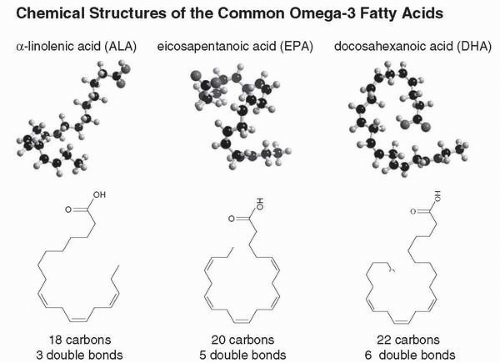
Figure 4.1 • Note the recurring double bonds at every third carbon atom. The presence of multiple double bonds in polyunsaturates, such as omega-3 fatty acids (n-3s) leads to a very kinked and flexible molecule. This flexibility permits increased molecular motion, which is why polyunsaturates (e.g., n-3 and n-6 oils) are liquid at room temperature and produce a more fluid cell membrane. Saturated fat, with no double bonds, is a straight and rigid molecule. Saturated fats are solids at room temperature and tend to produce less fluid cell membranes.


The major differences among the various omega-3 fatty acids are the length of the carbon chain and the number of double bonds (Fig. 4.1). DHA has a 22-carbon chain with six double bonds, EPA has a 20-carbon chain with five double bonds, and ALA has 18-carbon chain with three double bonds (1). In the omega-3 fatty acids, the double bonds recur every third carbon atom, and in vivo, humans can theoretically convert one omega-3 fatty acid into another. However, the rate-limiting step in the conversion process involves the enzyme Δ-6 desaturase (10), and the process of elongation is limited (10). Although controversial, the most recent data indicate that most people cannot adequately convert ALA into EPA and DHA to meet their nutritional needs (11, 12, 13). In one recent human study, less than 8% of ALA was converted into EPA and less than 0.5% of ALA was converted into DHA (12). Women convert more ALA into DHA than men, due to an estrogen-dependent up-regulation of hepatic DHA synthesis from EPA and ALA, particularly during pregnancy (14,15). Thus, it appears to be crucial for men to obtain EPA and DHA directly from marine or other sources. For women, it is not clear if the estrogen-mediated increase in DHA is adequate to meet the nutritional need for EPA and DHA, especially when not pregnant.


MEMBRANE EFFECTS OF THE OMEGA-3 FATTY ACIDS

Among the major biologic functions of the omega-3 fatty acids is their role in cell membranes. Omega-3 fatty acids, particularly DHA, are incorporated into the phospholipids that comprise the lipid bilayer surrounding every cell, as well as the organelles within cells (e.g., mitochondria and nuclear membranes) (1). The unique chemical structure of the omega-3 fatty acids is what produces their specific biologic effects within the membranes. The term polyunsaturated denotes the presence of two or more double bonds in the fatty acid molecule. The presence of multiple double bonds in the carbon chain produces a more highly folded and flexible molecule than those seen in more saturated fatty acids (Fig. 4.1) (16).

The inherent flexibility and other physical properties of PUFAs is why these oils are liquids at room temperature, in contrast to saturated fats, which are solids. This difference in melting point between the PUFAs and more saturated fatty acids explains why membranes with a high content of omega-3 fatty acids are more fluid at a given body temperature when compared with membranes containing high concentrations of saturated fatty acids (16). This difference in membrane fluidity is thought to have significant biologic consequences, particularly on the conformation and the activities of proteins intrinsic to the lipid membrane, such as neurotransmitter receptors and enzymes regulating signal transduction in neurons (17). However, it appears unlikely that the fluidizing effect on cell membranes is solely responsible for the clinical actions of omega-3 fatty acids in mood disorders. For example, omega-6 fatty acids are also polyunsaturates with fluidizing effects on cell membranes (18,19) yet appear to have no beneficial effects (or even deleterious effects) in mood disorders.


OMEGA-3 FATTY ACIDS AND SIGNAL TRANSDUCTION

Omega-3 fatty acid-containing phospholipids within the inner leaflet of the lipid bilayer appear to dampen abnormal intracellular signal transduction (20). They accomplish this by inhibiting the G-protein-mediated and phospholipase C-mediated hydrolysis of crucial membrane phospholipids, such as phosphatidylinositol (PI), into the second messenger molecules inositol triphosphate (IP3) and diacylglycerol (DAG) (20, 21, 22, 23). This effect has been
demonstrated in various cell types and tissues in the periphery (20,22), and it presumably occurs in brain as well. The established mood stabilizers, lithium and valproate also appear to inhibit different aspects of signal transduction related to the PI system (21,23). Thus, one possible mechanism for the apparent mood stabilizing and antidepressant action of the omega-3 fatty acids is this dampening effect on signal transduction.

Calcium is another crucial intracellular signaling agent, the concentration of which is tightly regulated by cells (20,24). Omega-3 fatty acids modulate the influx of calcium ions through the L-type calcium channel (25,26). This effect has been suggested to be the result of phospholipase A2-mediated hydrolysis of omega-3 fatty acid containing phospholipids into free omega-3 fatty acids. The free omega-3 fatty acids appear to interact with the L-type calcium channel, blocking the influx of calcium (25,26). L-type calcium channel blockers, such as verapamil and nimodipine, have been shown to have efficacy in most, but not all, studies in bipolar disorder (24,27). Thus, it is possible that omega-3 fatty acids exert their putative mood-stabilizing effect, in part, via this calcium flux inhibition.

Omega-3 fatty acids also directly reduce the activity of protein kinase C (PKC) in a number of different cell types and tissues (28, 29, 30). PKC can be considered a “third messenger” molecule, and is a ubiquitous enzyme, responsible for activating many crucial cellular responses (31). PKC inhibition is one of the several plausible mechanisms of action for valproate in bipolar disorder (21,23,32).


OMEGA-3 FATTY ACID ACTION IN IMMUNE AND INFLAMMATORY PATHWAYS

In 1991, Smith published his “Macrophage theory of depression” (33). Since that time, dozens of studies have examined various inflammatory pathways in patients with mood disorders. Most (34, 35, 36, 37, 38, 39, 40, 41), but not all (42,43), studies have observed abnormally excessive activity of inflammatory processes or elevated levels of inflammatory markers. In addition to the important regulatory role of omega-3 fatty acids in signal transduction and as a component of cell membranes, they are crucial to the normal functioning of inflammatory and immune pathways (44,45).

EPA, with its 20-carbon chain, is converted directly to the 20-carbon eicosanoids (1). Eicosanoid is an umbrella term for several classes of hormone-like cell-signaling molecules, including the prostaglandins, prostacyclins, thromboxanes, leukotrienes, and others. Eicosanoids are present in every organ system, including the central nervous system, and they help to regulate inflammatory and immune processes, in part, by modulating cytokine action (1,46).

Omega-6 fatty acids (particularly AA) generally are antagonistic to omega-3 (EPA) action in the immune and inflammatory systems (1). Evidence suggests that during human evolution, the dietary ratio of omega-6 to omega-3 fatty acids was somewhere between 1:1 and 4:1 (47, 48, 49, 50). Under these optimal conditions, the omega-6 fatty acid AA competes with the omega-3 fatty acid EPA to achieve balanced immune and inflammatory function. However, the widespread use of omega-6-containing vegetable oils and the dramatic decline in the consumption of foods with omega-3 fatty acids in the United States and other developed Western nations has resulted in a highly skewed ratio of omega-6 to omega-3 fatty acids (50, 51, 52, 53, 54). This overabundance of omega-6 fatty acids has shifted our eicosanoid-dependent inflammatory and immune pathways into a chronically activated, proinflammatory state. The various proposed mechanisms of action for the omega-3 fatty acids in mood disorders are summarized in Table 4.1.


TABLE 4.1 Potential Mechanisms of Action of Omega-3 Fatty Acids in Mood Disorders


Inhibition of signal transduction


EPA, acting in the phospholipid cell membrane’s inner leaflet, inhibits the action of the phosphatidylinositol-specific phospholipase C signal transduction pathway, reducing the generation of second messenger molecules, possibly in the serotonin system.


Suppression of eicosanoid-mediated inflammatory processes


EPA competes with arachidonic acid, its omega-6 counterpart, at multiple sites in the body. High amounts of EPA will reduce the synthesis of arachidonic acid-derived eicosanoids, thereby reducing the formation of specific proinflammatory cytokines. Certain cytokines (IL-2, IL-6, and others), as a component of an exaggerated inflammatory response, can induce depression through unknown mechanisms.


Increased membrane fluidity


DHA, and to a lesser extent EPA, are incorporated into the cell membrane, where they affect the physical structure and increase the fluidity of the membrane. This, in turn, leads to changes in the structure and function of various cell structures, particularly transmembrane proteins, such as neurotransmitter receptors and ion channels.


Modulation of ion channel activity


DHA, EPA, and alpha-linolenic acid all affect calcium, sodium, and potassium ion channels, which are crucial for regulating neuronal activity.


Modulation of receptor activity


EPA and various EPA-derived eicosanoids may directly affect mood via specific receptors in the brain.


Nuclear receptor effects


EPA, DHA, and eicosanoids can directly activate nuclear receptors, such as peroxisomal proliferator-activated receptors (PPARs), leading to changes in gene expression and energy balance, possibly affecting mood.


DHA, docosahexanoic acid; EPA, eicosapentanoic acid; IL, interleukin.


OMEGA-3 FATTY ACIDS IN BIPOLAR DISORDER


The Initial Double-Blind, Placebo-Controlled Trial

The authors’ group’s initial interest in omega-3 fatty acids for bipolar disorder was based on the similarity between the biochemical and pharmacologic actions of the omega-3 fatty acids and the established and putative mood stabilizers (21). The shared property of inhibition of specific signal transduction pathways was particularly striking, and our hypothesis that an omega-3 fatty acid formulation derived from fish oil would be an effective mood stabilizer was tested in a small pilot study (55). This 4-month, prospective, double-blind, parallel design, placebo-controlled trial compared high dose omega-3 fatty acids (6.2 g of EPA plus 3.4 g of DHA per day) to placebo (olive oil) in bipolar patients who had experienced a recent manic or hypomanic episode (55). This relatively high daily dose was chosen to match the effective dosage of omega-3 fatty acids in previously reported controlled studies for rheumatoid arthritis and other medical disorders (56, 57, 58). Other methodologic details are described in Table 4.2.


TABLE 4.2 Comparison of Controlled Treatment Studies of Omega-3s in Bipolar Disorder


Methodology


Stoll et al. 1999 (55)


Frangou et al. 2006 (59)


Keck et al. 2006 (60)


Stoll et al. 2007 (61)


Patient population


Adult outpatients with bipolar disorder, types I and II


Adult outpatients with bipolar disorder, types I and II


Adult outpatients with bipolar disorder, types I, II, or NOS


Adult outpatients with bipolar disorder type I only


Mood state at study entry and/or recent clinical course


Any mood state permitted, but subjects must have had a recent mood episode or have rapid cycling


Depressed


Depressed or rapid cycling


Euthymic or subsyndromal, but subjects must have had mood episode or have rapid cycling


Sample size


Total N=30 (14 omega-3, placebo)


Total N=75 (50 omega-3, 25 placebo)


Total N=116 (61 omega-3, 55 placebo)


Total N=60 (30 omega-3, 30 placebo)


Concomitant medications


Any mood stabilizer or antipsychotic agent was permitted; 22/30 subjects were receiving concomitant pharmacotherapy


All but one subject received ongoing antidepressants (mostly SSRIs)


All but two subjects (one in each group) received ongoing antidepressants (mostly SSRIs)


All subjects received lithium or valproate only


Study drugs


EPA 6.2 + DHA 3.4 g/day


EPA 1 vs. 2 g/day


EPA 6 g/day


EPA 6.2 + DHA 3.4 g/day


Placebo


Olive oil


Liquid paraffina


Liquid paraffina


Olive oil


Study duration


4 months


3 months


4 months


12 months


Primary outcome measures


Length of time before relapse or recurrence


Change in depression rating scale scores at endpoint


Change in depression rating scale scores at endpoint


Length of time before relapse or recurrence


Secondary outcome measures


Number of responders in each group


Change in global improvement and maniab ratings


Change in mania and global improvement


Number of responders in each group ratings


Results


Efficacy (primary outcome measure)


The omega-3 group had a significantly longer period of remission vs. the placebo groupc


The omega-3 group had a significantly greater improvement in depression scores vs. placebo group


No difference between the omega-3 and placebo groups was observed


No difference between the omega-3 and placebo groups was observed


Efficacy (secondary outcome measures)


There was a statistically significantly higher proportion of responders in the omega-3 group vs. the placebo groupc


The omega-3 group had a statistically significantly greater improvement in global scores vs. the placebo group


No difference between the omega-3 and placebo groups was observed


No difference between the omega-3 and placebo groups was observed


Overall efficacy


+


+


a The two EPA groups were found to have similar outcomes, and were combined for the final analysis.

b Mania scores were too low for both groups at baseline and end point, to make any meaningful comparison.

c The improvements in this trial were primarily due to a lessening of depressive symptoms.


The results of this preliminary double-blind, placebo-controlled trials were striking. For nearly every outcome measure, the omega-3 fatty acid group performed better than the placebo group. For example, for one of our primary outcome measures, a Kaplan-Meier survival analysis revealed that the duration of remission was significantly greater in the omega-3 fatty acid-treated group when compared to placebo group (P=0.002; Mantel-Cox). Methodologic strengths of this study include the prospective, double-blind, placebo-controlled design, as well as the 4-month study duration. All patients had a recent hypomanic or manic episode or were experiencing rapid cycling, creating a cohort of patients with more severe and/or more treatment-refractory bipolar disorder than a typical community-based sample. Methodologic problems in this study included the small sample size, the differing mood states permitted at study entry, lack of control of concomitant pharmacotherapy, and the use of both bipolar I and bipolar II patients, which added to the heterogeneity inherently present in any sample of bipolar patients.


Three New Double-Blind, Placebo-Controlled Trials

Despite these methodologic shortcomings, the results of the trial were highly encouraging for the use of omega-3 fatty acids in patients with bipolar disorder, at least as adjuncts to conventional mood stabilizers. Since this initial positive study was published, three additional double-blind, placebo-controlled trials of adjunctive omega-3 fatty acids in bipolar disorder have been completed (59, 60, 61). All four studies had the typical methodologic strengths and weaknesses of small, placebo-controlled studies. Furthermore, all four studies had important methodologic differences from one another, making any definitive conclusion regarding the efficacy of omega-3 fatty acids in bipolar disorder premature. Table 4.2 summarizes and compares the methodologies and the findings in these four studies. Only one of these three newer studies showed positive results (59), while two were unable to confirm any clinical benefit from omega-3 supplementation in patients with bipolar disorder (60,61).


Methodologic Issues That May Have Contributed to the Contradictory Findings

The four clinical trials described in Table 4.2 appear superficially similar. However, a closer examination reveals considerable methodologic differences that could possibly explain the
conflicting findings. For example, in three of the four studies, patients with different subtypes of bipolar disorder were included (55,59,60). The fourth study (61) recruited only patients with bipolar disorder type I. It is conceivable that patients with bipolar type I may respond differently to omega-3 fatty acids from patients with bipolar type II. Unfortunately, the sample sizes of these studies appear too small to be able to do valid analyses of bipolar types I and II separately. Other methodologic differences include differing mood states at baseline.

The design of the first study (55) permitted bipolar subjects in any mood state to be enrolled. The second study (59) only included subjects with bipolar depression, and the third study (60) included subjects with bipolar depression and rapid cycling symptoms. In contrast, the fourth study (61) required subjects to be euthymic or subsyndromal at the time of entry into the study, but they had to have been recently ill or have rapid cycling symptoms as well.

Concomitant pharmacotherapy was another potential source of unpredictable variability and added heterogeneity in these studies. It is very difficult to recruit adequate numbers of medication-free bipolar outpatients from the community to perform monotherapy treatment studies. The fourth study (61) attempted to better control for concomitant drug treatment by requiring subjects to be receiving either lithium or valproate monotherapy.

Further sources of variability were the formulations and dosages of the omega-3 fatty acid study drugs used in these studies. For example, subjects in the first and fourth studies (55,61) received nearly 10 g per day of concentrated fish oil containing both EPA and DHA in a ratio of approximately 2:1 (EPA to DHA). The second and third studies (59,60) used omega-3 formulations containing only EPA, but at differing dosages. Specifically, the second study (59) compared two dosages of EPA (1 and 2 g per day) to placebo, while the third study (60) compared a much higher dosage of EPA (6 g per day) to placebo.

It is not known to what extent, if any, the aforementioned methodologic differences contributed to the contradictory findings among the four studies. Several recently published meta-analyses of the controlled clinical trials of omega-3 fatty acids in mood disorders (bipolar disorder and unipolar major depression) attempted to combine the data from these studies to reach some overall conclusions regarding the efficacy of omega-3 fatty acids in mood disorders (62, 63, 64, 65, 66, 67). The majority of the authors of these meta-analyses concluded that the omega-3 fatty acids likely have antidepressant and/or mood-stabilizing action. However, each paper warned that the high degree of heterogeneity and the multiple sources of bias in these studies reduces the validity of such meta-analyses. Clearly, additional and better-designed clinical trials are required to establish definitively whether or not the omega-3 fatty acids are effective antidepressants and/or mood stabilizers in bipolar disorder.


Antidepressant Versus Antimanic Effects of Omega-3 Fatty Acids: Selection Bias

It is important to note that in the two “positive” studies (55,59), the clinical action of the omega-3 fatty acids appeared to be primarily antidepressant and mood stabilizing. However, both clinical trials were vulnerable to the expected, but important selection bias of recruiting large numbers of subjects either in or at-risk for a depressive episode. For the first study (55), this bias was probably due to the natural predominance of depressive states when compared to hypomanic and manic mood states in bipolar disorder. For the second study (59), the investigators compounded the intrinsic bias toward depressive states by exclusively recruiting subjects who were experiencing an episode of major depression at the time of entry to the study. Thus, during both studies there were likely to be many more opportunities for the omega-3 fatty acids to “encounter” depressed, rather than hypomanic or manic mood states. Only a specifically designed study will be able to definitively clarify the issue of whether omega-3 fatty acids have more antidepressant than antimanic effects.


Epidemiologic Evidence for an Omega-3 Fatty Acid Role in Bipolar Disorder

There are very limited epidemiologic data regarding associations between omega-3 fatty acids and bipolar disorder, especially when compared with the dozen or more reports regarding population-based associations between omega-3 fatty acid intake and unipolar major depression (see section on unipolar major depression, below). Nonetheless, a well-designed and well-executed epidemiologic study was recently published that compared the relationship of fish consumption to the prevalence of bipolar disorder subtypes among more than a dozen countries (68). The lifetime prevalence for each bipolar disorder subtype (bipolar type I, bipolar type II, and bipolar “spectrum”) was lower in countries that consume more fish, and higher in countries with lower rates of fish consumption. For example, Iceland had the lowest lifetime prevalence of bipolar spectrum diagnoses (approximately 0.2%) and consumes more than 200 pounds of fish per person per year. In contrast, Germany had the highest lifetime prevalence of bipolar spectrum among countries included in this study (approximately 6.6%), and consumes less than 30 pounds of fish per person per year. Of course, this type of epidemiologic association data does not establish a cause-and-effect relationship between fish consumption and the prevalence of bipolar disorder. Nonetheless, this study is compelling and bolsters the other lines of evidence connecting omega-3 fatty acids to the pathophysiology and treatment of bipolar disorder.


Clinical Observations

This section provides some unscientific clinical observations regarding omega-3 fatty acid treatment of patients with bipolar disorder. After directly treating scores of bipolar patients with omega-3 fatty acids and being involved more indirectly in the omega-3 fatty acid treatment of hundreds of other patients with bipolar disorder over the past 10 years, it is the author’s strong impression that the antidepressant actions of omega-3 fatty acids are more robust than any antimanic effects. In fact, the spectrum of activity of the omega-3 fatty acids in bipolar disorder seems to resemble that of lamotrigine, where the antidepressant action is substantially stronger than the antimanic effects (67).

Perhaps consistent with the antidepressant effects of the omega-3 fatty acids, there have been sporadic published and unpublished anecdotal cases of apparent hypomania or mania induction with omega-3 fatty acids (68, 69, 70). From the available evidence, it appears that ALA from flaxseed oil may be more likely to induce abnormal mood elevation than EPA, and EPA more likely than DHA (68, 69, 70). In the author’s clinical practice, only a handful of cases have been observed where hypomania or mania emerged soon after the omega-3 fatty acids were started. However, without controlled data, it is impossible to conclude definitively that the omega-3 fatty acids were responsible for the reported mood elevation, since patients with bipolar disorder are at risk for switching spontaneously, regardless of treatment (71). Nonetheless, it may be prudent to discuss this potential risk with patients and to monitor their clinical status as clinicians would for any somatic treatment.


OMEGA-3 FATTY ACIDS IN UNIPOLAR MAJOR DEPRESSION

The notion that omega-3 fatty acids may be effective antidepressants in unipolar depression predates the work in bipolar disorder by a number of years (7,33,72). There are now seven published double-blind, placebo-controlled trials of omega-3 fatty acids in patients with unipolar major depression. However, even before the first placebo-controlled trial was published, there were extensive and compelling data linking omega-3 fatty acids to the etiology, pathophysiology, and treatment of major depression. The following sections
review the old and the new lines of evidence supporting a role for the omega-3 fatty acids in major depression.


The Epidemiology of Unipolar Major Depression and Omega-3 Fatty Acids

In 1998, Hibbeln (73) published his findings of a strong relationship between the amount of fish a given country consumes per capita and the rates of major depression within that country. For example, countries such as Japan, Korea, and Taiwan have among the highest levels of fish consumption and the lowest prevalence of major depression in the world. The correlation between high fish consumption and low rates of major depression appears strong, despite a small sample size and potential confounding variables, such as possible neurobiologic differences among different ethnic groups and assertions of culture-specific underreporting of major depression in some Asian countries (74, 75, 76).

Hibbeln has also recently published a larger and more impressive data set indicating an even more robust association between the rates of postpartum depression and fish consumption among a large number of countries (77). High fish-consuming nations exhibited the lowest rates of postpartum depression. There is a sound scientific rationale for this association between low omega-3 fatty acid consumption and high rates of postpartum depression. High amounts of long-chain omega-3 fatty acids are required by the developing fetus and newborn, which they receive through the placenta and breast milk, respectively (78, 79, 80). The baby’s ability to import and incorporate omega-3 fatty acids appears to outweigh the mother’s ability to retain these compounds. If the mother has a diet low in omega-3 oils and a resultant low level of omega-3 fatty acids in her body, she is at risk for experiencing depletion of omega-3 fatty acids during the pregnancy and breast-feeding (81,82). A low omega-3 fatty acid content of the brain and body may lead to a greater risk of becoming depressed through some as yet unknown pathophysiologic mechanism.

Only gold members can continue reading. Log In or Register to continue

Stay updated, free articles. Join our Telegram channel

Sep 7, 2016 | Posted by in PSYCHIATRY | Comments Off on Omega-3 Fatty Acids in Mood Disorders: A Review of Neurobiologic and Clinical Actions

Full access? Get Clinical Tree

Get Clinical Tree app for offline access