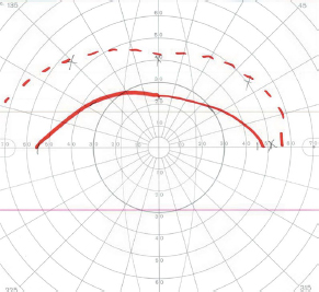
8Ophthalmology A patient presents with a central scotoma in the left eye and a superior temporal visual defect in the right eye. Where is the patient’s lesion? A. In the left inferior optic radiations B. In the right occipital pole C. In the orbital portion of the left optic nerve D. At the junction of the left optic nerve and chiasm E. In the optic chiasm A 10-month-old girl presents to clinic after her parents noticed that her eyes do not follow a moving object when her head is held in place. She is diagnosed with congenital oculomotor apraxia. What mechanism does she utilize to track objects? A. Optokinetic reflex B. Preserved convergence ability C. Vestibuloocular reflex D. Tonic labyrinthine reflex E. Ability to turn head A man presents with a ptosis of the right eye, anisocoria with the right pupil being smaller than the left, and a history of the right upper half of his face no longer sweating. Intraocular cocaine administration bilaterally dilates only the left eye, and Paredrine (amphetamine) administration has no effect on the right pupil. Where is the lesion causing the Horner syndrome? A. Brainstem B. Cervical spinal cord C. Sympathetic chain proximal to the superior cervical ganglion D. Sympathetic chain distal to the superior cervical ganglion but proximal to the off-take of the vasomotor fibers E. Sympathetic chain distal to the superior cervical ganglion and distal to the off-take of the vasomotor fibers A conjugate horizontal gaze palsy combined with an internuclear ophthalmoplegia (one-and-a-half syndrome) can be caused by a lesion involving the: A. Contralateral abducens nerve and the ipsilateral medial longitudinal fasciculus B. Bilateral paramedian pontine reticular formations C. Ipsilateral paramedian pontine reticular formation and the ipsilateral medial longitudinal fasciculus D. Contralateral paramedian pontine reticular formation and the ipsilateral medial longitudinal fasciculus E. Bilateral medial longitudinal fasciculi Optic nerve sheath meningiomas present with what characteristics? A. Arising from the dural layer of the meninges lining the intraorbital or intracanalicular optic nerve B. Painless, slowly progressive monocular vision loss C. Optociliary shunt vessels without optic atrophy D. Best treated with surgical resection while there is stability of the vision defect E. Most commonly affecting young women with an incidence three times that in men A patient incurs an infarction of the midbrain and develops a right trochlear nerve deficit as manifested on clinical exam. For what should the examiner look in the contralateral eye to localize the extent of the lesion? A. Mydriatic pupil B. Abducens nerve deficit C. Horner syndrome D. Oculomotor palsy A patient presents with a pupil that is tonically dilated with a very slow reaction to light. During accommodative testing, there is a more pronounced constriction. Syphilis and dorsal midbrain syndrome are ruled out through workup. Between what two neurons in the pupillary reflex pathway is the lesion? A. Prior to the first neuron B. Between the first and second neurons C. Between the second and third neurons D. Between the third and fourth neurons E. After the fourth neuron A 60-year-old woman presents with a 1-year history of progressive irritability, right-sided anosmia, and decreased right-sided vision. A funduscopic exam shows left-sided papilledema. What is the most likely lesion? A. A large, left parasagittal meningioma B. A right olfactory groove meningioma C. A suprasellar glioma D. A left sphenoid wing meningioma What factor predicts the occurrence of Terson syndrome following aneurysmal subarachnoid hemorrhage? A. Hunt-Hess score B. Ventriculomegaly C. Fisher grade D. Anterior communicating artery aneurysm A patient closes his eye forcefully, and it is noted that his eye slowly opens after several seconds despite his efforts to keep it closed. What is the likely diagnosis? A. Myasthenia gravis B. Hemifacial spasm C. Hemifacial paresis due to stroke D. Disinsertion of the levator palpebrae E. Botulinum toxin injection into the orbicularis oculi A 13-year-old girl presents to the emergency room for the third time in a year with an acute occurrence of a severe, bifrontal headache (worse on the right) with right orbital pain. She endorses photophobia, nausea, and diplopia. Exam reveals a right oculomotor nerve palsy that the patient states has occurred with past headaches. MRI and MR angiography of the brain are done and are unremarkable. What is the likely diagnosis? A. Multiple sclerosis B. Ophthalmoplegic migraine C. Myasthenia gravis D. Classic migraine E. Orbital cellulitis Which of the following is a characteristic of optic nerve gliomas? A. They most commonly are pilocytic astrocytomas, WHO grade IV. B. Seventy percent occur in the third decade of life. C. They present with optic atrophy and disk edema with an afferent pupillary defect. D. Surgical resection is the first-line treatment. E. They are associated with neurofibromatosis type 2. Where does the Goldman visual field test result shown in this image localize the patient’s lesion?
![]()
Stay updated, free articles. Join our Telegram channel


Full access? Get Clinical Tree


