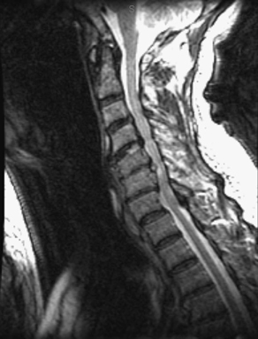
28 A 77-year-old Asian man fell, sustaining a central cord syndrome. His neurologic functioning improved except for some fine motor difficulty and continued myelopathy. After rehabilitation, he returned for operative planning. Magnetic resonance imaging (MRI) of the cervical spine (Figs. 28-1 and 28-2) shows cervical stenosis, most significantly at C5 and C6, with ossification of the posterior longitudinal ligament (OPLL) and cord signal change. Cervical stenosis with OPLL and cord signal change An anterior cervical decompression and fusion was accomplished. Ossification of the posterior longitudinal ligament, although typically seen in Japanese patients, can be associated with degenerative disease, diabetes, or ankylosing spondylitis. The posterior longitudinal ligament undergoes some combination of hypertrophy, ossification, and calcification. It can appear in separate segments, as a continual band, or as a mixture. Patients present with myeloradicular symptoms or central cord syndrome after a fall. Treatment options vary. Extensive literature has documented a variety of laminoplasty techniques. The posterior approach has the benefit of avoiding the pathology while providing room for the cervical cord. The anterior approach directly addresses the pathology but has a higher risk of unintended durotomy and possible neurologic damage. Neurophysiologic monitoring during the case is a worthwhile consideration.
Ossification of the Posterior Longitudinal Ligament (OPLL)
Presentation
Radiologic Findings
Diagnosis
Treatment
Discussion

Ossification of the Posterior Longitudinal Ligament (OPLL)
Only gold members can continue reading. Log In or Register to continue

Full access? Get Clinical Tree


