2 | Ossification of the Posterior Longitudinal Ligament: Posterior Cervical Approach |
 | Case Presentation |
A 61-year-old, obese (260 lb), and emphysematous male presented with 6 weeks of an increasing severe spastic myeloradicular syndrome (Nurick grade 4). Neurological findings included 3/5 upper/proximal lower extremity weakness, bilateral Babinski responses with hyperreflexia, a relative pin sensation loss below the C5 level, and decreased vibratory and position appreciation in the lower extremities. Plain x-rays revealed congenital stenosis, marked C2-5 hyperlordosis, and shingling of the C3 and C4 laminae. Dynamic films demonstrated 3 to 4 mm of motion between the C3 and C4 spinous processes. Magnetic resonance (MR) and computed tomographic (CT) studies both confirmed C2-5 stenosis, ossification of the posterior longitudinal ligament (OPLL), ossification of the yellow ligament (OYL), with hyperlordosis, whereas the T2-weighted MR additionally revealed an increased signal in the cord opposite the C3-4 level.
Following awake fiberoptic intubation/positioning, a “focal” C3-4 laminectomy, and C2-6 instrumented fusion were performed under continuous somatosensory evoked potential and electromyographic monitoring. Exposure of the laminae and facet joints from C2 to C6 was followed by laminectomy of C3 and C4 and undercutting of C2 and C5 for removal of OYL under microscope visualization. Dissection was confined to the lateral aspect of the spinal canal, avoiding placement of the rongeur beneath the lamina in the midline. The posterior fusion was performed utilizing braided titanium cable wires passed through the base of the C2, C3, C5, and C6 spinous processes, and then tightened in a “cerclage” fashion through bilateral eyelets affixed to titanium rods on either side of the spinous processes. Iliac autograft and allograft were applied over the decorticated facet joints. Following surgery, a hard cervicothoracic orthosis was placed, and the patient was extubated and moving all four extremities adequately. Postoperatively, he rapidly improved, demonstrating only mild residual radiculopathy (Nurick grade 0.5) within 1 week and no symptoms within 1 month. The cervical thoracic orthosis was worn until the CT and dynamic x-ray studies confirmed fusion 4 months later.
 | Background |
Laminectomy for dorsal decompression of the cervical spinal cord in the presence of an adequate lordotic curvature has long been utilized.1,2 With or without additional posterior arthrodesis, it is still a valuable adjunct for the management of patients with OPLL and hyperlordosis (without kyphosis). Laminectomy remains a safe, rapid, and effective operative alternative for carefully selected older individuals with their greater comorbidities.
Cervical Stenosis
Relative stenosis describes a canal of 13 mm, and absolute stenosis a canal measuring fewer than 10 mm in anterior/posterior diameter.3 Stenosis, OPLL, ossification of the anterior longitudinal ligament (OALL) and OYL, all contribute to “acquired stenosis.” Severe cord compression results in pathological gray and white matter atrophy, demyelination, and infarction.4 Typically, OPLL and stenosis contribute to slowly progressive cord damage, whereas in 10% of cases, an acute hyperextension injury can precipitate quadriparesis. Two major clinical grading systems for myelopathy include the Nurick scale and JOA scores.5–7
Radiological Findings
Candidates for either cervical laminectomy with/without fusion or laminoplasty should exhibit at least 10 degrees of cervical lordosis.6–9 With a dorsal approach, less than 7 mm of ventral OPLL should be present, and a minimum canal dimension of 17 mm should be created for the cord to sufficiently migrate away from ventrally situated pathology and be decompressed.10 MR examinations better define cord compression, myelomalacia/edema, cord atrophy, intramedullary cystic necrosis, or syrinx formation (Fig. 2–1).11,12 Alternatively, multiplanar CT examinations directly confirm the presence of hypertrophied (early) or ossified posterior longitudinal ligament.
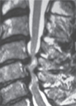
Figure 2–1 T2 midline sagittal magnetic resonance study (and the computed tomography) documents marked ventral ossification of the posterior longitudinal ligament compression at the C4-5 and C5-6 levels with more moderate intrusion at C6-7. Additionally, shingling of the C5 and C6 laminae and infolding of the hypertrophied/ossified yellow ligament (OYL) contribute to severe dorsal cord compression at the C4-5, C5-6, and C6-7 levels. Surgical intervention included a laminectomy of C5 and C6 with undercutting of C4 and C7 to remove OYL, followed by posterior C2-T2 fusion.
 | Methods of Surgical Management |
Laminectomy with or without Fusion
With 10 degrees of lordosis or hyperlordosis, the multilevel laminectomy with medial facetectomy/foraminotomy (with/without fusion) sufficiently releases axial cord tension, improving perfusion and facilitating dorsal cord migration in patients with OPLL3,13 When Hamanishi and Tanaka’s myelopathic patients had laminectomies performed, half with and half without fusion, comparable outcomes were demonstrated 3.5 years postoperatively.6 Keyhole laminoforaminotomy and microendoscopic posterior cervical laminoforaminotomy increased the risk of cord/root injury in OPLL patients.14 The untethering of dentate ligaments is contraindicated because it does not facilitate dorsal cord migration and is associated with multiple risks, including pneumocephalus, hematomas, cerebrospinal fluid (CSF) fistulas, and meningitis.
Laminectomy followed by Fusion
Instability is typically defined by > 3.5 mm of translation, > 20 degrees of angulation, and/or > 1 to 2 mm of motion between the tips of adjacent spinous processes on dynamic lateral x-rays. Laminectomy of three or more levels results in a 25% incidence of instability, with resection of 50% or more of individual facet joints largely accounting for destabilization.15,16
Posterior Fusion Techniques Utilizing Lateral Mass/Pedicle Screws
Posterior cervical lateral mass or pedicle screws combined with plates or rod fusions successfully stabilize the cervical spine following laminectomy, yielding an 80 to 97% incidence of good outcomes.17–22 In Houten and Cooper’s series, average 4.6-level laminectomies and posterior lateral mass cervical plates resulted in fusion and preserved lordosis, while proving superior to multilevel anterior cervical surgery, laminectomy alone, or laminoplasty.21 Employing lateral mass plates with pedicle screw fixation, 83% of Abumi and Kaneda’s postoperative laminectomy patients also fused without kyphosis.22 Although fusion rates with lateral mass plates following laminectomy approached 100% in some series, morbidity ranged from 0 to 1.8% for nerve root injury, 2.6% for cord injury, and up to 6.1% for screw malposition or pullout.19,21–26
Posterior Fusion Techniques Utilizing Cables/Rods
Spinous process wiring or rod/eyelet/cable techniques and facet fusions provide rapid, safe, and effective dorsal stabilization in different settings.7,17,27–29 Following failed anterior cervical fusions, posterior spinous process or facet wiring techniques result in 94 to 100% secondary fusion rates.27,28 Utilized solely for posterior fusion, a bilateral rod/eyelet construct (Vertex Reconstruction System, Medtronic Sofamor Danek, Memphis, TN) affixed to spinous processes with cerclage braided titanium cables provided excellent stability following focal one- to two-level laminectomies with extreme hyperlordosis (Fig. 2–2).29
The Skip Laminectomy without Fusion
The skip laminectomy, which must be differentiated from the focal laminectomy, is defined by a one-level laminectomy leaving an intact lamina between decompressed levels; the term skip refers to a process that can be repeated.30,31 Preserving muscular attachments to intact spinous processes theoretically maintains stability in this procedure without the need for fusion. Comparing the efficacy of the skip laminectomy in 43 patients versus laminoplasty (C3-7), skip laminectomy reduced the range of motion on dynamic x-rays (98% vs 61%) and yielded comparable JOA recovery rates (59.2% versus 61.0%).31
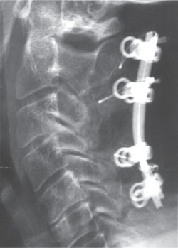
Figure 2–2 On the 6-month postoperative lateral x-ray, the rod/eyelet (Vertex Reconstruction System, Medtronic Sofamor Danek, Memphis, TN) and braided titanium cable construct is visualized. Note the wiring of the C2, C3, C5, and C6 levels following a complete laminectomy of C4 with undercutting of C2 and C5.


