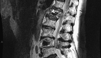
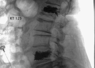
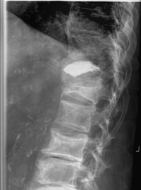
55 An 81-year-old woman fell from a standing position and developed intractable low back pain with radiculopathy. She was neurologically intact, but her pain increased with standing and walking. Her workup revealed focal lumbar stenosis with L1 and L4 osteoporotic compression fractures. Magnetic resonance imaging (MRI) of the lumbar spine demonstrated lumbar stenosis with L1 and L4 compression fractures (Fig. 55-1). Osteoporosis with spinal stenosis and compression fractures FIGURE 55-1 Sagittal T2-weighted MRI shows significant degenerative stenosis with associated compression fractures. The patient underwent percutaneous L1, L4, and L5 decompressions, followed by L1 and L4 kyphoplasties. The kyphoplasties were done in the operating room after the decompression, through a transpedicular route. The result can be seen in Fig. 55-2. Osteoporosis is a prevalent disease in which the cortical bone becomes too porous. It is estimated that over 50% of Caucasian women experience an osteoporotic fracture in their lifetime. In 2003 there were 1.5 million osteoporotic fractures in the United States alone, 50% of which involved the vertebral bodies and 25% involved the hip. The typical patient is an elderly Caucasian woman with a small habitus and positive family history. The use of certain medications, such as steroids, and some chemotherapy can accelerate osteoporosis. In addition, alcohol and tobacco use coupled with inadequate calcium intake can augment the severity of this bone loss. FIGURE 55-2 Lateral fluoroscopic image demonstrates the completion of the L1 and L4 kyphoplasties. FIGURE 55-3 One can see the results of a T12 kyphoplasty, and also an L1 compression fracture. The incidence of fractures from seemingly nontraumatic events increases exponentially in the eighth decade for women who have low bone density. While prevention, nutrition/exercise, and pharmaceutical treatment are the mainstay, surgical intervention also has a role. The treatment of an osteoporotic compression fracture has three key factors: (1) overall assessment of bone quality, (2) addressing the compression fracture, and (3) pharmaceuticals. The underlying pathology must be treated. A bone densitometry should be performed on all postmenopausal women with risk factors as well as women presenting with osteoporotic compression fractures. In addition, a bone and mineral specialist can help coordinate pharmaceuticals-calcium and vitamin D supplementation and the use of calcium “fusing” agents-and address lifestyle changes and how to decrease other risk factors. The compression factor itself has to be addressed. An acutely painful fracture is a surgical indication. Acute fractures have increased signal on T2 in the vertebral body from marrow edema, as well as active intake on bone scan. An MRI with gadolinium is needed to rule out other reasons for the fracture (i.e., neoplasm) and evaluate for neural compression. Chronic or painless fractures are not surgical indications, unless there is progressive kyphosis. If neural compression is the surgical indication, decompression can be done. Fusion hardware failure is a real risk in this group because of bone quality, previous medications, and age. Kyphoplasty has been successful in treating the painful compression fractures of osteoporosis. This procedure, usually done percutaneously, involves restoring vertebral body height and alignment by inflating a balloon, and then securing the correction by filling the space with bone cement. Patients report pain reduction and higher activity tolerance. This can also be an outpatient procedure. Vertebroplasty is less expensive but has a higher rate of cement extravasation due to the high-pressure injection. Furthermore, it does not reduce the fracture or restore normal spine mechanics. Prophylactic treatment is a source of debate but makes biomechanical sense, at least at the thoracolumbar junction. Both kyphoplasty and vertebroplasty have potential complications, usually related to the effects of cement extravasation or access issues. The results of a T12 kyphoplasty and also an L1 compression fracture are demonstrated on x-ray (Fig. 55-3). The L1 fracture developed months after the kyphoplasty. The question is whether the fracture is procedure related or disease-process related. The methylmethacrylate has a greater density than osteoporotic bone, and this may stress an already compromised level. In addition, the natural history of osteoporosis dictates additional fracture development. Although the clinical course can give some idea as to the etiology, adjacent level disease should be monitored. Because this new fracture was not painful, no invasive treatment was done. How these procedures relieve pain is debatable. Pain relief is thought to result from stabilization of the fractured vertebrae. Reduction of postural kyphosis (more so following kyphoplasty) seems to further enhance the mechanics of the spine, decrease delayed deformity, and improve pain. It is speculated that the exothermic reaction generated by methylmethacrylate injection causes denervation of the periosteal nerve endings. This seems to contribute to pain control. Furthermore, thermal injury by methylmethacrylate is cytotoxic to tumor cells when used for contained spine metastases. Percutaneous bone augmentation techniques cannot be offered in cases of traumatic fractures unrelated to osteoporosis, or in the presence of a three-column injury, or when retropulsion with neurologic compromise is identified. The presence of a vertebral body arteriovenous malformation can pose a bleeding risk when performing this procedure. Similarly, the presence of an old “scarred-down” fracture or severe scoliosis can create technical difficulty. Phillips FM, Ho E, Campbell-Hupp M, McNally T, Wetzel F, Gupta P. Early radiographic and clinical results of balloon kyphoplasty for the treatment of osteoporotic vertebral compression fractures. Spine 2003;28:2260–2265 Ryu KS, Park CK, Kim MC, Kang JK. Dose-dependent epidural leakage of polymethylmethacrylate after percutaneous vertebroplasty in patients with osteoporotic vertebral compression fractures. J Neurosurg 2002;96:56–61
Osteoporosis
Presentation
Radiologic Findings
Diagnosis
Treatment
Discussion
SUGGESTED READINGS
< div class='tao-gold-member'>
Osteoporosis
Only gold members can continue reading. Log In or Register to continue


Full access? Get Clinical Tree


