CHAPTER 36 Overview of Petroclival Meningiomas
INTRODUCTION
Clivus meningiomas in particular have until recently been uniformly lethal. The outlook for such patients must be improved by achieving an earlier and more accurate diagnosis, by improving surgical techniques, and by developing a better understanding the pathological anatomy.1
Posterior fossa meningiomas account for only 10% to 15% of intracranial meningiomas, and petroclival meningiomas account for only 3% to 10% of posterior fossa meningiomas.1–8 This is consistent with the incidence of 1.7% reported by Cushing and Eisenhardt in their series of 295 meningiomas.2 Owing to their rarity, crucial location, insidious growth, and relentless natural progression toward fatality, clival and petroclival meningiomas remain the most formidable of all meningiomas and the most challenging in the neurosurgical realm. The simple definition of these tumors and accurate characterization has yet to be settled and universally followed.
The natural history of nonoperative patients harboring clival meningiomas is that of relentless progression to an eventually fatal outcome.1–3,9 A review of the early literature shows a cumulating operative mortality that exceeds 50%.1,9,10 Before 1970, only one successful total removal was reported.4 Hence, these tumors were generally considered inoperable.3,11
With the advent of refined skull-base approaches, microsurgical techniques, intraoperative monitoring, advanced imaging, and modern anesthesia and postoperative care, the previously dismal surgical outcome has been largely overcome, with reports signifying a major decrease in operating mortality and morbidity, a higher incidence of total removal, and an improved clinical outcome1,5,10,12–17 (Table 36-1). The size of the lesion is a significant factor influencing surgical morbidity and mortality.15,18,19
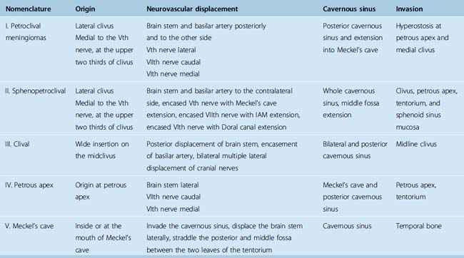
TABLE 36-1 Author’s classification of petroclival meningiomas based on origin and anatomical displacement20

Meningiomas are benign tumors whose treatment objective is total resection. This is best achieved during the first operation when arachnoid membranes are intact, facilitating dissection of neurovascular structures and resection of the tumor.20 The recurrence rate is directly related to the resection grade, with a 89% recurrence rate of meningiomas that were not totally removed.21,22 Although surgeons should pursue total removal with skill and zeal, their judgment should not be averted from the goal of preserving or improving neurologic function. Consequently, surgeons may be forced at times to accept subtotal removal. However, the concept of decompressive removal to lessen surgical complications followed by fractionated radiation therapy has proven ineffective. For long-term control, clinical recurrence rates after 15 years of subtotal resection and radiation therapy have reached 75% with a complication rate of 56%.23 The advent of radiosurgery has revived this concept, with authors resorting to subtotal removal followed by adjuvant radiosurgery.24–26 All of these series report a relatively short follow-up that is insufficient to determine the real late recurrence rate, and reports exist on delayed failure of radiosurgery and its aggressive recurrence pattern.27 The concern is the lost opportunity of curative removal at the initial presentation and the risks and difficulties in managing these recurrences.28,29
DEFINITION AND CLASSIFICATION
Petroclival meningiomas are thought to arise from the region of the spheno-occipital synchondrosis.2,9,30 Over the years there has been ongoing debate as to what the anatomic term “petroclival” includes. Cushing and Eisenhardt included five tumors in their gasseropetrosal group.2 Recognizing that these lesions were unique in their involvement of both the supratentorial and infratentorial compartments simultaneously, they aptly wrote, “… in these cases the fully grown tumors sits like a saddle astride the anterior end of the petrous ridge which demarcates the middle cranial fossa, where the growth organizes from the cerebellar of posterior fossa, where it does its chief damage.”2
Since Cushing and Eisenhardt’s description of the gasseropetrosal meningioma, multiple classification systems have been introduced in the literature. Unfortunately these early classifications were somewhat arbitrary and at best confusing and inconsistent.1–4,9,10,31–35 Castellano and Ruggiero, reviewing autopsy data, described meningiomas of the posterior fossa based on the site of their dural attachment.3 They divided the tumors into five groups: cerebellar convexity, tentorium, posterior surface of the petrous bone, clivus, and foramen magnum. In an appendix to the group, they added those “meningiomas of the cavum Meckelii extending into the posterior fossa.” Although Castelanno and Ruggiero believed these should be middle fossa tumors, they conceded that they should be classified as clival tumors for anatomic, pathologic, and radiologic reasons.3
Yaşargil and colleagues differentiated the posterior fossa meninigiomas based on intraoperative observations.1 The absence of a purely medial clival origin, as well as the absence of associated hyperostosis along the middle region, impressed them. They described these meningiomas as “attached at any of the lateral sites along the petroclival borderline where the sphenoid, petrous, and occipital bones meet.” Based on their findings, they classified the posterior fossa locations as clival, petroclival, sphenopetroclival, foramen magnum, and cerebellopontine angle.1 Therefore, based on surgical anatomy, natural history, and results, most authors use the term “petroclival” to describe meningiomas located along the superior two thirds of the clivus and medial to the Vth cranial nerve arising from the spheno-occipital synchondrosis.1,2,5,9,30,36,37 Meningiomas arising from the lower one third of the clivus are typically classified as foramen magnum and are not addressed in this chapter.
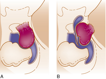
We believe the site of origin must be the basis of classification because of its effect on the pathologic anatomy and the subsequent displacement of the neurovascular structure with a profound influence on the surgical intervention and its outcome.20 The presence of multiarachnoid layers that facilitate safe neurovascular dissection is influenced by a few millimeters difference in the origin (Fig. 36-1).
This principle was recently stressed and presented also by others with a report of their classification.38
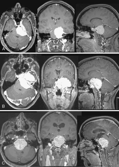
Table 36-1 shows the classification relating the tumor origin with the corresponding pathologic anatomy, tumor invasion, and the influence selection of the surgical approach (Fig. 36-2).
CLINICAL FINDINGS
The typical presentation of a petroclival meningioma is that of an insidious onset. Most studies report that the period of symptoms before diagnosis averages between 2.5 and 5 years, ranging from 1 month to 17 years.1,4,5,8–10,19,39 The tumor’s inconsistent and variable presentation testifies to this diagnostic dilemma, especially before the introduction of modern neuroradiologic techniques. The clinical presentations can be divided into four groups based on involvement of the cranial nerves, mass effect on the cerebellum, brain stem compression and increased intracranial pressure, and whether the source of the symptoms is the tumor mass or obstructive hydrocephalus.9,10
Cranial neuropathies are commonly associated with petroclival meningiomas. The Vth and VIIIth cranial nerves are the most frequently involved, up to 70% in some series. Facial nerve involvement occurs in about half of the patients.1,5,10,16,19,36 The lower cranial nerves are involved in about one third of cases, usually associated with the larger tumors extending inferiorly.5,10 Interestingly, despite their proximity and in many cases intimate involvement, cranial nerves III, IV, and VI are involved in fewer than half of the cases.1,5,10 In the series reported by Hakuba and colleagues, the incidence of abducens neuropathy was 40% and oculomotor paresis was present in 27%.10 The true incidence of trochlear nerve involvement in these patients is not well documented owing to the lack of preoperative formal examinations.
Cerebellar signs are the most frequently identified clinical findings in these patients.1,5,8,10,18,36,39 Hakuba and colleagues reported ataxia in 70% of their patients.10 Mayberg and Symon’s series supported this finding.36 Headaches are present in about 70% of patients. Long tract signs and somatosensory deficits, consistent with brain stem compression, are variable. Spastic paresis has been reported in 15% to 57% of patients and somatosensory deficits are identified in 15% to 20%.1,5,8,10,18,36,39,40 Despite occasional reports of false localizing signs in meningiomas of the posterior fossa, most series report findings appropriate to the tumor’s location.1,3,10,36,41–43 A clival meningioma seldom produces a syndrome of basilar insufficiency.4
PREOPERATIVE EVALUATION
Clinical Examination
A comprehensive preoperative evaluation is critical in choosing the treatment modality and the surgical approach. Complete clinical, neurologic, audiologic, endocrinologic, and ophthalmologic examinations are performed. The audiogram results will not only provide the clinician with a useful baseline of the patient’s cranial nerve VIII function, it will also give insight into the limitations of the surgical approach. As in acoustic schwannoma patients who elect to postpone surgery, serial examinations of these patients with regard to hearing preservation may provide the impetus for resection of the tumor. As stated earlier, hearing loss can be clinically present in up to three quarters of these patients.1,5,10,19,36 In addition, neuron-ophthalmologic examination preoperatively detects cranial neuropathies involving CN III, IV, and/or VI.1,5,10 As expected, sphenopetroclival meningiomas and petroclival meningiomas extending into the cavernous sinus have a higher rate of extraocular motor neuropathies. In cases of suprasellar tumor extension in patients with sphenopetroclival meningiomas, formal visual field testing preoperatively will reveal the corresponding defect.
Additional testing is dictated by the size and extension of the tumor. In patients with a large supratentorial extension, especially into the suprasellar space, pituitary axis evaluation may be necessary. Patients with evidence of hypothyroidism or hypocortisol levels should be treated preoperatively. Patients with significant tumor extension into the area of the medulla are at risk for lower cranial difficulties. Preoperative deficits involving the lower cranial nerves (IX, X, and XI) occur in up to one third of patients and warrants formal evaluation with endoscopic visualization of the vocal cords and video swallowing test.5,10
Stay updated, free articles. Join our Telegram channel

Full access? Get Clinical Tree


