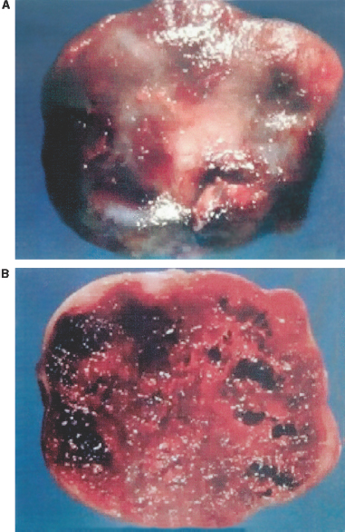
3 Huan Wang and Meena Gujrati Cavernous malformations are one of the distinct and common clinicopathologic categories of vascular malformations, characteristically seen as a well-circumscribed lesion, consisting of enlarged vascular channels with no intervening parenchyma. They were first described by Luschka in 1853,1 and Virchow2 gave a detailed description in 1863. Until recently, there has been considerable confusion regarding the nomenclature. Cavernous malformations have previously been variably referred to as cavernous hemangiomas, cavernous angiomas, and cavernomas, all of which imply a neoplastic process, but these lesions are considered hamartomas and, therefore, cavernous malformation is the preferred and accepted term. They have also been included in the descriptions of angiographically cryptic or occult vascular malformations because cavernous malformations cannot be routinely detected on angiography. They were frequently mislabeled as arteriovenous malformations, especially when mixed features of vascular malformations were not recognized.3 The neurosurgical importance of cavernous malformations has been increasingly emphasized, and computed tomography (CT) scanning and magnetic resonance imaging (MRI) have revolutionized their diagnosis. In large autopsy series, cavernous malformations were estimated to range from 0.02 to 0.13% of the general population.4,5 However, with the introduction of MRI, these lesions were found to be more common than previously thought, ranging from 0.2 to 0.4%.6,7 Cavernous malformations appear to occur in every age group (range, 4 months to 84 years; mean, 34.6 years) with an approximately equal male-to-female ratio.5,6 Most occur sporadically, but ~10 to 30% are part of a familial syndrome inherited as an autosomal dominant pattern with variable penetrance.5,8 A gene responsible for familial cases has been mapped to chromosome 7q using linkage analysis.9 A higher incidence of the familial form has been reported in a patient population of Mexican descent.10 The majority (80%) of cavernous malformations are reported to be located supratentorially, but up to 15% occurred infratentorially, and 5% occurred in the spinal cord.5,8,11 These frequencies are proportional to the volume of the CNS tissue in these different regions. In the cerebrum, the majority of cavernous malformations are cortical or subcortical with characteristic locations around the Rolandic fissure, but they can also occur in the basal ganglia, the periventricular white matter, corpus callosum, internal capsule, and lateral and third ventricles.5 The most usual site in the posterior fossa is the brain stem, particularly the pons.5 Cavernous malformations form 5 to 16% of all spinal vascular malformations. Most of these lesions involve vertebral bodies or epidural space; in contrast, intramedullary lesions are rare and tend to be distributed evenly throughout.11–15 An association of spinal cord cavernous malformation with multiple malformations in the neuraxis has been well described.16 Multiple lesions are common, ranging from 26 to 50% of patients.10 The familial form of the disease is more typically characterized by the presence of multiple lesions.10 In rare cases, cavernous malformations have been reported to involve cranial nerve nuclei or cranial nerves, commonly in the optic nerve and chiasma, III, VII, and VIII nerves.17 Intracranial extracerebral cavernous malformations are also very rare and have been reported in the cavernous sinus and in the region of cerebellopontine angle.17–19 Dura-based cavernous malformations also occur and are usually seen in the middle cranial fossa.17 The precise mechanisms by which cavernous malformations develop remain elusive. Some are genetically transmitted through autosomal dominant patterns, but most appear to be sporadic in nature. Cavernous malformations have traditionally been presumed to be congenital lesions,20 and reported cases of cavernous malformations in neonates support a congenital origin.21,22 It is well documented that lesions may increase in size with time due to small repeated hemorrhages, gradual thrombosis, or thickening and hyalinization of the walls of the vascular channels.23,24 Recently, de novo development of cavernous malformations has been observed in both sporadic and familial cases by serial MRI.25 De novo cavernous malformations have also been reported after radiation therapy, surgical biopsy, and chemotherapy.25–28 Spontaneous lesions have also been reported in association with pregnancy and were presumed to be hormone-related, but hormone receptors could not be demonstrated in the tissue by immunohistochemistry.29 Infection with the polyoma virus has been shown to induce the formation of multiple cavernous malformations in nude mice.30 Thus, viral infections that cause hemorrhages followed by reactive angiogenesis resulting in the formation of cavernous malformations may explain their apparent de novo origins in some patients. Capillary telangiectases have also been suggested as precursors of cavernous malformations.31 Cavernous malformations are not benign entities as previously thought. The rate of hemorrhage has been reported as 13% per patient per year or 2% per lesion per year,32 and only ~11 to 20% of cases may be truly asymptomatic.33 Clinical presentation depends on anatomic location. Seizures, headaches, and progressive neurologic deficits are the three most common presentations.32,33 Infratentorial lesions present more commonly with progressive neurologic deficits because of the space constraints.5
Pathology of Cerebral Cavernous Malformations
 Incidence of Cavernous Malformations
 Prevalence by Anatomic Location
 Etiology
 Clinical Presentation

Pathology of Cerebral Cavernous Malformations
Only gold members can continue reading. Log In or Register to continue

Full access? Get Clinical Tree


