, Nima Etminan1 and Daniel Hänggi1, 2
(1)
Neurochirurgische Klinik, Universitätsklinikum Düsseldorf, Düsseldorf, Germany
(2)
Medical Art Christine Opfermann-Rüngeler, Zentrum für Anatomie Heinrich Heine Universität, Düsseldorf, Germany
2.1 Terminal Versus Lateral Aneurysms
More than 90 % of all saccular aneurysms arise from bifurcations or appear at the origin of small side branches (percentages are approximate) [1, 2]:
Middle cerebral artery main bifurcation (15–30 %)
Anterior communicating artery (20–30 %)
Internal carotid–posterior communicating artery (15 %)
Basilar tip (5 %)
Rare bifurcation aneurysm locations:
Internal carotid artery bifurcation (2 %)
Internal carotid–anterior choroidal artery (1–2 %)
Pericallosal–callosomarginal artery (2 %)
Basilar–superior cerebellar artery (2 %)
Vertebral artery–posterior inferior cerebellar artery (2 %)
Vertebrobasilar junction (1 %)
Basilar–anterior inferior cerebellar artery (0.5–1 %)
The reported frequency at different sites varies somewhat among different publications and also between series of ruptured and unruptured aneurysms. In particular, aneurysms at the anterior communicating artery are more frequent in series of ruptured aneurysms. In contrast, in pediatric and familial cases, aneurysms appear to occur rarely at the anterior communicating artery.
The distinction between terminal aneurysms and lateral, side branch-related aneurysms is somewhat artificial (Fig. 2.1). In fact, some asymmetry of the bifurcation is common also with terminal aneurysms in that one of the two branches is larger than the other. If the difference in the diameter of the branches is significant, the smaller branch appears as a side branch. The diameters of the side branches also correlate with the angle between the afferent artery and the branches. In case of a large asymmetry between the branches, the larger branch continues more or less in the direction of the afferent artery, while the smaller branch originates more or less at a 90° angle.
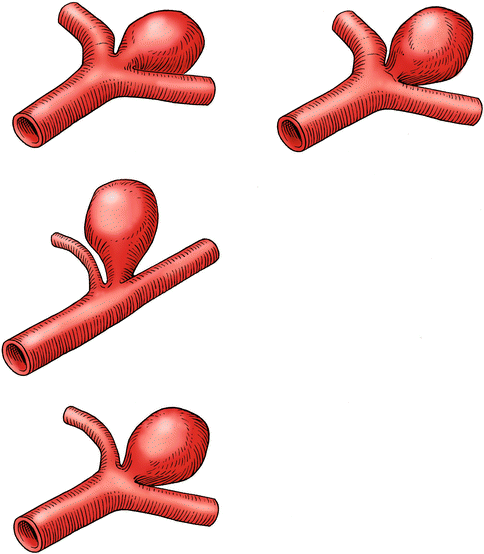
Fig. 2.1
Terminal versus lateral aneurysms. Some asymmetry of the bifurcation or the aneurysm projection is necessary for flow in the aneurysm. If the diameter of the branches differs significantly, the smaller branch appears as a side branch. An aneurysm in relation to a small side branch is called a lateral aneurysm, whereas an aneurysm between branches of similar diameter is called a terminal aneurysm. The diameters of the side branches also correlate with the angle between the afferent artery and the branches. If there is a large asymmetry between the branches, the larger branch continues more or less in the direction of the afferent artery, and the small branch originates more or less at a 90° angle
2.2 Geometry of Bifurcations
Extracranial arterial bifurcations often show an acute angle between the branches, as in the case of the abdominal aorta, with an acute angle between the common iliac arteries. In contrast, intracranial arteries most often develop a blunt angle between the branches. The reason for this peculiarity is not obvious, but everyday experience confirms this rule. The most logical explanation is that the angle is related to the shape of the arterial environment. If an artery passes through a longitudinal organ such as an extremity, the bifurcations have acute angles, but if arteries pass through an essentially round organ such as the brain, the bifurcations are T-shaped.
In principle, bifurcations of cerebral arteries and the projection of aneurysms could assume any three-dimensional configuration; afferent and efferent arteries need not necessarily lie in a flat plane. In reality, however, there is always a smooth transition from the direction of the afferent vessel to the direction of the branches (Fig. 2.2). Therefore, it is best to imagine the bifurcations as lying on a curved plane.
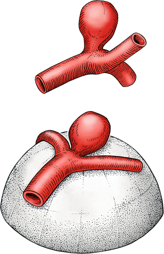
Fig. 2.2
Geometry of bifurcations. Intracranial arterial bifurcations most often have a blunt angle between the branches. In principle, bifurcations of cerebral arteries and the projection of aneurysms could assume any three-dimensional configuration, but in reality, the bifurcations can be imagined as lying on a curved plane. The projection of an aneurysm, however, is not confined to this plane
The diameters of the branches are related, given that the flow velocities in the cerebral arteries vary little. Therefore, in the case of equal branches, the relative diameter of the branches in comparison to the afferent artery corresponds to the square root of 0.5 (i.e., 0.7). If the branches are unequal, the sum of the cross-sectional areas of the branches adds to the cross section of the afferent artery. Figure 2.3 shows some examples of size relations of afferent and efferent arteries.
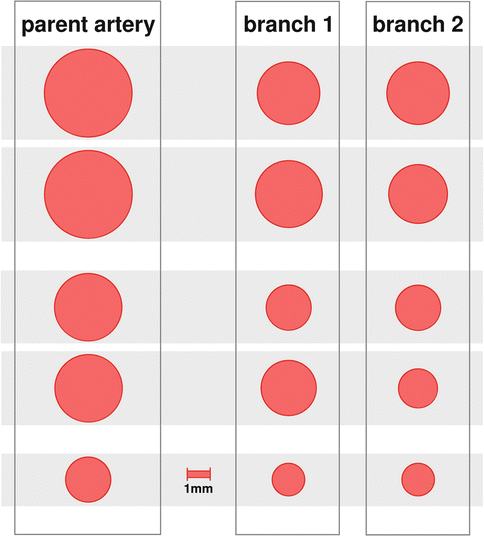
Fig. 2.3
The diameters of cerebral artery branches are related. The area of inflow equals approximately the sum of the areas of the branches. This illustration shows some examples of size relations of afferent and efferent arteries
The intracranial internal carotid artery (ICA) has two typical configurations (Fig. 2.4). The terminal bifurcation regularly assumes the typical T shape, but with variable asymmetry of the diameters of the middle cerebral artery (MCA) and the anterior communicating artery (Acom), the relative size of the posterior communicating artery (Pcom) determines the direction of the distal ICA. The orifice of the Pcom usually is located in the ventral midline of the ICA. In approximately 70 % of individuals, the Pcom is small and constitutes a typical side branch; the distal ICA continues in the direction of the proximal ICA. In about 30 %, however, the Pcom is larger than 2 mm and supplies the posterior cerebral artery, leading to a dorsal deviation of the distal ICA. The diameter of the distal ICA in this situation is also smaller than the diameter of the proximal ICA. The anterior choroidal artery uniformly has a small diameter and is a typical side branch of the ICA.
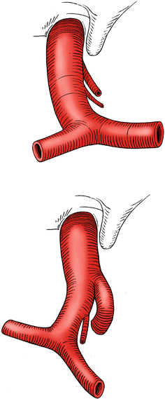
Fig. 2.4
The intracranial internal carotid artery (ICA) has two typical configurations, depending on the size of the posterior communicating artery (Pcom). If the Pcom is small and constitutes a typical side branch, the distal ICA continues in the direction of the proximal ICA. If the Pcom is large and supplies the posterior cerebral artery, the distal ICA turns upward. The anterior choroidal artery uniformly has a small diameter and is a typical side branch of the ICA
The anterior cerebral artery bifurcation often displays a few features in patients with aneurysms at the anterior communicating artery (Acom) that differ from the norm (Fig. 2.5). Most often the size of the ACAs in patients with ACA aneurysms is clearly asymmetric. Furthermore, progressive elongation of the ACAs leads to distortion of the ACA, so that one A2 segment lies more anteriorly than the other. In about 80 % of these cases, the A2 segment on the side of the dominant A1 lies more posteriorly than the contralateral A2 [3]. With a frontolateral approach to the Acom aneurysm, this predominant configuration (also called the open configuration) allows for easy visualization of the entire Acom complex and particularly both A2 segments. In contrast, in approximately 20 % of patients with an Acom aneurysm, the Acom is turned the other way, so that the A2 on the side of the dominant A1 lies anteriorly. This configuration (also called the closed configuration) is problematic for an anterolateral approach because the contralateral A2 origin hides behind the ipsilateral A2.
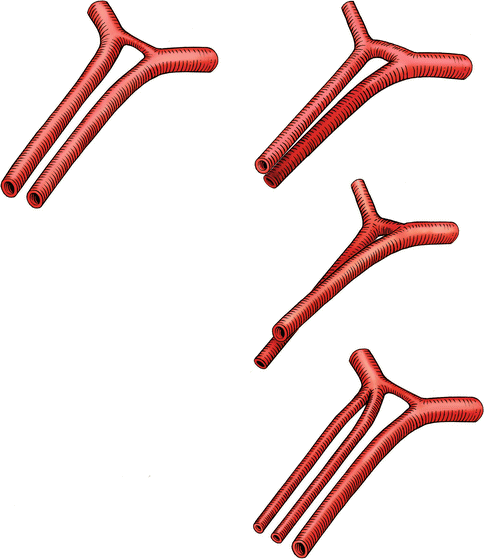
Fig. 2.5
Variations of the anterior communicating artery (Acom) complex. Most often, the size of the A1 segments in patients with ACA aneurysms is clearly asymmetric. Furthermore, progressive elongation of the A1s leads to rotation of the ACA, resulting usually in a more posterior origin of the A2 segment on the side of the dominant A1. The other variant, resulting in a more posterior position of the contralateral A2, is rare. Sometimes, an A2 divides early into two main trunks, resulting in three A2 segments. If the surgeon is unaware of this special configuration, the risk of inadvertently occluding the third branch with the aneurysm clip becomes significant
A further particular anatomical property of the Acom complex is important. In a small percentage of patients, the A2 divides early into two main trunks, resulting in three A2 segments. If the surgeon is unaware of this special configuration, the risk of inadvertently occluding the third branch with the aneurysm clip becomes significant. On the other hand, an unpaired pericallosal artery (azygos A2) is a constellation associated with aneurysms at the origin of the callosomarginal arteries. On average, the azygos A2 variant is seen in approximately 3 % of angiograms [4].
The MCA bifurcation in aneurysm patients also exhibits some typical features that are relevant for microsurgical treatment. The MCA bifurcation usually is a typical T-shaped bifurcation. A trifurcation is rare. As in the case of the anterior cerebral artery, the M1 segment often has undergone substantial elongation during life, leading to a high-lying and curved course. The distal M1 then actually can lie behind the bifurcation when the MCA bifurcation is approached through the Sylvian fissure (Fig. 2.6). In this situation, we recommend approaching and exposing the distal M1 between the M2 trunks.
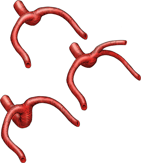
Fig. 2.6
The MCA bifurcation is usually a typical T-shaped bifurcation. A trifurcation is rare. As in the case of the anterior cerebral artery, the M1 segment often has undergone substantial elongation during life, leading to a high-lying and curved course of the M1. The distal M1 then can lie behind the bifurcation if the MCA bifurcation is approached through the Sylvian fissure
Cerebral aneurysms are commonly associated with anomalies of the circle of Willis. The most frequently seen combination is the Acom aneurysm associated with hypoplasia of one anterior cerebral artery. According to various authors, the first segments of the anterior cerebral arteries (A1) are clearly asymmetric in approximately 80 % of patients with an aneurysm at the Acom, whereas the prevalence of unequal A1 segments in healthy individuals is estimated to be only 10–15 % [2, 5, 6]. Furthermore, Acom aneurysms arise consistently at the crotch between the dominant A1 and the communicating artery.
Thrombosis or therapeutic ligation of the internal carotid artery also constitutes a well-known risk factor for subsequent formation of saccular aneurysms [7]. Occlusion of other cerebral arteries has also been associated with an increased incidence of aneurysms of the patent blood channels.
Another category of geometrical variations associated with aneurysms is arteriovenous malformations (AVMs). Between 10 and 30 % of AVM patients are reported to have associated feeder aneurysms [8, 9]. Aneurysms associated with AVMs are located either on the feeder vessels or at common sites of the circle of Willis. The aneurysm-promoting effect of arterial hypoplasia, occlusion, and AVMs is named “hemodynamic stress” [10].
Related posts:


Stay updated, free articles. Join our Telegram channel

Full access? Get Clinical Tree


