Head injury: initial assessment and management for neurosurgical consultants
Basic minimum acute data
History
• Time of injury
• Mechanism
–Kinematics; energy, speed, height
–Struck head?
• Best exam at scene
–Loss of/level of consciousness
–GCS
–Asymmetry (pupils, motor)
– Moved legs?
• Exacerbators
–Apnea
–Shock
–Prolonged extraction
–Other injuries
–Resuscitation
Exam
• Via standardized tool (GCS, motor, pupils, other)
Imaging
• Time of imaging
• Is there a mass lesion causing symptoms or significant tissue shift now or with predictable worsening?
Swelling-prone (deterioration- prone) injuries
Nonswelling-prone injuries
Acute subdural hematoma (not in atrophic brain)
“Pure” diffuse axonal injury
Multifocal/large contusions
Isolated focal lesion not near brainstem, falx
Temporal or posterior fossa lesions
Chronic subdural hematoma
High energy contact injury
Atrophic/encephalomalacic brain
Gunshot wound
+ Exacerbators
There are several features of the history with particular relevance in children. The exact mechanism of injury relevant to the child is of great importance in predicting the type and severity of injury; it is insufficient, for instance, to just know the child was a passenger in a car crash. Was the child restrained? In what way? Was there intrusion into the vehicle? Was the child ejected? For all injury mechanisms, if the event was witnessed or caregivers arrived at the scene promptly, it is helpful to try to create a “mental video” of events as they unfolded. If the mechanism was a fall, what was the head to ground height? What position was the child in when discovered, and what happened next? What was the initial and the best exam at the scene—was the child unconscious initially, was the child ever see to be crying, speaking, eyes open, moving all extremities with full strength and good excursion? Were there asymmetries in the exam? Was there any evidence of apnea, or seizure activity? Was there a need for resuscitation , and for what indications? What kind of immobilization was performed, what kind of airway protection was required, and what drugs and doses were given, and when? The latter is critical in interpreting the examination on arrival—is this child still pharmacologically sedated and/or paralyzed? (Having a twitch monitor and reversal agents readily available in the Emergency Department can be extremely helpful in early assessment when pharmacologic interference is uncertain.)
In addition, it is important to document, as much as possible, who are the parents/guardians, and if there are additional victims of the injury scenario, what is their relation to the child. First responders may not know past medical history, and it needs to be determined as efficiently as possible who knows the child and is able to provide this information. Often it is best to dispatch a member of the team to interact with or contact relatives who can determine whether the child has allergies, was premature or has other preexisting neurologic or general medical problems, and what other aspects of the child’s baseline are relevant. This is particularly true for very young children—does this child normally talk? Ambulate? With whom does the child reside? Additionally, since there can be many variations in family structure and custody arrangements, and since injury to a child can be a volatile issue among family members, early involvement of the social work team can be invaluable in obtaining this information, providing initial support to the distraught family, and providing a communication and “social management ” plan early in the course of treatment. This is helpful to the entire team, but also to the clinicians who must interact promptly with the family to provide information about the injuries and treatment plan, to set the stage for an ongoing positive care team/family relationship, and to involve the correct members of the family appropriately in decision-making. Making assumptions that an adult who gives consent for an intervention is the legal guardian of the child can be the source of later difficulties.
Initial Resuscitation
Airway considerations follow general pediatric trauma resuscitation guidelines. In a significantly injured pediatric polytrauma patient, the airway should be secured by an experienced clinician. Often Pediatric Emergency Medicine physicians, Trauma team members from General Surgery/Pediatric Surgery, and Pediatric Anesthesia participants are present very early in the acute care course, and whose job it is to secure the airway should be determined by established protocol to avoid confusion or inefficiency. Use of a checklist has been shown to increase resuscitation efficiency [14]. In young children, significant air accumulation in the stomach can lead to physiologic compromise and so needs to be avoided or relieved promptly if it occurs.
Because of the small blood volume of young children but robust compensatory mechanisms, the risk of shock is increased in specific situations (outlined below) and can become manifest suddenly when decompensation occurs. Use of isotonic or, sometimes, hypertonic solutions typically enables prompt and assertive fluid resuscitation with minimal risk of fluid overload in most children with good baseline cardiopulmonary function, and is more likely to protect against rather than exacerbate neural injury [15]. When in doubt, slight over-resuscitation is generally better tolerated than under-resuscitation in most polytrauma settings in children from the neurotrauma point of view.
Neurologic Assessment
Older children and adolescents with a clearly decreased level of consciousness are examined similar to adults, using a systematic approach to determine level of responsiveness and presence of focal neurologic deficits. Once the pharmacologic status has been determined (especially, if a child might be under the influence of a paralytic agent but not a sedative), children should be approached calmly by first speaking clearly and close to the ear (shouting is not necessary), asking questions using the child’s name, and then using progressive levels of stimulation as needed. Some children have a paradoxical response to the application of painful stimulation, which will make them more withdrawn and less responsive, so a gentler approach to the exam can be helpful. Manually opening the eyelids, sometimes accompanied by slight manual movement of the head (depending on the level of concern for cervical injuries,) can be an effective and painless way to encourage alerting and interaction. It should be remembered that Horner’s syndrome, which can reflect cervical or cerebrovascular injury, is manifested as failure of the affected pupil to dilate in dim light, so in an appropriate clinical context, this should be specifically assessed. Trapezius pinch and nail bed pressure can be used as needed to check for general level of alertness and to examine the strength in each extremity; the latter also can be assessed by observation of spontaneous activity during maneuvers such as blood draw.
Despite decades of use, aspects of the basic neurotrauma exam are sometimes implemented variably and inconsistently. Although the Glasgow Coma Scale does include a “motor” component, it is designed to assess level of consciousness, and does not include a specific strength exam, which must be performed separately. Rather, the GCS motor score assesses the ability of the brain to recognize input and respond with appropriate motor patterns, which must be tested systematically. As one common example, “localization” on the GCS is an important finding with considerable prognostic and acute management significance, and assessment for this feature should be performed by a standard and reproducible protocol, upon arrival and in serial exams. For children older than about 18 months, the author prefers bending the elbow and placing the forearm at the patient’s waist. Localization is confirmed if a pinch of the anterior trunk elicits flexion of the arm so that the hand moves toward the noxious stimulus, and a second pinch at the lower trunk or thigh results in extension of the arm toward that stimulation. Nonspecific reaching for an endotracheal tube can be an unreliable indicator of the ability of the injured/recovering brain to accurately identify the specific source of noxious stimulation, and should not be used as part of a GCS determination.
Children and adolescents with less severe brain injuries who can be moved safely are most effectively examined by turning on the lights, removing bedcovers, speaking gently to the child , and when possible, sitting the child up on his or her own power, which aids in alerting. When allowable, a cold drink or ice chips can help a child awaken so that the neurologic status can be separated from the lingering effects of deeper stages of sleep. Considerable clinical information can be gleaned from this type of approach rather than applying noxious stimulation as the first maneuver.
Several clinical presentations can appear to represent behavioral issues in children presenting with head injury when in fact they arise from neurologic dysfunction. First is behavioral disinhibition and irritability. Head-injured patients, particularly adolescents, may alternate between obtunded and explosive, combative, and rude, even in youngsters who are calm and polite at baseline. This can cause both involuntary emotional judgments and failure of recognition of neurologic dysfunction on the part of clinicians as well as consternation of parents, and should be both recognized and explained as something that reflects consequences of the injury. Similarly, extreme irritability, especially when coupled with arching or opisthotonic posturing, can indicate incipient herniation in younger children.
A second scenario is unrecognized aphasia. This occurs most often in children with dominant temporal lobe contusions, and can appear superficially to reflect uncooperative or disinhibited behavior, or decreased mental status. If a patient appears alert but does not follow command, aphasia should be considered.
An additional acute assessment pitfall in children is cortical blindness. This can occur in otherwise relatively alert-appearing children, typically after an impact to the back of the head (such as falling from a swing). Young children may be very frightened by their transient blindness and yet unable to express what is wrong, so may appear inconsolable or combative. Cortical blindness almost always resolves in the first day or two, and should be kept in mind in the setting of a confusing presenting exam.
Infants and young children present their own assessment difficulties. A number of analogues of the GCS for preverbal children have been developed over past decades, and most of these have never been fully validated as predictors of outcome, although they can be useful in older babies. Younger infants can be particularly challenging, as the GCS contains behaviors that are not part of the normal infant repertoire (such as localizing painful stimulation). For this reason, the Infant Face Scale (IFS) was designed as a 3–15 point scale analogous to the GCS but with specific assessment of age-dependent normal and abnormal response patterns after traumatic brain injury [16]. The scale pays particular attention to the fact that very young infants can appear superficially “awake” even with major cortical damage, due to the persistence of brainstem modulation of behavioral patterns. The severity of injury can be identified, however, by diminution in grimacing or crying to noxious stimulation; this can be assessed even while a child is intubated. The scale also takes into account the fact that infants can demonstrate movements that superficially may appear voluntary but in fact reflect seizure activity, which is common after trauma in this age group.
One should not wait for a change in the GCS or IFS score to recognize deterioration. These scales reflect the best that a patient can do, but do not reflect subtle changes that an experienced provider, such as the bedside nurse, will recognize. These subtle findings include longer latency between stimulus and response, increasing requirement to repeat the request or stimulation to garner a response, impersistence of the response, and a lower excursion of movement. In general, a patient who “sleeps unless stimulated” is a patient with the potential to worsen, and should be observed especially closely.
Imaging
Radiation doses to which children are exposed in an acute polytrauma evaluation are considerable, and the smaller and younger the child, the greater is the potential for damage. Both increased risk of malignancy and potential cognitive injury have been reported from CT scans in children [17–19]. For this reason, much effort has gone into reducing unnecessary examinations and reducing the dose of those undertaken (for instance, the Image Gently campaign) [20]. Both head and extracranial CT protocols exist with reduced radiation, and these typically are sufficient for initial assessment. However, for further assessment of both significant brain and spine injuries, MRI has more sensitivity and specificity for parenchymal assessment, especially in comparison to reduced-dose CT scans in which resolution is lost. In addition, for assessment of vascular injuries, while both CTA and MRA can be performed, MRA is often sufficient with no additional radiation exposure, although this should be decided on a case-by-case basis.
Rapid MRI techniques as well as tailored examinations can be performed with little or no sedation in many clinical scenarios, and often give additional information about acute injury as well as unfolding pathophysiology of injury. Thus, MRI is used increasingly both in the Emergency Department and as the follow-up imaging modality of choice in many pediatric settings [21]. The National Institutes of Health, along with other government agencies, sponsored the creation of Common Data Elements for Traumatic Brain Injury, including parameters for both CT and MR imaging in children, which can be used to guide imaging decisions, protocols, and interpretation in children [22, 23].
Spine Evaluation
The level of suspicion for spine injury in children is dependent on patient age, mechanism, history, and initial exam [24]. Specific scenarios are described in more detail below. As a general rule, the spine should be appropriately immobilized until it can be adequately assessed either on clinical or radiologic grounds. Even young children usually can be assessed via appropriate clinical examination, and not every child requires imaging. When imaging is necessary, it should be kept in mind that preadolescent children have a higher risk of ligamentous, rather than bony, injury, and often are optimally assessed with MRI depending on the specific scenario, though this evolving area continues to evoke considerable controversy [25–27]. From a systems point of view, having a coordinated spine trauma service, typically involving pediatric neurosurgery and orthopedic surgery, is helpful in management decisions. While in infants , ligamentous injuries may heal spontaneously with appropriate immobilization, the trend has been toward early instrumentation in older children with unstable spine injuries. Distraction injuries are a special category in young children, and are discussed in more detail below.
Risks Associated with Sedation
Since an association between neurotoxicity and exposure to common anesthetic agents in immature animals and possibly in children was discovered, parents and clinicians have expressed increasing concern about possible deleterious effects from sedation for procedures, imaging studies, or as part of medical care [28–30]. For this reason, specific choices of agents may be preferred by pediatric anesthesia providers and critical care specialists. As a general rule, procedures and imaging studies requiring sedation should be as efficient, tailored, and as brief as possible, and sedation in the emergency department or intensive care unit likewise should be as brief and as safe as possible. Using sedation and sedating analgesics only as needed and at the lowest doses possible also enables serial neurologic exams, which can be of great help in following the neurologic status during injury progression.
General Algorithm for Tailored Acute Triage and Initial Management
While many “guidelines” for acute management of head injury have been utilized, most of these are based on the GCS at presentation and have been applied primarily to those at the severe end of the injury spectrum. In “severe” pediatric head injury, there are few topics on which management can be recommended at a high level of evidentiary certainty, with the majority of attempts to compile literature-based recommendations for management existing at the “option” level of evidence [31]. Because of limitations in the GCS in capturing all the elements that go into decision-making about acute management, there has been an effort in the head injury community to better tailor management to other features of the specific injury, including the pathoanatomic type of injury and other exacerbating injury or host factors [10]. In addition, algorithms need to include patients with less severe injury presentations, but who may be at risk for deterioration, including children who may be difficult to assess. To this end, a characterization of injury based not only on severity, but also on history, mechanism, examination findings, physiologic stressors, host factors, and imaging findings has been developed, which uses these various features to characterize injuries as “swelling/deterioration-prone” versus “nonswelling/deterioration-prone” injury patterns (Table 29.1). This characterization is then the starting point for a simple algorithm to help guide initial triage and management , in order to promptly attend to immediate problems and treat evolving patterns before deterioration (Fig. 29.1). While some of these principles may be obvious to the experienced neurosurgeon, the algorithm can serve as an initial guide for communication among team members as to why different patients with similar clinical appearances with respect to GCS score may be handled in different ways. The general principle is to intervene proactively to prevent or promptly manage those problems which are known to evolve in the acute and subacute management epochs under specific types of scenarios, and can be associated with increased morbidity or mortality. The tools include surgical intervention, medical management, and continuous close observation and monitoring. This algorithm can be applied to pediatric or adult patients [32].
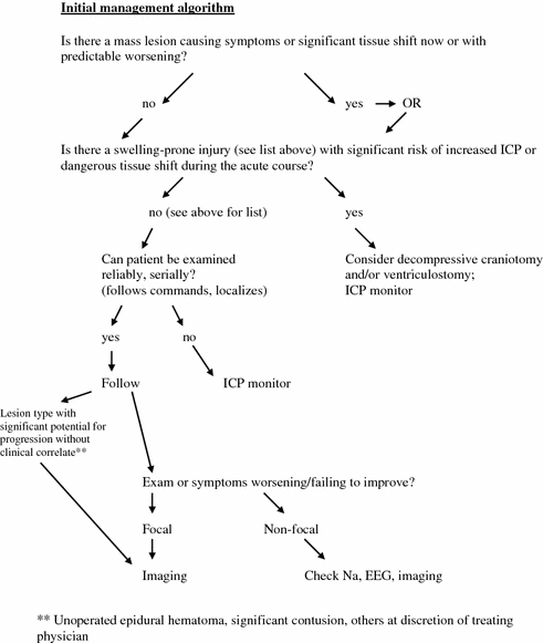
Fig. 29.1
Injury characterization is used in an algorithm designed to prevent specific types of complications, and to match the tools needed to monitor these changes with the specific clinical injury pattern. This approach was designed to be applicable to all ages of patients. Adapted from [1], with permission
Communication Among Team Members/Specialties and Families
The family of an injured child is a family in crisis, and often relatives are unable to fully process the initial information provided. Also, many parents are preoccupied with a sense of failed responsibility to protect their child, concern for additional injured family members, or with anger at others who they see as responsible for the injury.
In the author’s experience, it is helpful to sit down with the family after the initial assessment, along with the other care providers (e.g., trauma surgeon, orthopedic surgeon, emergency and/or critical care physician, social worker, etc.), to provide a simple overview of what the overall status is, who is who on the team and who is “in charge” of what issues, and what are the planned next steps in evaluation and management . It can be helpful to address immediately typical concerns like the child’s level of pain or anxiety, and what family members can do (or do not need to worry about doing) to be of help. Additionally, it can help to express explicitly that most families cannot fully remember or make sense of all they are told, and that the team expects them to have additional questions and will be available should questions or new information arise. Determining which family members will be the points of contact with the extended family can be useful at this initial meeting. Finally, it can be reassuring to explicitly plan a meeting for the next day, where the situation will be clearer, and family members will have a chance to ask more detailed questions.
Specific Injury Scenarios—Recognition, Acute Management , and Pitfalls
As there are a number of common scenarios involving specific mechanisms associated with pathoanatomic patterns of injury, we will outline some of the mechanism patterns and acute management concerns which may be helpful to recognize in children of different ages.
“Missile” motor vehicle occupants This injury scenario occurs when young children (infants , preschool, and early school age) are unrestrained inside a vehicle involved in a high-speed crash. Because they are mobile, they can become essentially airborne inside the vehicle, often moving head first, and can sustain major contact injuries to the head as it strikes an unyielding surface (Fig. 29.2). Because of significant scalp and skull injuries, the ability to expand the elastic scalp with subgaleal hematoma, and a relatively low blood volume at baseline, young children may present with shock just from skull and subgaleal hemorrhage, even without major scalp laceration or concomitant somatic injury. This problem is exacerbated if there are accompanying lacerations, long bone fractures, or visceral hemorrhages. Because children have robust cardiovascular compensatory abilities, at initial presentation they may appear relatively well from a hemodynamic point of view, but are prone to “crashing” into instability as blood loss reaches a critical threshold, taking the unwary clinician by surprise. This scenario can be complicated by the fact that a motor vehicle crash involving young children may be associated with multiple patient/occupant arrivals simultaneously, and so can predispose to distractions for the care team from an initially relatively well-appearing child . Thus, the management mantra in this setting is to pay careful attention to volume replacement early, typically with isotonic fluids, and to continue close and frequent monitoring during the acute postinjury period.

 Interventional Radiology in the Civilian Neurotrauma Setting
Communication Between Teams and Multidisciplinary Rounds and Single Primary POC for Family Communication—Lessons Learned and Who’s in Charge?
Coagulopathy in Traumatic Brain Injury
Nutrition, Antibiotics, and Post-traumatic Seizure Prophylaxis
Interventional Radiology in the Civilian Neurotrauma Setting
Communication Between Teams and Multidisciplinary Rounds and Single Primary POC for Family Communication—Lessons Learned and Who’s in Charge?
Coagulopathy in Traumatic Brain Injury
Nutrition, Antibiotics, and Post-traumatic Seizure Prophylaxis
 Mechanical Ventilation in Traumatic Brain Injury
 Craniofacial Reconstruction in the Polytrauma Patient


Related posts:
Interventional Radiology in the Civilian Neurotrauma Setting
Communication Between Teams and Multidisciplinary Rounds and Single Primary POC for Family Communication—Lessons Learned and Who’s in Charge?
Coagulopathy in Traumatic Brain Injury
Nutrition, Antibiotics, and Post-traumatic Seizure Prophylaxis
Mechanical Ventilation in Traumatic Brain Injury
Craniofacial Reconstruction in the Polytrauma Patient
Stay updated, free articles. Join our Telegram channel
Full access? Get Clinical Tree
