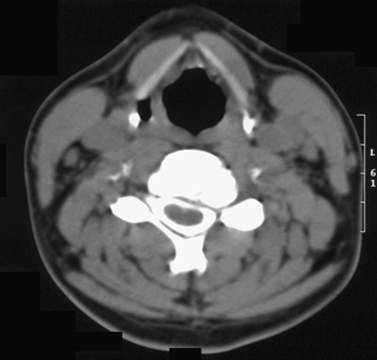
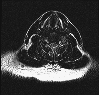
21 A 40-year-old woman presented with neck pain radiating down her left upper extremity. She had mild left biceps weakness with hyperreflexia. Axial computed tomography (CT) myelogram of the cervical spine (Fig. 21-1) shows a left lateral disc herniation with osteophytes at C5–C6. This is seen again on axial T2-weighted magnetic resonance imaging (MRI) (Fig. 21-2). This patient had multiple similar levels (C4–C7). FIGURE 21-1 Axial cut of postmyelogram CT demonstrates left eccentric foraminal compression. Cervical myeloradiculopathy with foraminal compression A three-level, left-sided, percutaneous microlaminotomy and foraminotomy procedure was done.
Percutaneous Cervical Decompression
Presentation
Radiologic Findings

Diagnosis
Treatment

Percutaneous Cervical Decompression
Only gold members can continue reading. Log In or Register to continue

Full access? Get Clinical Tree


