Peripheral nervous system
The peripheral nervous system consists of nerve endings, peripheral nerve trunks, plexuses and ganglia, which link the CNS with other parts of the body. Most of the neurones in the peripheral nervous system are, therefore, either afferent or efferent with respect to the CNS.
Muscle
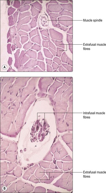
All behaviour depends on the ability to control the activity of skeletal muscles, which maintain posture and permit movement. Such control is subserved by a rich innervation of muscle with both motor and sensory neurones. Individual muscle cells (fibres) run parallel to the main axis of the muscle and fall into two main functional groups, namely extrafusal and intrafusal (Fig. 3.1).
Extrafusal muscle cells are by far the more numerous, constituting the bulk of the muscle and conferring its contractile strength. Extrafusal muscle fibres are innervated by alpha motor neurones, the cell bodies of which lie in the ventral horn of the spinal cord grey matter and in the motor cranial nerve nuclei of the brainstem. The axon of an individual alpha motor neurone typically branches within the target muscle to innervate a number of muscle fibres; the combination of a single motor neurone and the muscle fibres that it innervates is known as a motor unit. Motor units consist of relatively small numbers of muscle fibres in those muscles with which delicate, precise movements can be made, such as the muscles of the hand and the extraocular muscles. In contrast, the motor units of large postural muscles – such as the quadriceps, for example – are comprised of relatively larger numbers of muscle cells. Intrafusal muscle fibres are highly specialised cells that act as sensory receptors. They occur in groups known as muscle spindles. Intrafusal muscle fibres bear sensory endings that signal muscle stretch and tension to the CNS. They also receive a motor innervation from gamma motor neurones, whose cells of origin, like those of alpha motor neurones, lie in the ventral horn of the spinal cord and the motor cranial nerve nuclei of the brainstem. Gamma motor neurones function to control the sensitivity of the sensory endings on intrafusal muscle fibres. Intrafusal muscle fibres and their gamma motor neurone innervation are crucially important in mediation of the stretch reflex and, thus, in the control of muscle tone (pp 75-77).
Nerve endings
There are various, overlapping, conventions for the classification of nerve endings. Overall, they may be classified as either afferent or efferent. Afferent nerve endings respond to mechanical, thermal or chemical stimulation (mechanoreceptors, thermoreceptors or chemoreceptors, respectively). The nerve fibres to which they belong conduct action potentials to the CNS. If the afferent information reaches a conscious level, then the pathway is termed sensory. Efferent (effector) nerve endings innervate muscle or secretory cells and, under control from the CNS, influence muscular contraction or cellular secretion. Nerve endings that induce movement are called motor; those that induce secretion are sometimes called secretomotor.
Afferent nerve endings
The sensory systems, or modalities, are broadly divided into the general and the special senses. The special senses are olfaction, vision, hearing, balance and taste and these are considered elsewhere.
Functionally, there are three types of general sensory ending:
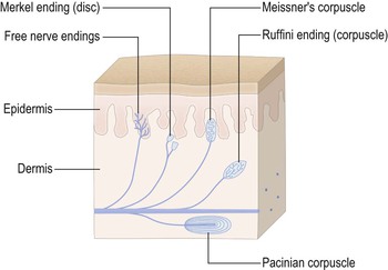
Structurally, sensory nerve endings may be either unencapsulated or encapsulated (Fig. 3.3). Unencapsulated endings, or free nerve endings, consist of the terminal branches of sensory nerve fibres lying freely in the innervated tissue. These are the most abundant type of sensory ending, occurring widely in the integument and also within muscles, joints, viscera and other structures. In the skin, they mediate thermal and painful sensations. The parent nerve fibres are designated physiologically as group Aδ (III) finely myelinated and group C unmyelinated fibres. These are of relatively small diameter and slow conducting. Merkel endings (discs) are located near the border of the epidermis. These are slowly adapting receptors that respond to touch/pressure. The axons of origin are large and myelinated.

Encapsulated nerve endings are surrounded by a structural specialisation of non-neural tissue, the combination of nerve and its encapsulation often being referred to as a corpuscle. Meissner’s corpuscles occur in the dermal papillae of the skin and are especially numerous in the fingertips. They respond with great sensitivity to touch. They are rapidly adapting receptors and are responsible for fine, or discriminative, touch. Pacinian corpuscles occur in skin and in deep tissues, e.g. surrounding joints and in mesentery. The largest are a few millimetres long and they are associated with group Aα (I) large diameter, myelinated axons. Pacinian corpuscles are rapidly adapting and respond to mechanical distortion, especially vibration. Ruffini endings (corpuscles) are slowly adapting mechanoreceptors occurring in the dermis of the skin.
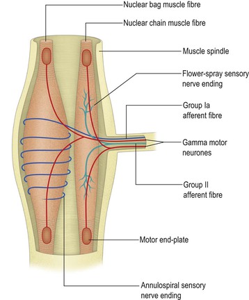
Within skeletal muscles, intrafusal muscle fibres, which in small groups constitute muscle spindles, act as stretch receptors (Fig. 3.4). There are two types of intrafusal muscle fibre, referred to as nuclear bag and nuclear chain fibres. Intrafusal muscle fibres bear two types of sensory ending, which become activated when the muscle in which they lie is stretched. Annulospiral endings (Fig. 3.5) are associated with fast conducting group Ia afferent fibres and flower-spray endings are associated with slower conducting group II afferents. Muscle spindles are particularly abundant in muscles capable of fine, skilled movements. They are important in kinaesthesia and in the control of muscle tone, posture and movement. Their functional importance in motor control is considered in more detail in Chapter 8.

< div class='tao-gold-member'>
Stay updated, free articles. Join our Telegram channel

Full access? Get Clinical Tree


